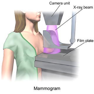
Mammography is the process of using low-energy X-rays to examine the human breast for diagnosis and screening. The goal of mammography is the early detection of breast cancer, typically through detection of characteristic masses or microcalcifications.
The American College of Radiology (ACR), founded in 1923, is a professional medical society representing nearly 40,000 diagnostic radiologists, radiation oncologists, interventional radiologists, nuclear medicine physicians and medical physicists.

Breast cancer screening is the medical screening of asymptomatic, apparently healthy women for breast cancer in an attempt to achieve an earlier diagnosis. The assumption is that early detection will improve outcomes. A number of screening tests have been employed, including clinical and self breast exams, mammography, genetic screening, ultrasound, and magnetic resonance imaging.

Molecular breast imaging (MBI), also known as scintimammography, is a type of breast imaging test that is used to detect cancer cells in breast tissue of individuals who have had abnormal mammograms, especially for those who have dense breast tissue, post-operative scar tissue or breast implants.

Tomosynthesis, also digital tomosynthesis (DTS), is a method for performing high-resolution limited-angle tomography at radiation dose levels comparable with projectional radiography. It has been studied for a variety of clinical applications, including vascular imaging, dental imaging, orthopedic imaging, mammographic imaging, musculoskeletal imaging, and chest imaging.
Daniel B. Kopans, MD, FACR is a radiologist specializing in mammography and other forms of breast imaging.
Thomas M. Kolb, M.D., is an American radiologist specializing in the detection and diagnosis of breast cancer in young, predominantly high-risk premenopausal women. He has served as an assistant clinical professor of Radiology at Columbia University College of Physicians and Surgeons from 1994–2010. Dr. Kolb is double board certified, having received his training in pediatrics at the Albert Einstein College of Medicine in Bronx, New York, and in diagnostic radiology at the Columbia-Presbyterian Medical Center in New York.

Radiologists without Borders is a 401(c)(3) non-profit organization that delivers humanitarian aid to developing countries, in the form of radiological services and equipment. Radiologists without Borders was founded in 2008, by New York-based radiologist Tariq Gill. The organization is composed entirely of volunteers, and its mission statement is "to bring life saving diagnostic imaging solutions to medically underserved populations worldwide."
Automated whole-breast ultrasound (AWBU) is a medical imaging technique used in radiology to obtain volumetric ultrasound data of the entire breast.

H. Gilbert Welch is an American academic physician and cancer researcher. He was an internist at the Veterans Administration Medical Center in White River Junction, Vermont, as well as a professor of medicine at the Dartmouth Institute for Health Policy and Clinical Practice. In September 2018, Welch resigned from Dartmouth College after a 20-month long research misconduct investigation at Dartmouth concluded he had committed plagiarism.
The Canadian National Breast Screening Study, sometimes abbreviated as CNBSS or NBSS, was a randomized trial conducted with the aim of evaluating whether mammography reduced breast cancer incidence or mortality among women who underwent screening. The trial was initiated in 1980, and was conducted in fifteen screening centers in six different Canadian provinces. It was the first study designed to determine whether mammography was effective among women between the ages of 40 and 49.

In medicine, breast imaging is a sub-speciality of diagnostic radiology that involves imaging of the breasts for screening or diagnostic purposes. There are various methods of breast imaging using a variety of technologies as described in detail below. Traditional screening and diagnostic mammography uses x-ray technology and has been the mainstay of breast imaging for many decades. Breast tomosynthesis is a relatively new digital x-ray mammography technique that produces multiple image slices of the breast similar to, but distinct from, computed tomography (CT). Xeromammography and galactography are somewhat outdated technologies that also use x-ray technology and are now used infrequently in the detection of breast cancer. Breast ultrasound is another technology employed in diagnosis and screening that can help differentiate between fluid filled and solid lesions, an important factor to determine if a lesion may be cancerous. Breast MRI is a technology typically reserved for high-risk patients and patients recently diagnosed with breast cancer. Lastly, scintimammography is used in a subgroup of patients who have abnormal mammograms or whose screening is not reliable on the basis of using traditional mammography or ultrasound.
Marjorie Clare Dalgarno (1901–1983) was an Australian radiologist and a pioneer of mammography. She performed the first mammogram in Australia at the Rachel Forster Hospital and demonstrated the benefits of mammography as a breast cancer screening tool.
Nola M. Hylton is an American oncologist who is Professor of Radiology and Director of the Breast Imaging Research Group at the University of California, San Francisco. She pioneered the usage of magnetic resonance imaging for the detection, diagnosis, and staging of breast cancer by using MRIs to locate tumors and characterize the surrounding tissue.
HB 2102, also known as "Henda's Law", is a breast density (BD) notification law approved in 2011 by the FDA that mammography patients be provided educational materials on dense breast tissue can hide abnormalities, including breast cancer, from traditional screening. Henda's Law aims to promote patient doctor discussion as well as reduce the rate of false negatives, as mammography may not detect abnormalities in dense breasts.
Vicky Goh is a professor, chair of clinical cancer imaging, and head of cancer imaging department at the King's College London, England, United Kingdom. She joined King's College London in 2011. She is also a consultant radiologist at Guy's and St Thomas' Hospital in London.
Etta Driscoll Pisano is an American breast imaging researcher. She is a professor in residence of radiology at the Beth Israel Deaconess Medical Center and chief research dean at the American College of Radiology. In 2008, she was elected a member of the National Academy of Medicine.

Henrietta Elizabeth Banting or “Lady Banting” was a Canadian physician and the second wife of Sir Frederick Banting. Banting was the Director of Women's College Hospital's Cancer Detection Clinic from 1958-1971. While working at the Cancer Detection Clinic, she conducted a research study on mammography to measure its effectiveness as a diagnostic tool for breast cancer.
Elizabeth Margaret Forbes was a Canadian radiologist. Forbes was the Chief of Radiology at Toronto's Women's College Hospital (WCH) from 1955 to 1975. She is remembered for co-authoring “one of the first Canadian papers on mammography” with WCH's Henrietta Banting.
Dense breast tissue, also known as dense breasts, is a condition of the breasts where a higher proportion of the breasts are made up of glandular tissue and fibrous tissue than fatty tissue. Around 40–50% of women have dense breast tissue and one of the main medical components of the condition is that mammograms are unable to differentiate tumorous tissue from the surrounding dense tissue. This increases the risk of late diagnosis of breast cancer in women with dense breast tissue. Additionally, women with such tissue have a higher likelihood of developing breast cancer in general, though the reasons for this are poorly understood.


