Related Research Articles

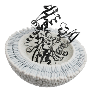
A biological membrane, biomembrane or cell membrane is a selectively permeable membrane that separates the interior of a cell from the external environment or creates intracellular compartments by serving as a boundary between one part of the cell and another. Biological membranes, in the form of eukaryotic cell membranes, consist of a phospholipid bilayer with embedded, integral and peripheral proteins used in communication and transportation of chemicals and ions. The bulk of lipids in a cell membrane provides a fluid matrix for proteins to rotate and laterally diffuse for physiological functioning. Proteins are adapted to high membrane fluidity environment of the lipid bilayer with the presence of an annular lipid shell, consisting of lipid molecules bound tightly to the surface of integral membrane proteins. The cell membranes are different from the isolating tissues formed by layers of cells, such as mucous membranes, basement membranes, and serous membranes.

Phospholipids are a class of lipids whose molecule has a hydrophilic "head" containing a phosphate group and two hydrophobic "tails" derived from fatty acids, joined by an alcohol residue. Marine phospholipids typically have omega-3 fatty acids EPA and DHA integrated as part of the phospholipid molecule. The phosphate group can be modified with simple organic molecules such as choline, ethanolamine or serine.

The fluid mosaic model explains various characteristics regarding the structure of functional cell membranes. According to this biological model, there is a lipid bilayer in which protein molecules are embedded. The phospholipid bilayer gives fluidity and elasticity to the membrane. Small amounts of carbohydrates are also found in the cell membrane. The biological model, which was devised by Seymour Jonathan Singer and Garth L. Nicolson in 1972, describes the cell membrane as a two-dimensional liquid that restricts the lateral diffusion of membrane components. Such domains are defined by the existence of regions within the membrane with special lipid and protein cocoon that promote the formation of lipid rafts or protein and glycoprotein complexes. Another way to define membrane domains is the association of the lipid membrane with the cytoskeleton filaments and the extracellular matrix through membrane proteins. The current model describes important features relevant to many cellular processes, including: cell-cell signaling, apoptosis, cell division, membrane budding, and cell fusion. The fluid mosaic model is the most acceptable model of the plasma membrane. In this definition of the cell membrane, its main function is to act as a barrier between the contents inside the cell and the extracellular environment.
Mycoplasma pneumoniae is a very small cell wall-less bacterium in the class Mollicutes. It is a human pathogen that causes the disease mycoplasma pneumonia, a form of atypical bacterial pneumonia related to cold agglutinin disease. M. pneumoniae is characterized by the absence of a peptidoglycan cell wall and resulting resistance to many antibacterial agents. The persistence of M. pneumoniae infections even after treatment is associated with its ability to mimic host cell surface composition.

The plasma membranes of cells contain combinations of glycosphingolipids, cholesterol and protein receptors organised in glycolipoprotein lipid microdomains termed lipid rafts. Their existence in cellular membranes remains controversial. Indeed, Kervin and Overduin imply that lipid rafts are misconstrued protein islands, which they propose form through a proteolipid code. Nonetheless, it has been proposed that they are specialized membrane microdomains which compartmentalize cellular processes by serving as organising centers for the assembly of signaling molecules, allowing a closer interaction of protein receptors and their effectors to promote kinetically favorable interactions necessary for the signal transduction. Lipid rafts influence membrane fluidity and membrane protein trafficking, thereby regulating neurotransmission and receptor trafficking. Lipid rafts are more ordered and tightly packed than the surrounding bilayer, but float freely within the membrane bilayer. Although more common in the cell membrane, lipid rafts have also been reported in other parts of the cell, such as the Golgi apparatus and lysosomes.
Biomedicine is a branch of medical science that applies biological and physiological principles to clinical practice. Biomedicine stresses standardized, evidence-based treatment validated through biological research, with treatment administered via formally trained doctors, nurses, and other such licensed practitioners.
The Davson–Danielli model was a model of the plasma membrane of a cell, proposed in 1935 by Hugh Davson and James Danielli. The model describes a phospholipid bilayer that lies between two layers of globular proteins, which is both trilaminar and lipoprotinious. The phospholipid bilayer had already been proposed by Gorter and Grendel in 1925; however, the flanking proteinaceous layers in the Davson–Danielli model were novel and intended to explain Danielli's observations on the surface tension of lipid bi-layers.
Seymour Jonathan Singer was an American cell biologist and professor of biology, emeritus, at the University of California, San Diego.
Sulfatide, also known as 3-O-sulfogalactosylceramide, SM4, or sulfated galactocerebroside, is a class of sulfolipids, specifically a class of sulfoglycolipids, which are glycolipids that contain a sulfate group. Sulfatide is synthesized primarily starting in the endoplasmic reticulum and ending in the Golgi apparatus where ceramide is converted to galactocerebroside and later sulfated to make sulfatide. Of all of the galactolipids that are found in the myelin sheath, one fifth of them are sulfatide. Sulfatide is primarily found on the extracellular leaflet of the myelin plasma membrane produced by the oligodendrocytes in the central nervous system and in the Schwann cells in the peripheral nervous system. However, sulfatide is also present on the extracellular leaflet of the plasma membrane of many cells in eukaryotic organisms.

Molecular biophysics is a rapidly evolving interdisciplinary area of research that combines concepts in physics, chemistry, engineering, mathematics and biology. It seeks to understand biomolecular systems and explain biological function in terms of molecular structure, structural organization, and dynamic behaviour at various levels of complexity. This discipline covers topics such as the measurement of molecular forces, molecular associations, allosteric interactions, Brownian motion, and cable theory. Additional areas of study can be found on Outline of Biophysics. The discipline has required development of specialized equipment and procedures capable of imaging and manipulating minute living structures, as well as novel experimental approaches.
Exosomes, ranging in size from 30 to 150 nanometers, are membrane-bound extracellular vesicles (EVs) that are produced in the endosomal compartment of most eukaryotic cells. In multicellular organisms, exosomes and other EVs are found in biological fluids including saliva, blood, urine and cerebrospinal fluid. EVs have specialized functions in physiological processes, from coagulation and waste management to intercellular communication.
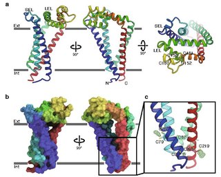
CD9 is a gene encoding a protein that is a member of the transmembrane 4 superfamily also known as the tetraspanin family. It is a cell surface glycoprotein that consists of four transmembrane regions and has two extracellular loops that contain disulfide bonds which are conserved throughout the tetraspanin family. Also containing distinct palmitoylation sites that allows CD9 to interact with lipids and other proteins.
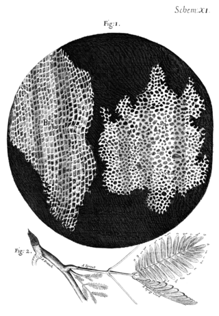
Cell theory has its origins in seventeenth century microscopy observations, but it was nearly two hundred years before a complete cell membrane theory was developed to explain what separates cells from the outside world. By the 19th century it was accepted that some form of semi-permeable barrier must exist around a cell. Studies of the action of anesthetic molecules led to the theory that this barrier might be made of some sort of fat (lipid), but the structure was still unknown. A series of pioneering experiments in 1925 indicated that this barrier membrane consisted of two molecular layers of lipids—a lipid bilayer. New tools over the next few decades confirmed this theory, but controversy remained regarding the role of proteins in the cell membrane. Eventually the fluid mosaic model was composed in which proteins “float” in a fluid lipid bilayer "sea". Although simplistic and incomplete, this model is still widely referenced today.

The cell membrane is a biological membrane that separates and protects the interior of a cell from the outside environment. The cell membrane consists of a lipid bilayer, made up of two layers of phospholipids with cholesterols interspersed between them, maintaining appropriate membrane fluidity at various temperatures. The membrane also contains membrane proteins, including integral proteins that span the membrane and serve as membrane transporters, and peripheral proteins that loosely attach to the outer (peripheral) side of the cell membrane, acting as enzymes to facilitate interaction with the cell's environment. Glycolipids embedded in the outer lipid layer serve a similar purpose. The cell membrane controls the movement of substances in and out of a cell, being selectively permeable to ions and organic molecules. In addition, cell membranes are involved in a variety of cellular processes such as cell adhesion, ion conductivity, and cell signalling and serve as the attachment surface for several extracellular structures, including the cell wall and the carbohydrate layer called the glycocalyx, as well as the intracellular network of protein fibers called the cytoskeleton. In the field of synthetic biology, cell membranes can be artificially reassembled.
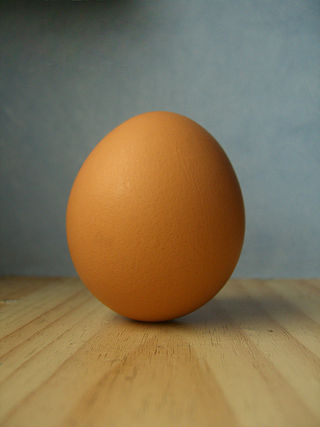
Chickens and their eggs have been used extensively as research models throughout the history of biology. Today they continue to serve as an important model for normal human biology as well as pathological disease processes.
Sarah Spiegel is professor and chair of the Department of Biochemistry and Molecular Biology at Virginia Commonwealth University (VCU). In the mid-1990s she discovered the sphingosine-1-phosphate (S1P) molecule, a lipid which has been identified as a signaler for the spread of cancer, inflammation, and cardiovascular disease. Her research continues to focus on S1P.
Before the emergence of electron microscopy in the 1950s, scientists did not know the structure of a cell membrane or what its components were; biologists and other researchers used indirect evidence to identify membranes before they could actually be visualized. Specifically, it was through the models of Overton, Langmuir, Gorter and Grendel, and Davson and Danielli, that it was deduced that membranes have lipids, proteins, and a bilayer. The advent of the electron microscope, the findings of J. David Robertson, the proposal of Singer and Nicolson, and additional work of Unwin and Henderson all contributed to the development of the modern membrane model. However, understanding of past membrane models elucidates present-day perception of membrane characteristics. Following intense experimental research, the membrane models of the preceding century gave way to the fluid mosaic model that is accepted today.
Kai Simons is a Finnish professor of biochemistry and cell biology and physician, living and working in Germany. He introduced the concept of lipid rafts, and coined the term trans-Golgi network. He is the co-founder and co-organizer of the European Molecular Biology Laboratory and European Molecular Biology Organization, and initiated the foundation of Max Planck Institute of Molecular Cell Biology and Genetics.

Anne Jacqueline Ridley is professor of Cell Biology and Head of School for Cellular and Molecular Medicine at the University of Bristol. She was previously a professor at King's College London.
John A. Hickman is a British-French cancer pharmacologist.
References
- 1 2 "Curriculum Vitae GARTH L. NICOLSON". The Institute for Molecular Medicine. February 20, 2010. Archived from the original on August 8, 2021. Retrieved August 6, 2014.
- 1 2 "Professor Emeritus Garth L. Nicolson" (PDF). The Institute for Molecular Medicine. Archived from the original (PDF) on January 23, 2019. Retrieved August 7, 2014.
- 1 2 Nicolson, Garth L; Nicolson, Nancy L. "About the authors". Project Day Lily. Archived from the original on August 12, 2014. Retrieved August 6, 2014.
- ↑ "IMM – Faculty and Associate Faculty". The Institute for Molecular Medicine. Retrieved August 7, 2014.
- 1 2 Singer, S. J.; Nicolson, G. L. (1972). "The Fluid Mosaic Model of the Structure of Cell Membranes". Science. 175 (4023): 720–731. Bibcode:1972Sci...175..720S. doi:10.1126/science.175.4023.720. JSTOR 1733071. PMID 4333397. S2CID 83851531.
- 1 2 Cherry, Richard (1991). New Techniques of Optical Microscopy and Microspectroscopy. Boca Raton, Florida: CRC Press, Inc. p. 199. ISBN 978-0-8493-7117-2. ISSN 0265-4377.
{{cite book}}:|journal=ignored (help) - 1 2 Luckey, Mary (2014). Membrane Structural Biology: With Biochemical and Biophysical Foundations (2 ed.). Cambridge (UK): Cambridge University Press. p. 6. ISBN 978-1-107-72933-9.
- 1 2 Jacobson, K; Sheets, E.; Simson, R (1995). "Revisiting the fluid mosaic model of membranes". Science. 268 (5216): 1441–1442. Bibcode:1995Sci...268.1441J. doi:10.1126/science.7770769. PMID 7770769.
- ↑ "Garth Nicolson Prof". iHealthTube. February 7, 2013. Retrieved August 6, 2014.
- ↑ "Cancer and Metastasis Reviews".
- ↑ Martin, Laura. "The Fluid Mosaic Model of the Cell Membrane – The Mosaic". oepnstax cnx. Retrieved August 7, 2014.
- ↑ Ash, Michael (August 28, 2012). "A Special Note from Garth Nicolson PhD Why Lipid Replacement Therapy(LRT®) is Key to our Health". Clinical Education. Nutri-Link Ltd. Retrieved August 7, 2014.
- ↑ Wiśniewska, A; Draus, J; Subczynski, WK (2003). "Is a fluid-mosaic model of biological membranes fully relevant? Studies on lipid organization in model and biological membranes". Cellular & Molecular Biology Letters. 8 (1): 147–159. PMID 12655369.
- ↑ Engelman, Donald M. (2005). "Membranes are more mosaic than fluid". Nature. 438 (7068): 578–580. Bibcode:2005Natur.438..578E. doi: 10.1038/nature04394 . PMID 16319876. S2CID 4416823.
- ↑ Kusumi, Akihiro; Fujiwara, Takahiro K.; Chadda, Rahul; Xie, Min; Tsunoyama, Taka A.; Kalay, Ziya; Kasai, Rinshi S.; Suzuki, Kenichi G.N. (2012). "Dynamic organizing principles of the plasma membrane that regulate signal transduction: commemorating the fortieth anniversary of Singer and Nicolson's fluid-mosaic model". Annual Review of Cell and Developmental Biology. 28 (1): 215–250. doi:10.1146/annurev-cellbio-100809-151736. PMID 22905956.
- ↑ Nicolson, Garth (2013). "Update of the 1972 Singer-Nicolson Fluid-Mosaic Model of Membrane Structure". Discoveries. 1 (1): e3. doi: 10.15190/d.2013.3 . PMC 7159824 . PMID 32309537.
- ↑ Nicolson, Garth L. (2014). "The Fluid—Mosaic Model of Membrane Structure: Still relevant to understanding the structure, function and dynamics of biological membranes after more than 40 years". Biochimica et Biophysica Acta (BBA) - Biomembranes. 1838 (6): 1451–1466. doi: 10.1016/j.bbamem.2013.10.019 . PMID 24189436.
- ↑ Nicolson, GL; Nicolson, NL (1997). "The eight myths of Operation 'Desert Storm' and Gulf War syndrome". Medicine, Conflict, and Survival. 13 (2): 140–6. doi:10.1080/13623699708409329. PMID 9178600.
- ↑ Nicolson, GL; Bruton DM, Jr; Nicolson, NL (1996). "Chronic fatigue illness and Operation Desert Storm". Journal of Occupational and Environmental Medicine. 38 (1): 14–6. doi: 10.1097/00043764-199601000-00003 . PMID 8871324.
- ↑ Nicolson, GL; Nicolson, NL (1997). "The eight myths of Operation 'Desert Storm' and Gulf War syndrome". Medicine, Conflict, and Survival. 13 (2): 140–146. doi:10.1080/13623699708409329. PMID 9178600.
- ↑ Blair, Mike (July 3, 1995). "The Government Is Lying To You -- And Ill Veterans -- About Gulf War Syndrome". The Spotlight. Archived from the original on February 24, 2016. Retrieved August 9, 2014.
- ↑ Nicolson, G (2001). "Continuing research into Gulf War illness". Science. 292 (5518): 853b–853. doi:10.1126/science.292.5518.853b. PMID 11341275. S2CID 19886150.
- ↑ Nicolson, Garth L (November 19, 1998). "WRITTEN TESTIMONY OF Dr. Garth L. Nicolson Special Oversight Board for Department of Defense Investigations of Gulf War Chemical and Biological Incidents". U. S. Senate Hart Office Building SH-216. Retrieved August 9, 2014.
- ↑ Gray, GC; Kaiser, KS; Hawksworth, AW; Watson, HL (1999). "No serologic evidence of an association found between Gulf War service and Mycoplasma fermentans infection". The American Journal of Tropical Medicine and Hygiene. 60 (5): 752–7. doi:10.4269/ajtmh.1999.60.752. PMID 10344648. S2CID 32534013.
- ↑ Lo, SC; Levin, L; Ribas, J; Chung, R; Wang, RY; Wear, D; Shih, JW (2000). "Lack of serological evidence for Mycoplasma fermentans infection in army Gulf War veterans: a large scale case-control study". Epidemiology and Infection. 125 (3): 609–16. doi:10.1017/s0950268800004891. PMC 2869645 . PMID 11218212.
- ↑ Nicolson, Garth L.; Nasralla, Marwan Y.; Haier, Joerg; Pomfret, John (2002). "High frequency of systemic mycoplasmal infections in Gulf War veterans and civilians with Amyotrophic Lateral Sclerosis (ALS)". Journal of Clinical Neuroscience. 9 (5): 525–529. doi:10.1054/jocn.2001.1075. PMID 12383408. S2CID 24870742.