Related Research Articles

In anatomy, the temporomandibular joints (TMJ) are the two joints connecting the jawbone to the skull. It is a bilateral synovial articulation between the temporal bone of the skull above and the condylar process of mandible below; it is from these bones that its name is derived. The joints are unique in their bilateral function, being connected via the mandible.

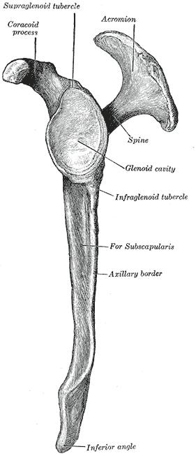
The scapula, also known as the shoulder blade, is the bone that connects the humerus with the clavicle. Like their connected bones, the scapulae are paired, with each scapula on either side of the body being roughly a mirror image of the other. The name derives from the Classical Latin word for trowel or small shovel, which it was thought to resemble.

The coracoid process is a small hook-like structure on the lateral edge of the superior anterior portion of the scapula. Pointing laterally forward, it, together with the acromion, serves to stabilize the shoulder joint. It is palpable in the deltopectoral groove between the deltoid and pectoralis major muscles.

The human shoulder is made up of three bones: the clavicle (collarbone), the scapula, and the humerus as well as associated muscles, ligaments and tendons.

The nasal cavity is a large, air-filled space above and behind the nose in the middle of the face. The nasal septum divides the cavity into two cavities, also known as fossae. Each cavity is the continuation of one of the two nostrils. The nasal cavity is the uppermost part of the respiratory system and provides the nasal passage for inhaled air from the nostrils to the nasopharynx and rest of the respiratory tract.

Plesiosaurus is a genus of extinct, large marine sauropterygian reptile that lived during the Early Jurassic. It is known by nearly complete skeletons from the Lias of England. It is distinguishable by its small head, long and slender neck, broad turtle-like body, a short tail, and two pairs of large, elongated paddles. It lends its name to the order Plesiosauria, of which it is an early, but fairly typical member. It contains only one species, the type, Plesiosaurus dolichodeirus. Other species once assigned to this genus, including P. brachypterygius, P. guilielmiimperatoris, and P. tournemirensis have been reassigned to new genera, such as Hydrorion, Seeleyosaurus and Occitanosaurus.
The shoulder girdle or pectoral girdle is the set of bones in the appendicular skeleton which connects to the arm on each side. In humans, it consists of the clavicle and scapula; in those species with three bones in the shoulder, it consists of the clavicle, scapula, and coracoid. Some mammalian species have only the scapula.

The glenoid fossa of the scapula or the glenoid cavity is a bone part of the shoulder. The word glenoid is pronounced or and is from Greek: gléne, "socket", reflecting the shoulder joint's ball-and-socket form. It is a shallow, pyriform articular surface, which is located on the lateral angle of the scapula. It is directed laterally and forward and articulates with the head of the humerus; it is broader below than above and its vertical diameter is the longest.
The inferior transverse ligament is a weak membranous band, situated behind the neck of the scapula and stretching from the lateral border of the spine to the margin of the glenoid cavity.

The hepatogastric ligament or gastrohepatic ligament connects the liver to the lesser curvature of the stomach. It contains the right and the left gastric arteries. In the abdominal cavity, it separates the greater and lesser sacs on the right. It is sometimes cut during surgery in order to access the lesser sac. The hepatogastric ligament consists of a dense cranial portion and the caudal portion termed the pars flaccida.
Shoulder surgery is a means of treating injured shoulders. Many surgeries have been developed to repair the muscles, connective tissue, or damaged joints that can arise from traumatic or overuse injuries to the shoulder.

The muscles of respiration are the muscles that contribute to inhalation and exhalation, by aiding in the expansion and contraction of the thoracic cavity. The diaphragm and, to a lesser extent, the intercostal muscles drive respiration during quiet breathing. The elasticity of these muscles is crucial to the health of the respiratory system and to maximize its functional capabilities.

The skeletal system of the horse is a skeletal system of a horse that has three major functions in the body. It protects vital organs, provides framework, and supports soft parts of the body. Horses typically have 205 bones. The pelvic limb typically contains 19 bones, while the thoracic limb contains 20 bones.

A humerus fracture is a break of the humerus bone in the upper arm. Symptoms may include pain, swelling, and bruising. There may be a decreased ability to move the arm and the person may present holding their elbow. Complications may include injury to an artery or nerve, and compartment syndrome.

Wadiasaurus is an extinct genus of dicynodont from the family Kannemeyeria, that lived in herds from the early to Middle Triassic. Substantial fossorial evidence of W. indicus was recovered from Yerrapalli Formation of the Pranhita-Godavari valley, India, and it is so far the only Kannemeyeriid known for certain from India. The Kannemeyeriiformes underwent a significant diversification during the middle Triassic, with roughly 40 known species distributed worldwide. All Kannemeyeriiformes were medium to large bodied, graviportal herbivores with relatively erect posture and gait. Wadiasaurus indicus is currently the only known species of Wadiasaurus.
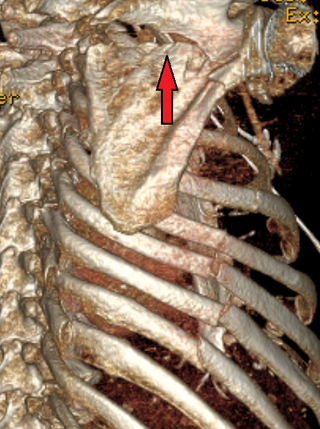
A scapular fracture is a fracture of the scapula, the shoulder blade. The scapula is sturdy and located in a protected place, so it rarely breaks. When it does, it is an indication that the individual was subjected to a considerable amount of force and that severe chest trauma may be present. High-speed vehicle accidents are the most common cause. This could be anywhere from a car accident, motorcycle crash, or high speed bicycle crash but falls and blows to the area can also be responsible for the injury. Signs and symptoms are similar to those of other fractures: they include pain, tenderness, and reduced motion of the affected area although symptoms can take a couple of days to appear. Imaging techniques such as X-ray are used to diagnose scapular fracture, but the injury may not be noticed in part because it is so frequently accompanied by other, severe injuries that demand attention. The injuries that usually accompany scapular fracture generally have the greatest impact on the patient's outcome. However, the injury can also occur by itself; when it does, it does not present a significant threat to life. Treatment involves pain control and immobilizing the affected area, and, later, physical therapy.

Mandibular fracture, also known as fracture of the jaw, is a break through the mandibular bone. In about 60% of cases the break occurs in two places. It may result in a decreased ability to fully open the mouth. Often the teeth will not feel properly aligned or there may be bleeding of the gums. Mandibular fractures occur most commonly among males in their 30s.

Ahshislepelta is a monospecific genus of ankylosaur dinosaur from New Mexico that lived during the Late Cretaceous in what is now the Hunter Wash Member of the Kirtland Formation. The type and only species, Ahshislepelta minor, is known only from an incomplete postcranial skeleton of a small subadult or adult individual. It was named in 2011 by Michael Burns and Robert M. Sullivan. Based on the size of the humerus, Ahshislepelta is larger than Pinacosaurus mephistocephalus but smaller than Talarurus and Pinacosaurus grangeri.
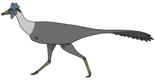
Bonapartenykus is a monospecific genus of alvarezsauroid dinosaur from Argentina that lived during the Late Cretaceous (Campanian-Maastrichtian) in what is now the upper Allen Formation of the Río Negro Province. The type and only species, Bonapartenykus ultimus, is known from a nearly articulated but partial skeleton that was found in close association to two incomplete eggs and several clusters of eggshells belonging to the oogenus Arriagadoolithus. Bonapartenykus was named in 2012 by Federico L. Agnolin, Jaime E. Powell, Fernando E. Novas and Martin Kundrát. Bonapartenykus has an estimated length of 2.5 m (8.2 ft) and weight of 72 kg (159 lb), making it the largest member of the clade Alvarezsauroidea.
Bone malrotation refers to the situation that results when a bone heals out of rotational alignment from another bone, or part of bone. It often occurs as the result of a surgical complication after a fracture where intramedullary nailing (IMN) occurs, especially in the femur and tibial bones, but can also occur genetically at birth. The severity of this complication is often neglected due to its complexity to detect and treat, yet if left untreated, bone malrotation can significantly impact regular bodily functioning, and even lead to severe arthritis. Detection throughout history has become more advanced and accurate, ranging from clinical assessment to ultrasounds to CT scans. Treatment can include an osteotomy, a major surgical procedure where bones are cut and realigned correctly, or compensatory methods, where individuals learn to externally or internally rotate their limb to compensate for the rotation. Further research is currently being examined in this area to reduce occurrences of malrotation, including detailed computer navigation to improve visual accuracy during surgery.
References
- Bestard, E.A. et al. (1986). "Glenoplasty in the management of recurrent shoulder dislocations". Contemporary Orthopaedics12, 47.
- Romero, J. et al. (2001). "Scapular neck fracture - the influence of permanent malalignment of the glenoid neck on clinical outcome". Archives of Orthopaedic and Trauma Surgery121, 313–316.
| | This human musculoskeletal system article is a stub. You can help Wikipedia by expanding it. |
| | This standards- or measurement-related article is a stub. You can help Wikipedia by expanding it. |