Histopathologic diagnosis of dermatitis

Last updated

Histopathology of dermatitis can be performed in uncertain cases of inflammatory skin condition that remain uncertain after history and physical examination. [1]

Contents

Sampling

Generally a skin biopsy:

A superficial or shave biopsy is regarded as insufficient. [2]

Fixation

Staining

Generally 3 sections for H&E staining and one section with periodic acid Schiff (PAS) [notes 1] [2]

Microscopic evaluation

One approach is to classify into mainly either of the following, primarily based on depth of involvement: [2]

  • Vesiculobullous lesions
  • Pustular dermatosis
  • Non vesicullobullous, non-pustular
  • With epidermal changes
  • Without epidermal changes. These characteristically have a superficial perivascular inflammatory infiltrate, and can be classified by type of cell infiltrate: [2]
  • Lymphocytic (most common)
  • Lymphoeosinophilic
  • Lymphoplasmacytic
  • Mast cell
  • Lymphohistiocytic
  • Neutrophilic

Continue in corresponding section:

Non vesicullobullous, non-pustular lesions with epidermal changes

Spongiotic dermatitis

It is characterized by epithelial intercellular edema. [2]

CharacteristicsMicrographPhotograph
AcuteSubacuteChronic
Generally/Not otherwise specified [notes 2] Typical findings: [2]
  • Variable degree of epidermal spongiosis and vesicle formation, filled with proteinaceous fluid containing lymphocytes and histiocytes.
  • Usually superficial dermal edema with perivascular lymphocytic infiltrate, with exocytosis.
  • No acanthosis or parakeratosis.
Typical findings: [2]
  • Mild to moderate spongiosis and exocytosis of inflammatory cells
  • Irregular acanthosis and parakeratosis.
  • Superficial dermal perivascular lymphohistiocytic infiltrate
  • Swelling of endothelial cells
  • Papillary dermal edema are present
Typical findings: [2]
  • The spongiosis is mild to absent
  • Pronounced irregular acanthosis, hyperkeratosis, and parakeratosis
  • Minimal dermal inflammation and exocytosis of inflammatory cells are present.
  • Possibly fibrosis of papillary dermis

PAS stain is essential to exclude fungal infection. [2]

Micrograph of subacute spongiotic dermatitis.jpg Subacute
Allergic/contact dermatitis or atopic dermatitisAs above. Eosinophils may be present in the dermis and epidermis (eosinophilic spongiosis). [2] Spongiotic dermatitis from drug allergy.jpg Allergic dermatitis Eczema (14100950936).jpg Atopic dermatitis
Seborrheic dermatitisTypical findings: [5]
  • Focal, usually mild, spongiosis with overlying scale crust, with a few neutrophils
  • The crust is often centered on a follicle
  • The papillary dermis is generally mildly edematous
  • Dilated blood vessels in the superficial vascular plexus
  • Mild superficial perivascular infiltrate of lymphocytes, histiocytes and occasional neutrophils. There is some exocytosis of inflammatory cells but not as prominent as in nummular dermatitis
Typical findings: [5]
  • Psoriasiform hyperplasia, initially slight, with mild spongiosis
  • Usually numerous yeast-like organisms in the surface keratin
  • Same changes as seen in acute stage.
Typical findings: [5]
  • More pronounced psoriasiform hyperplasia
  • Only minimal spongiosis
  • Presence of scaling crusts in a folliculocentric distribution, distinguishes from psoriasis.
Seborrhoeic dermatitis example.jpg

In addition to above, an unspecific spongiotic dermatitis can be consistent with nummular dermatitis, dyshidrotic dermatitis, Id reaction, dermatophytosis, miliaria, Gianotti-Crosti syndrome and pityriasis rosea. [2] [notes 2]

Interface dermatitis

These are sorted into either: [2]

  • Interface dermatitis with vacuolar change
  • Interface dermatitis with lichenoid inflammation
Interface dermatitis with vacuolar change
Causes of vacuolar interface dermatitis edit
Main conditions [6] CharacteristicsMicrographPhotograph
Generally/Not otherwise specifiedTypical findings, called "vacuolar interface dermatitis": [6]
  • Mild inflammatory cell infiltrate along the dermoepidermal junction (black arrow in image)
  • Vacuolization within the basal keratinocytes (white arrow in image)
  • Often necrotic, predominantly basal, individual keratinocytes, manifesting as colloid or Civatte bodies
Vacuolar interface dermatitis, annotated.jpg
Acute graft-versus-host-disease
  • Vacuolar alteration of various severity, from focal or diffuse vacuolation of the basal keratinocytes (grade I), to separation at the dermoepidermal junction (grade III)
  • Involvement of the hair follicle [6]
  • Rarely eosinophils [6]
Micrographs of grades of skin graft-versus-host-disease.jpg
Allergic drug reaction
  • Rarely involvement of hair follicles. [6]
  • Frequently eosinophils [6]
Spongiotic dermatitis from drug allergy.jpg
Lichen sclerosus Hyperkeratosis, atrophic epidermis, sclerosis of dermis and dermal lymphocytes. [7] Micrograph of lichen sclerosus.jpg
Erythema multiforme
Lupus erythematosisTypical findings in systemic lupus erythematosus: [8]
  • Fibrinoid necrosis at the dermoepidermal junction
  • Liquefactive degeneration and atrophy of the epidermis
  • Mucin deposition in the reticular dermis
  • Edema, small hemorrhages
  • Mild and mainly lymphocytic infiltrate in the upper dermis
  • Fibrinoid material in the dermis around capillary blood vessels, on collagen and in the interstitium
  • In non-bullous cases, perivascular and interstitial neutrophils are sometimes present in the upper dermis, with damage to blood vessels
Histopathology of systemic lupus erythematosus.jpg Butterfly rash of lupus erythematosus.jpg

An interface dermatitis with vacuolar alteration, not otherwise specified, may be caused by viral exanthems, phototoxic dermatitis, acute radiation dermatitis, erythema dyschromicum perstans, lupus erythematosus and dermatomyositis. [2]

Interface dermatitis with lichenoid inflammation
Main conditions [2] CharacteristicsMicrographPhotograph
Generally/Not otherwise specifiedTypical findings: [2]
  • In the papillary dermis: a confluent, band-like, dense inflammation of mainly small lymphocytes and a few histiocytes, along or hugging the dermoepidermal junction.
  • Often vacuolar degeneration of basal keratinocytes and apoptotic bodies (colloid or Civatte bodies).
Lichen planusIrregular epidermal hyperplasia with a jagged “sawtooth” appearance, compact hyperkeratosis or orthokeratosis, foci of wedge-shaped hypergranulosis, basilar vacuolar degeneration, slight spongiosis in the spinous layer, and squamatization. The dermal papillae between the elongated rete ridges are frequently dome shaped. Necrotic keratinocytes can be observed in the basal layer of the epidermis and at the dermal-epidermal junction. Eosinophilic remnants of anucleate apoptotic basal cells may also be found in the dermis and are referred to as “colloid or civatte bodies”. Whickham striae are usually seen in the areas of hypergranulosis. Vacuolar degeneration at the basal layer may be noted leading to focal subepidermal clefts (Max Joseph spaces). Squamatization occurs as a result of maturation and flattening of cells in the basal layer. It happens in areas of marked hypergranulosis with prominence of the sawtooth pattern of rete ridges. Wedge-shaped hypergranulosis can occur in the eccrine ducts (acrosyringia) or hair follicles (acrotrichia). In the hypertrophic subtype, the associated hyperkeratosis, parakeratosis, hypergranulosis, papillomatosis, acanthosis, and hyperplasia markedly increased with thicker collagen bundles forming in the dermis. Moreover, the rete ridges are more elongated and rounded as opposed to the typical sawtooth pattern. In atrophic LP, loss of the rete ridges and dermal fibrosis is prominent. In vesiculobullous LP, the disease progression is quicker. Hence, some of the distinctive features such as hyperkeratosis, hypergranulosis, or dense lymphocytic dermal-epidermal infiltrate may not be present. LP lesion may resolve with residual hyperpigmentation caused by a persistent increase in the number of melanophages in the papillary dermis. [9] Histopathology of lichen planus.jpg Lichen planus 1.jpg
Lichenoid drug reaction

Can virtually be indistinguishable from cutaneous LP both clinically and histopathologically.

  • Typically, lesions have a photodistribution in the absence of oral mucosal involvement. [9]
  • Characteristically parakeratosis, a dermal eosinophilic infiltrate, and a perivascular lymphocytic infiltrate affecting the reticular dermis.
  • Epidermal changes are less common in lichenoid drug eruptions when compared to classic lichen planus. However, a higher concentration of necrotic keratinocyte and eosinophils in the infiltrate can be helpful in distinguishing lichenoid drug reaction from cutaneous lichen planus. A lengthy interval between the commencement of drug therapy and the onset of lesions does not exclude a diagnosis of lichenoid drug reaction. Resolution of the lesions often occurs within weeks to months after discontinuation of the offending drug. [9]
Histopathology of lichenoid drug reaction.jpg
Lichen nitidus
  • Localized granulomatous lymphohistiocytic infiltrate in an expanded dermal papilla
  • Thinning of overlying epidermis and downward extension of the rete ridges at the lateral margin of the infiltrate, resulting in a typical "claw clutching a ball" appearance. [10]
Histopathology of lichen nitidus.jpg Photography of lichen nitidus.jpg
Lichen amyloidosusPresence of amyloid, possibly with direct immunofluorescence and Congo red staining. [11]
Congo red. Histopathology of lichen amyloidosis with Congo red.jpg
Congo red.

Interface dermatitis with lichenoid inflammation, not otherwise specified, can be caused by lichen planus-like keratosis, lichenoid actinic keratosis, lichenoid lupus erythematosus, lichenoid GVHD (chronic GVHD), pigmented purpuric dermatosis, pityriasis rosea, and pityriasis lichenoides chronica. [2] Unusual conditions that can be associated with a lichenoid inflammatory cell infiltrate are HIV dermatitis, syphilis, mycosis fungoides, urticaria pigmentosa, and post-inflammatory hyperpigmentation. [2] In cases of post-inflammatory hyperpigmentation, it is important to exclude potentially harmful mimics such as a regressed melanocytic lesion or lichenoid pigmented actinic keratosis. [2]

Psoriaform dermatitis

Examining multiple deeper levels is recommended if initial cuts do not correlate well with the clinical history. [2]

Psoriaform dermatitis typically displays: [2]

  • Regular epidermal hyperplasia, elongation of the rete ridges, hyperkeratosis, and parakeratosis.
  • Usually:A superficial perivascular inflammatory infiltrate
  • Often: Thinning of epidermal cells overlying the tips of dermal papillae (suprapapillary plates), and dilated, tortuous blood vessels within these papillae

Further histopathologic diagnosis is performed by the following parameters:

Psoriasiform dermatitis [2]
ConditionHyperkeratosisParakeratosisAcanthosisSuprapapillary plateGranular cell layer changesSpinous cell layer changesBasal cell layer changesOther distinctive featureMicrographPhotograph
PsoriasisPresentDiffuseRegularThinDecreased or absentIncreased mitoses; minimal spongiosis
Clubbed rete pegs [12] [13]
Absent
  • Microabscesses
Micrograph of psoriasis vulgaris.jpg Psoriasis on elbow.jpg
Psoriasiform drug reactionPresentFocalRegular and irregularNormal or thickNormalSpongiosis; eosinophilic infiltrateInflammatory cells; Civatte bodies
Chronic allergic/contact and atopic dermatitisPresentFocal; crust may be presentIrregularNormal or thickNormalSpongiosis; eosinophilic infiltrateAbsent
Fungal infectionCompactFocal; crust may be presentIrregularNormal or thickNormalOccasional neutrophiles;Absent
Lichen simplex chronicusPresentFocal; thick crustRegular or irregularThin or thickThickened; hypergranulosis±minimal inflammatory infiltrateAbsent
ScabiesPresentFocal or diffuseIrregularNormal or thickNormalInflammatory infiltrate; eosinophilic spongiosisAbsent
Seborrheic dermatitis and HIV dermatitisPresentFocalIrregularNormal or thickNormalSpongiosis; lymphocytic and neutrophilic infiltrateAbsent
Pityriasis rubra pilarisCompactShoulder parakeratosis; [notes 3] alternating orthokeratosis and parakeratosisRegular or irregularNormal or thickNormalSpongiosis; lymphocytic infiltrate; rare acantholysisOccasional vacuolar change
Pityriasis roseaPresentFocalIrregularNormal or thickNormalSmall foci of spongiosis; lymphocytic infiltrateOccasional necrotic keratinocytes
SyphilisPresentFocalRegular or irregularNormal or thickNormalLymphocytes and neutrophilsInterface change
Pityriasis lichenoides chronicaPresentCaps of parakeratosisIrregularNormalNormalMild spongiosis, lymphocytic infiltrate; necrotic keratinocytesNecrotic keratinocytes
Mycosis fungoidesPresentFocalRegular or irregularNormalNormalMinimal or no spongiosis; ±Pautrier microabscessAtypical lymphoid cells lining the dermo–epidermal junction
Pautrier microabscesses Histopathology of Pautrier microabscesses.jpg
Pautrier microabscesses

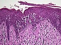
Non vesicullobullous, non-pustular lesions without epidermal changes

Lymphocytic infiltrate

Main conditions [2] CharacteristicsMicrographPhotograph
Urticaria, lymphocyte predominantPerivascular location. Mast cells are relatively sparse, potentially demonstrated with special stains, preferably tryptase stain. Extravasated erythrocytes are present in about 50% of the cases. No vasculitis. [14] Micrograph of urticaria.jpg Dermal edema [solid arrows in (A,B)] and a sparse superficial predominantly perivascular and interstitial infiltrate of lymphocytes and eosinophils without signs of vasculitis (dashed arrow). [15] Urticaria near navel.jpg
Fungal skin infection Often visible fungus. Other signs depend on fungus species. [16]
Pigmented purpuric dermatosis
  • Perivascular infiltrate, but may involve the dermis, further away from blood vessels. [17]
  • Sometimes tendency for lichenoid infiltrate [notes 4] [17]
  • Mild vascular damage, mainly endothelial swelling and focal karyorrhectic debris. [17]
  • Red blood cell extravasation. [17]
  • The epidermis may be normal or may exhibit spongiosis, focal parakeratosis, exocytosis and/or vacuolar change. [17]
Histopathology of Schamberg disease.jpg Schambergdisease-26male.png
Erythema annulare centrifugum
  • Superficial types: [18]
  • Mild spongiosis, parakeratosis and microvesiculation.
  • "Coat-sleeve anomaly": tight lymphohistiocytic infiltrate surrounding superficial vessels

Deep lesions: Sharply demarcated perivascular mononuclear cell infiltrate in middle to deep dermis [18]

Micrograph of erythema annulare centrifugum.jpg Erythema annulare centrifugum on arms and legs.jpg
Not otherwise specified [notes 2] A lesion with superficial lymphocytic infiltrate without additional histopathologic characteristics can be due to for example drug reactions and insect bites. [2] [notes 2]

Lymphoeosinophilic infiltrate

Main conditions [2] CharacteristicsMicrographPhotograph
Urticaria, lymphocyte predominantPerivascular location. Mast cells are relatively sparse, potentially demonstrated with special stains, preferably tryptase stain. Extravasated erythrocytes are present in about 50% of the cases. No vasculitis. [14] Micrograph of urticaria.jpg Dermal edema (solid arrows) and a sparse superficial predominantly perivascular and interstitial infiltrate of lymphocytes and eosinophils (dashed arrow) Urticaria near navel.jpg
Prevesicular stage of bullous pemphigoidImage at right shows influx of inflammatory cells including eosinophils and neutrophils in the dermis (solid arrow) and blister cavity (dashed arrows), and deposition of fibrin (asterisks). [15] However, the diagnosis of bullous pemphigoid consist of at least 2 positive results out of 3 criteria: [19]
  • Pruritus and/or predominant cutaneous blisters
  • Linear IgG and/or C3c deposits (in an n- serrated pattern) by direct immunofluorescence microscopy (DIF)
  • Positive epidermal side staining by indirect immunofluorescence microscopy on human salt-split skin (IIF SSS) on a serum sample.
Micrograph of infiltrate in bullous pemphigoid.jpg
Not otherwise specified [notes 2] A lesion with superficial lymphoeosinophilic infiltrate without additional histopathologic characteristics can be due to for example drug reactions and insect bites. [2] [notes 2]

Lymphoplasmacytic infiltrate

Main conditions [2] CharacteristicsMicrographPhotograph
RosaceaTypically enlarged, dilated capillaries and venules located in the upper dermis, angulated telangiectasias, perivascular and perifollicular lymphocytic infiltration, and superficial dermal edema. [20] Micrograph of rosacea.jpg Rosacea.jpg
Secondary syphilisVarious, but often one or a combination of: [21]
  • Psoriasiform hyperplasia with superficial neutrophils
  • Lichenoid tissue reaction, epidermal apoptosis and exocytosis of neutrophils
  • Superficial and deep chronic infiltrate in the dermis
  • Often numerous plasma cells in about 1/3 of cases
  • Often endothelial swelling.
Micrograph of secondary syphilis, HE.jpg Secondary stage syphilis sores (lesions) on the soles of the feet. Plantar lesions-CDC.jpg
Erythema migransTypically a superficial and deep perivascular lymphocytic infiltrate. [22] Plasma cells are typically located at the periphery of the lesion, whereas eosinophils are in the center. [22]
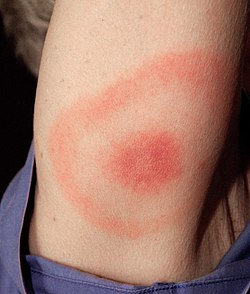
 Erythema migrans - erythematous rash in Lyme disease - PHIL 9875.jpg
Kaposi’s sarcoma in patch stageThe patch stage typically shows irregular proliferation of jagged vascular channels in the dermis below an integral epidermis. The so-called promontory sign is sometimes found in patch stage lesions and denotes vascular spaces surrounding pre-existing blood (see image). [23]

vessels

Micrograph of promontory sign of kaposi's sarcoma.jpg Patch stage Kaposi's sarcoma.jpg
Not otherwise specified [notes 2] A lesion with superficial lymphoplasmacytic infiltrate without additional histopathologic characteristics can be due to for example trauma, ulceration, scar and early cutaneous connective tissue diseases. [2] [notes 2]

Mastocytosis

Main conditions [2] CharacteristicsMicrographPhotograph
Urticaria pigmentosaMastocytosis with a clinical picture of darkish spots. Histopathology of urticaria pigmentosa.jpg Urticaria pigmentosa lesions on a child.jpg
Not otherwise specified [notes 2] Includes the rare disease of primary mastocytosis. [2] [notes 2]

Lymphohistiocytic infiltrate

Leprosy Histopathology of leprosy.jpg
Leprosy

These include bacterial infections including leprosy, and the sample should therefore be stained with Ziel-Neelsen, acid fast stains, Gomori methenamine silver, PAS, and Fite stains. [2] If negative, an unspecific lymphohistocytic dermatosis may be caused by drug reactions and viral infections. [2] [notes 2]

Neutrophilic infiltrate

Main conditions [2] CharacteristicsMicrographPhotograph
Urticaria, neutrophil predominant
  • Interstitial location [14]
  • Relatively dense infiltrate [14]
  • Mast cells are relatively sparse, potentially demonstrated with special stains, preferably tryptase stain. [14]
  • Extravasated erythrocytes are present in about 50% of the cases [14]
  • No vasculitis. [14]
Micrograph of neutrophilic urticarial dermatosis.jpg Neutrophilic urticarial dermatosis.jpg
Dermatitis herpetiformis
  • Subepidermal vesicles and blisters associated with accumulation of neutrophils at the papillary tips. [24]
  • Sometimes presence of eosinophils, giving an appearance similar to bullous pemphigoid. [24]
  • The histopathology is unspecific in approximately 35%–40% of the cases, [24] and direct immunofluorescence is needed, showing deposition of IgA in the papillary dermis in a granular or fibrillar pattern. [25]
Micrograph of dermatitis herpetiformis.jpg Dermatitis herpetiformis.jpg
Early linear IgA bullous dermatosisSubepidermal blister formation. [26] Micrograph of linear IgA bullous dermatosis.jpg Linear IgA bullous dermatosis.jpg
Early febrile neutrophilic dermatosis (Sweet's syndrome)Neutrophilic and lymphohistiocytic infiltrate and edema. [27] Sweet's syndrome pathology.jpg Sweet's syndrome Crohn's disease.gif
Connective tissue disorders
  • Usually associated with epidermal changes. [2] (Systemic lupus erythematosis pictured)
Histopathology of systemic lupus erythematosus.jpg Butterfly rash of lupus erythematosus.jpg
Cutaneous small-vessel vasculitis
  • Neutrophils with nuclear dust (dashed arrows in image), with high affinity for postcapillary venules. [28]
  • Features of vascular injury: fibrinoid necrosis (asterisks) and erytrocyt extravasation (solid arrows)
Micrograph of cutaneous small-vessel vasculitis.jpg Cutaneous small-vessel vasculitis.jpg

Multinucleated giant cells

Suture granuloma, with multinucleated giant cells surrounding (grey) suture material. Histopathology of suture granuloma.jpg
Suture granuloma, with multinucleated giant cells surrounding (grey) suture material.
  • Foreign bodies indicate a foreign body granuloma.
  • Specific forms of multinucleated giant cells include the Touton giant cell, which contains a ring of nuclei surrounding a central homogeneous cytoplasm, with foamy cytoplasm surrounding the nuclei. [29] [30] The central cytoplasm (surrounded by the nuclei) may be both amphophilic and eosinophilic. [31]

Notes

  1. PAS is for evaluation of the epidermal basement membrane, blood vessels, and the presence of fungal organisms
  2. 1 2 3 4 5 6 7 8 9 10 11 In "not otherwise specified" cases, a diagnosis may be reached by a review of the medical history and physical examination, based upon the potential conditions at hand.
  3. Parakeratotic mounds at the edge of follicular ostia.
  4. Pigmented purpuric dermatitis of Gougerot and Blum particularly have a tendency for lichenoid infiltrate.

Reference list

  1. "Eczema". University of Maryland Medical Center. Archived from the original on 27 July 2016.
  2. 1 2 3 4 5 6 7 8 9 10 11 12 13 14 15 16 17 18 19 20 21 22 23 24 25 26 27 28 29 30 31 32 33 34 35 36 Alsaad, K O (2005). "My approach to superficial inflammatory dermatoses". Journal of Clinical Pathology. 58 (12): 1233–1241. doi: 10.1136/jcp.2005.027151 . ISSN   0021-9746. PMC   1770784 . PMID   16311340.
  3. 1 2 Katarzyna Lundmark, Krynitz, Ismini Vassilaki, Lena Mölne, Annika Ternesten Bratel. "Handläggning av hudprover – provtagningsanvisningar, utskärningsprinciper och snittning (Handling of skin samples - Instructions for sampling, cutting and incision" (PDF). KVAST (Swedish Society of Pathology). Archived from the original (PDF) on 2019-11-08. Retrieved 2019-09-09.{{cite web}}: CS1 maint: multiple names: authors list (link)
  4. Page 678 in: Chhabra, Seema; Minz, RanjanaWalker; Saikia, Biman (2012). "Immunofluorescence in dermatology". Indian Journal of Dermatology, Venereology and Leprology. 78 (6): 677–691. doi: 10.4103/0378-6323.102355 . ISSN   0378-6323. PMID   23075636. S2CID   1331564.
  5. 1 2 3 Mowafak Hamodat. "Skin inflammatory (nontumor) > Spongiotic, psoriasiform and pustular reaction patterns > Seborrheic dermatitis". PathologyOutlines.com. Topic Completed: 1 August 2011. Revised: 26 March 2019
  6. 1 2 3 4 5 6 Unless else specified in boxes, reference is: Alsaad, K O (2005). "My approach to superficial inflammatory dermatoses". Journal of Clinical Pathology. 58 (12): 1233–1241. doi:10.1136/jcp.2005.027151. ISSN   0021-9746.
  7. Lisa K Pappas-Taffer. "Lichen Sclerosus". Medscape. Updated: May 17, 2018
  8. Mowafak Hamodat. "Skin inflammatory (nontumor) > Lichenoid and interface reaction patterns > Lupus: systemic lupus erythematosus (SLE)". PathologyOutlines. Topic Completed: 1 August 2011. Revised: 26 March 2019
  9. 1 2 3 Gorouhi, Farzam; Davari, Parastoo; Fazel, Nasim (2014). "Cutaneous and Mucosal Lichen Planus: A Comprehensive Review of Clinical Subtypes, Risk Factors, Diagnosis, and Prognosis". The Scientific World Journal. 2014: 1–22. doi: 10.1155/2014/742826 . ISSN   2356-6140. PMC   3929580 . PMID   24672362.
    - Attribution 3.0 Unported (CC BY 3.0)
  10. Al-Mutairi N, Hassanein A, Nour-Eldin O, Arun J (2005). "Generalized lichen nitidus". Pediatr Dermatol. 22 (2): 158–60. doi:10.1111/j.1525-1470.2005.22215.x. PMID   15804308. S2CID   6281902.
  11. Shenoi, SD; Balachandran, C; Mehta, VandanaRai; Salim, T (2005). "Lichen amyloidosus: A study of clinical, histopathologic and immunofluorescence findings in 30 cases". Indian Journal of Dermatology, Venereology and Leprology. 71 (3): 166–169. doi: 10.4103/0378-6323.16230 . ISSN   0378-6323. PMID   16394404.
  12. Kunz M, Ibrahim SM (2009). "Cytokines and cytokine profiles in human autoimmune diseases and animal models of autoimmunity". Mediators of Inflammation. 2009: 1–20. doi: 10.1155/2009/979258 . PMC   2768824 . PMID   19884985.
  13. Raychaudhuri SK, Maverakis E, Raychaudhuri SP (January 2014). "Diagnosis and classification of psoriasis". Autoimmunity Reviews. 13 (4–5): 490–5. doi:10.1016/j.autrev.2014.01.008. PMID   24434359.
  14. 1 2 3 4 5 6 7 Barzilai, Aviv; Sagi, Lior; Baum, Sharon; Trau, Henri; Schvimer, Michael; Barshack, Iris; Solomon, Michal (2017). "The Histopathology of Urticaria Revisited—Clinical Pathological Study". The American Journal of Dermatopathology. 39 (10): 753–759. doi:10.1097/DAD.0000000000000786. ISSN   0193-1091. PMID   28858880. S2CID   45809524.
  15. 1 2 Giang, Jenny; Seelen, Marc A. J.; van Doorn, Martijn B. A.; Rissmann, Robert; Prens, Errol P.; Damman, Jeffrey (2018). "Complement Activation in Inflammatory Skin Diseases". Frontiers in Immunology. 9: 639. doi: 10.3389/fimmu.2018.00639 . ISSN   1664-3224. PMC   5911619 . PMID   29713318.
  16. Guarner, J.; Brandt, M. E. (2011). "Histopathologic Diagnosis of Fungal Infections in the 21st Century". Clinical Microbiology Reviews. 24 (2): 247–280. doi: 10.1128/CMR.00053-10 . ISSN   0893-8512. PMC   3122495 . PMID   21482725.
  17. 1 2 3 4 5 Stephen Lyle. "Pigmented purpuric dermatoses". Dermpedia.org. Retrieved 2019-11-05.
  18. 1 2 "Histology of erythema annulare centrifugum". DermNet NZ. Retrieved 2019-11-05.
  19. Meijer JM, Diercks GF, de Lang EW, Pas HH, Jonkman MF (December 2018). "Assessment of diagnostic strategy for early recognition of bullous and nonbullous variants of pemphigoid". JAMA Dermatol. 155 (2): 158–165. doi:10.1001/jamadermatol.2018.4390. PMC   6439538 . PMID   30624575.
  20. Celiker, Hande; Toker, Ebru; Ergun, Tulin; Cinel, Leyla (2017). "An unusual presentation of ocular rosacea". Arquivos Brasileiros de Oftalmologia. 80 (6): 396–398. doi: 10.5935/0004-2749.20170097 . hdl: 11424/241569 . ISSN   0004-2749. PMID   29267579.
  21. Assoc Prof Patrick Emanuel (2013). "Syphilis pathology". Dermnet NZ.
  22. 1 2 Wilson, Thomas C.; Legler, Allison; Madison, Kathi C.; Fairley, Janet A.; Swick, Brian L. (2012). "Erythema Migrans". The American Journal of Dermatopathology. 34 (8): 834–837. doi:10.1097/DAD.0b013e31825879be. ISSN   0193-1091. PMID   22722463.
  23. Soyer, H. Peter; Jakob, Lena; Metzler, Gisela; Chen, Ko-Ming; Garbe, Claus (2011). "Non-AIDS Associated Kaposi's Sarcoma: Clinical Features and Treatment Outcome". PLOS ONE. 6 (4): e18397. Bibcode:2011PLoSO...618397J. doi: 10.1371/journal.pone.0018397 . ISSN   1932-6203. PMC   3075253 . PMID   21533260.
  24. 1 2 3 Antiga, Emiliano; Caproni, Marzia (2015). "The diagnosis and treatment of dermatitis herpetiformis". Clinical, Cosmetic and Investigational Dermatology. 8: 257–265. doi: 10.2147/CCID.S69127 . ISSN   1178-7015. PMC   4435051 . PMID   25999753.
  25. Huma A. Mirza; Amani Gharbi; William Gossman. (2022). "Dermatitis Herpetiformis". StatPearls at National Center for Biotechnology Information. PMID   29630215.{{cite journal}}: CS1 maint: multiple names: authors list (link) Last Update: July 11, 2019.
  26. Saleem, Maryam; Iftikhar, Hassaan (2019). "Linear IgA Disease: A Rare Complication of Vancomycin". Cureus. 11 (6): e4848. doi: 10.7759/cureus.4848 . ISSN   2168-8184. PMC   6684112 . PMID   31410331.
  27. Casarin Costa, Jose Ricardo; Virgens, Anangelica Rodrigues; de Oliveira Mestre, Luisa; Dias, Natasha Favoretto; Samorano, Luciana Paula; Valente, Neusa Yuriko Sakai; Festa Neto, Cyro (2017). "Sweet Syndrome: Clinical Features, Histopathology, and Associations of 83 Cases". Journal of Cutaneous Medicine and Surgery . 21 (3): 211–216. doi:10.1177/1203475417690719. ISSN   1203-4754. PMID   28300447. S2CID   40797905.
  28. Giang, Jenny; Seelen, Marc A. J.; van Doorn, Martijn B. A.; Rissmann, Robert; Prens, Errol P.; Damman, Jeffrey (2018). "Complement Activation in Inflammatory Skin Diseases". Frontiers in Immunology. 9: 639. doi: 10.3389/fimmu.2018.00639 . ISSN   1664-3224. PMC   5911619 . PMID   29713318.
    • "Figures - available via license: CC BY 4.0"
  29. Grant-Kels, Jane (2007). Color Atlas of Dermatopathology. City: Informa Healthcare. pp. 107, 119. ISBN   978-0-8493-3794-9.
  30. Carmen Gómez-Mateo, Maria; Monteagudo, Carlos (2013). "Nonepithelial skin tumors with multinucleated giant cells". Seminars in Diagnostic Pathology. 30 (1): 58–72. doi:10.1053/j.semdp.2012.01.004. PMID   23327730.
  31. Sequeira, Fiona; Gandhi, Suneil (2012). "Named cells in dermatology". Indian Journal of Dermatology, Venereology and Leprology. 78 (2): 207–16. doi: 10.4103/0378-6323.93650 . PMID   22421663.

Sources

Definition of Free Cultural Works logo notext.svg  This article incorporates text from a free content work. Licensed under Creative Commons Attribution 4.0 International (CC BY 4.0) license.( license statement/permission ). Text taken from Dermatitis , Patholines.

Related Research Articles

<span class="mw-page-title-main">Lichen planus</span> Human chronic inflammatory disease

Lichen planus (LP) is a chronic inflammatory and autoimmune disease that affects the skin, nails, hair, and mucous membranes. It is not an actual lichen, but is named for its appearance. It is characterized by polygonal, flat-topped, violaceous papules and plaques with overlying, reticulated, fine white scale, commonly affecting dorsal hands, flexural wrists and forearms, trunk, anterior lower legs and oral mucosa. The hue may be gray-brown in people with darker skin. Although there is a broad clinical range of LP manifestations, the skin and oral cavity remain as the major sites of involvement. The cause is unknown, but it is thought to be the result of an autoimmune process with an unknown initial trigger. There is no cure, but many different medications and procedures have been used in efforts to control the symptoms.

<span class="mw-page-title-main">Telogen effluvium</span> Medical condition

Telogen effluvium is a scalp disorder characterized by the thinning or shedding of hair resulting from the early entry of hair in the telogen phase. It is in this phase that telogen hairs begin to shed at an increased rate, where normally the approximate rate of hair loss is 125 hairs per day.

<span class="mw-page-title-main">Pemphigus</span> Blistering autoimmune diseases

Pemphigus is a rare group of blistering autoimmune diseases that affect the skin and mucous membranes. The name is derived from the Greek root pemphix, meaning "blister".

<span class="mw-page-title-main">Nummular dermatitis</span> Medical condition

Nummular dermatitis is one of the many forms of dermatitis. It is characterized by round or oval-shaped itchy lesions. The name comes from the Latin word "nummus," which means "coin."

<span class="mw-page-title-main">Seborrheic keratosis</span> Skin disease

A seborrheic keratosis is a non-cancerous (benign) skin tumour that originates from cells, namely keratinocytes, in the outer layer of the skin called the epidermis. Like liver spots, seborrheic keratoses are seen more often as people age.

<span class="mw-page-title-main">Mycosis fungoides</span> Most common form of cutaneous T-cell lymphoma

Mycosis fungoides, also known as Alibert-Bazin syndrome or granuloma fungoides, is the most common form of cutaneous T-cell lymphoma. It generally affects the skin, but may progress internally over time. Symptoms include rash, tumors, skin lesions, and itchy skin.

<span class="mw-page-title-main">Bullous pemphigoid</span> Autoimmune disease of skin and connective tissue characterized by large blisters

Bullous pemphigoid is an autoimmune pruritic skin disease that typically occurs in people aged over 60, that may involve the formation of blisters (bullae) in the space between the epidermal and dermal skin layers. It is classified as a type II hypersensitivity reaction, which involves formation of anti-hemidesmosome antibodies, causing a loss of keratinocytes to basement membrane adhesion.

<span class="mw-page-title-main">Blaschko's lines</span> Anatomical structure

Blaschko's lines, also called the lines of Blaschko, are lines of normal cell development in the skin. These lines are only visible in those with a mosaic skin condition or in chimeras where different cell lines contain different genes. These lines may express different amounts of melanin, or become visible due to a differing susceptibility to disease. In such individuals, they can become apparent as whorls, patches, streaks or lines in a linear or segmental distribution over the skin. They follow a V shape over the back, S-shaped whirls over the chest and sides, and wavy shapes on the head. Not all mosaic skin conditions follow Blaschko's lines.

<span class="mw-page-title-main">White piedra</span> Medical condition

White piedra is a mycosis of the hair caused by several species of fungi in the genus Trichosporon. It is characterized by soft nodules composed of yeast cells and arthroconidia that encompass hair shafts.

<span class="mw-page-title-main">Skin biopsy</span> Removal of skin cells for medical examination

Skin biopsy is a biopsy technique in which a skin lesion is removed to be sent to a pathologist to render a microscopic diagnosis. It is usually done under local anesthetic in a physician's office, and results are often available in 4 to 10 days. It is commonly performed by dermatologists. Skin biopsies are also done by family physicians, internists, surgeons, and other specialties. However, performed incorrectly, and without appropriate clinical information, a pathologist's interpretation of a skin biopsy can be severely limited, and therefore doctors and patients may forgo traditional biopsy techniques and instead choose Mohs surgery.

<span class="mw-page-title-main">Dermatitis herpetiformis</span> Chronic autoimmune disorder leading to blistering skin

Dermatitis herpetiformis (DH) is a chronic autoimmune blistering skin condition, characterised by intensely itchy blisters filled with a watery fluid. DH is a cutaneous manifestation of coeliac disease, although the exact causal mechanism is not known. DH is neither related to nor caused by herpes virus; the name means that it is a skin inflammation having an appearance similar to herpes.

<span class="mw-page-title-main">Spongiosis</span> Accumulation of fluid in the outermost layer of skin (epidermis)

Spongiosis is mainly intercellular edema in the epidermis, and is characteristic of eczematous dermatitis, manifested clinically by intraepidermal vesicles, "juicy" papules, and/or lichenification. It is a severe case of eczema that affects the epidermis, dermis or subcutaneous skin tissues. The three types of spongiotic dermatitis are acute, subacute and chronic. A dermatologist can diagnose acute spongiotic dermatitis by examining the skin during an office visit, but a biopsy is needed for an accurate diagnosis of the type.

Unilateral nevoid telangiectasia presents with fine thread veins, typically over a segment of skin supplied by a particular nerve on one side of the body. It most frequently involves the trigeminal, C3 and C4, or nearby areas. The condition was named in 1970 by Victor Selmanowitz.

Cutaneous meningioma, also known as heterotopic meningeal tissue, and rudimentary meningocele is a developmental defect, and results from the presence of meningocytes outside the calvarium.

Erosive pustular dermatitis of the scalp presents with pustules, erosions, and crusts on the scalp of primarily older Caucasian females, and on biopsy, has a lymphoplasmacytic infiltrate with or without foreign body giant cells and pilosebaceous atrophy.

<span class="mw-page-title-main">Pigmented purpuric dermatosis</span> Medical condition

Pigmented purpuric dermatosis refers to one of the three major classes of skin conditions characterized by purpuric skin eruptions.

Postinflammatory hypopigmentation is a cutaneous condition characterized by decreased pigment in the skin following inflammation of the skin.

<span class="mw-page-title-main">Vacuolar interface dermatitis</span>

Vacuolar interface dermatitis is a dermatitis with vacuolization at the dermoepidermal junction, with lymphocytic inflammation at the epidermis and dermis.

Cutaneous manifestations of COVID-19 are characteristic signs or symptoms of the Coronavirus disease 2019 that occur in the skin. The American Academy of Dermatology reports that skin lesions such as morbilliform, pernio, urticaria, macular erythema, vesicular purpura, papulosquamous purpura and retiform purpura are seen in people with COVID-19. Pernio-like lesions were more common in mild disease while retiform purpura was seen only in critically ill patients. The major dermatologic patterns identified in individuals with COVID-19 are urticarial rash, confluent erythematous/morbilliform rash, papulovesicular exanthem, chilbain-like acral pattern, livedo reticularis and purpuric "vasculitic" pattern. Chilblains and Multisystem inflammatory syndrome in children are also cutaneous manifestations of COVID-19.