Hypothermia cap

Last updated

A hypothermia cap (also referred to as cold cap or cooling cap) is a therapeutic device used to cool the human scalp. Its most prominent medical applications are in preventing or reducing alopecia in chemotherapy, and for preventing cerebral palsy in babies born with neonatal encephalopathy caused by hypoxic-ischemic encephalopathy (HIE). It can also be used to provide neuroprotection after cardiac arrest, to inhibit stroke paralysis, and as cryotherapy for migraine headaches.

Contents

Worn tight on the head, hypothermia caps are typically made of a synthetic such as neoprene, silicone or polyurethane, and filled with a coolant agent such as ice or gel which is either frozen to a very cold temperature (−25 °C to −30 °C, or −13 °F to −22 °F) before application or continuously cooled by an auxiliary control unit.

In the United States a course of treatment may cost US$1,500 to US$3,000. [1]

Medical uses

Neonatal encephalopathy

Hypoxic ischemic encephalopathy (HIE) is a condition that occurs when the brain is deprived of an adequate oxygen supply, and is most commonly observed in newborn babies due to birth asphyxia. It is the leading cause of cerebral palsy, an irreversible neonatal brain injury that can result in long-term cognitive, motor, and visual impairments. About 10,000 babies are born each year with cerebral palsy.

In such cases, by slowing down cell metabolism and body functions, a hypothermic cap can be used to lessen a baby's need for oxygen. Research throughout the late 1990s and 2000s demonstrated that for every degree a baby's body temperature is lowered, its body functions and demand for energy slow down by 10 to 15 percent. Therefore, slowing metabolic demands through hypothermia therapy can rectify a mismatch between oxygen supply and cell need, lowering the risk for cerebral palsy. [2]

Among babies who meet the criteria for hypothermia therapy—full-term with no known pre-existing conditions, having neonatal distress, and an abnormal neurological exam [2] —cooling must begin within six hours of birth and body temperature must be maintained at 33–34 °C (91–93 °F) for 72 hours before being gradually warmed again. An alternative to hypothermia caps involves pumping cold water through a specially adapted blanket, cooling the whole body. [3]

Induced pediatric hypothermia was approved in the U.S. by the FDA in March 2007. The most prominent such hypothermia cap which utilizes a cooling unit, a control unit and temperature probes to maintain a steady flow of cool water through a cap covering the head. [4]

Cardiac arrest

A 2008 trial demonstrated that the pre-hospital induction of therapeutic hypothermia after cardiac arrest as soon as possible after return of spontaneous circulation (ROSC) can achieve optimal neuroprotective benefit. The hypothermia cap was applied to 20 patients after out-of-hospital cardiac arrest, with a median of 10 min after ROSC. The median time between initiation of cooling and hospital admission was 28 minutes. No side effects related to the hypothermia cap were observed. The study concluded that "prehospital use of hypothermia caps is a safe and effective procedure to start therapeutic hypothermia after cardiac arrest. This approach is rapidly available, inexpensive, non-invasive, easy to learn and applicable in almost any situation." [5]

Chemotherapy-induced hair loss

Hypothermia caps appear useful to prevent hair loss during some kinds of chemotherapy, specifically when taxanes or anthracyclines are used. [6] It should not be used when cancer is present in the skin of the scalp or in people with lymphoma or leukemia. [7] There are generally only minor side effects from treatment. [8]

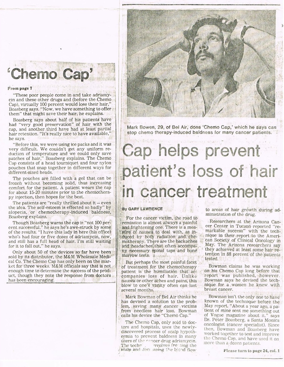
The first U.S. patent, filed in 1979 and granted in 1984, [9] [10] was for Mark Barron's "Chemo Cap", which consisted of resizable gel-filled nylon pouches that were frozen and worn for 15 to 20 minutes prior to treatment. [11] However, studies throughout the 1980s and early 1990s were not sufficiently encouraging, [6] and the patent expired in 1998. An analysis of 53 studies from 1995 through 2003, however, showed an average success rate of 73%. [6]

Today there are two types of caps; Manual cold caps sometimes referred to as 'Cold Caps/Cold Capping' & machine cooling often referred to as 'Scalp Cooling' However they can be used interchangeably.

There are three major providers of scalp cooling; Penguin Cold Cap, Paxman & Dignitana

Other uses

The use of hypothermia caps has also shown promise in inhibiting stroke paralysis. Studies are underway testing a combination treatment consisting of four drugs plus a hypothermia cap to try to slow the cell death that is triggered by an ischemic stroke. [12] Ischemic strokes are caused when a clot blocks blood flow to the brain, and comprise roughly 80% of all strokes. [12] The slowing of cell death is theorized to give the brain time to find an alternate blood supply through unblocked arteries, meaning patients may potentially avoid physical and speech impairments caused by ischemic strokes. [13]

Numerous studies have also suggested that therapeutic hypothermia can provide safe and effective adjunctive treatment for migraine headaches. For instance, a 1989 study in Headache: The Journal of Head and Face Pain showed 64.5% of 45 patients with migraine or migraine plus chronic daily headache evaluated use of a cold wrap for 20 to 30 minutes as mildly, moderately or completely effective. [14] In a 1984 study using a frozen gel pack, 80% of migraine patients reported the pack was effective. Numerous over-the-counter hypothermia caps today offer therapy for headaches. [15]

Side effects

In tests, minor cardiac arrhythmias occurred slightly more often in cooled infants, however the effect was not unexpected because mild sinus bradycardia is known to be associated with hypothermia. In tests, all cases were resolved with appropriate therapy. The cold cap system also increased the incidence of scalp edema; however, all cases were resolved prior to or after completion of treatment. [16]

See also

Related Research Articles

Clinical death is the medical term for cessation of blood circulation and breathing, the two criteria necessary to sustain the lives of human beings and of many other organisms. It occurs when the heart stops beating in a regular rhythm, a condition called cardiac arrest. The term is also sometimes used in resuscitation research.

<span class="mw-page-title-main">Cerebral edema</span> Excess accumulation of fluid (edema) in the intracellular or extracellular spaces of the brain

Cerebral edema is excess accumulation of fluid (edema) in the intracellular or extracellular spaces of the brain. This typically causes impaired nerve function, increased pressure within the skull, and can eventually lead to direct compression of brain tissue and blood vessels. Symptoms vary based on the location and extent of edema and generally include headaches, nausea, vomiting, seizures, drowsiness, visual disturbances, dizziness, and in severe cases, death.

<span class="mw-page-title-main">Ischemia</span> Restriction in blood supply to tissues

Ischemia or ischaemia is a restriction in blood supply to any tissue, muscle group, or organ of the body, causing a shortage of oxygen that is needed for cellular metabolism. Ischemia is generally caused by problems with blood vessels, with resultant damage to or dysfunction of tissue i.e. hypoxia and microvascular dysfunction. It also implies local hypoxia in a part of a body resulting from constriction. Ischemia causes not only insufficiency of oxygen, but also reduced availability of nutrients and inadequate removal of metabolic wastes. Ischemia can be partial or total blockage. The inadequate delivery of oxygenated blood to the organs must be resolved either by treating the cause of the inadequate delivery or reducing the oxygen demand of the system that needs it. For example, patients with myocardial ischemia have a decreased blood flow to the heart and are prescribed with medications that reduce chronotrophy and ionotrophy to meet the new level of blood delivery supplied by the stenosed vasculature so that it is adequate.

Vasospasm refers to a condition in which an arterial spasm leads to vasoconstriction. This can lead to tissue ischemia and tissue death (necrosis). Cerebral vasospasm may arise in the context of subarachnoid hemorrhage. Symptomatic vasospasm or delayed cerebral ischemia is a major contributor to post-operative stroke and death especially after aneurysmal subarachnoid hemorrhage. Vasospasm typically appears 4 to 10 days after subarachnoid hemorrhage.

<span class="mw-page-title-main">CADASIL</span> Medical condition

CADASIL or CADASIL syndrome, involving cerebral autosomal dominant arteriopathy with subcortical infarcts and leukoencephalopathy, is the most common form of hereditary stroke disorder, and is thought to be caused by mutations of the Notch 3 gene on chromosome 19. The disease belongs to a family of disorders called the leukodystrophies. The most common clinical manifestations are migraine headaches and transient ischemic attacks or strokes, which usually occur between 40 and 50 years of age, although MRI is able to detect signs of the disease years prior to clinical manifestation of disease.

<span class="mw-page-title-main">Cerebral hypoxia</span> Oxygen shortage of the brain

Cerebral hypoxia is a form of hypoxia, specifically involving the brain; when the brain is completely deprived of oxygen, it is called cerebral anoxia. There are four categories of cerebral hypoxia; they are, in order of increasing severity: diffuse cerebral hypoxia (DCH), focal cerebral ischemia, cerebral infarction, and global cerebral ischemia. Prolonged hypoxia induces neuronal cell death via apoptosis, resulting in a hypoxic brain injury.

<span class="mw-page-title-main">Alopecia totalis</span> Medical condition

Alopecia totalis is the loss of all hair on the head and face. Its causes are unclear, but believed to be autoimmune. Research suggests there may be a genetic component linked to developing alopecia totalis; the presence of DRB1*0401 and DQB1*0301, both of which are human leukocyte antigens (HLA), were found to be associated with long-standing alopecia totalis.

<span class="mw-page-title-main">Intrauterine hypoxia</span> Medical condition when the fetus is deprived of sufficient oxygen

Intrauterine hypoxia occurs when the fetus is deprived of an adequate supply of oxygen. It may be due to a variety of reasons such as prolapse or occlusion of the umbilical cord, placental infarction, maternal diabetes and maternal smoking. Intrauterine growth restriction may cause or be the result of hypoxia. Intrauterine hypoxia can cause cellular damage that occurs within the central nervous system. This results in an increased mortality rate, including an increased risk of sudden infant death syndrome (SIDS). Oxygen deprivation in the fetus and neonate have been implicated as either a primary or as a contributing risk factor in numerous neurological and neuropsychiatric disorders such as epilepsy, attention deficit hyperactivity disorder, eating disorders and cerebral palsy.

<span class="mw-page-title-main">Brain ischemia</span> Medical condition

Brain ischemia is a condition in which there is insufficient bloodflow to the brain to meet metabolic demand. This leads to poor oxygen supply or cerebral hypoxia and thus leads to the death of brain tissue or cerebral infarction/ischemic stroke. It is a sub-type of stroke along with subarachnoid hemorrhage and intracerebral hemorrhage.

Deep hypothermic circulatory arrest (DHCA) is a surgical technique in which the temperature of the body falls significantly and blood circulation is stopped for up to one hour. It is used when blood circulation to the brain must be stopped because of delicate surgery within the brain, or because of surgery on large blood vessels that lead to or from the brain. DHCA is used to provide a better visual field during surgery due to the cessation of blood flow. DHCA is a form of carefully managed clinical death in which heartbeat and all brain activity cease.

Targeted temperature management (TTM) previously known as therapeutic hypothermia or protective hypothermia is an active treatment that tries to achieve and maintain a specific body temperature in a person for a specific duration of time in an effort to improve health outcomes during recovery after a period of stopped blood flow to the brain. This is done in an attempt to reduce the risk of tissue injury following lack of blood flow. Periods of poor blood flow may be due to cardiac arrest or the blockage of an artery by a clot as in the case of a stroke.

<span class="mw-page-title-main">Neurointensive care</span> Branch of medicine that deals with life-threatening diseases of the nervous system

Neurocritical care is a medical field that treats life-threatening diseases of the nervous system and identifies, prevents, and treats secondary brain injury.

The Arctic Sun Temperature Management System is a non-invasive targeted temperature management system. It modulates patient temperature by circulating chilled water in pads directly adhered to the patient's skin. Using varying water temperatures and a computer algorithm, a patient's body temperature can be better controlled. It is produced by Medivance, Inc. of Louisville, Colorado.

Anagen effluvium is the pathologic loss of anagen or growth-phase hairs. Classically, it is caused by radiation therapy to the head and systemic chemotherapy, especially with alkylating agents.

Mild total body hypothermia, induced by cooling a baby to 33-34°C for three days after birth, is nowadays a standardized treatment after moderate to severe hypoxic ischemic encephalopathy in full-term and near to fullterm neonates. It has recently been proven to be the only medical intervention which reduces brain damage, and improves an infant's chance of survival and reduced disability.

<span class="mw-page-title-main">Athetoid cerebral palsy</span> Type of cerebral palsy associated with basal ganglia damage

Athetoid cerebral palsy, or dyskinetic cerebral palsy, is a type of cerebral palsy primarily associated with damage, like other forms of CP, to the basal ganglia in the form of lesions that occur during brain development due to bilirubin encephalopathy and hypoxic–ischemic brain injury. Unlike spastic or ataxic cerebral palsies, ADCP is characterized by both hypertonia and hypotonia, due to the affected individual's inability to control muscle tone. Clinical diagnosis of ADCP typically occurs within 18 months of birth and is primarily based upon motor function and neuroimaging techniques. While there are no cures for ADCP, some drug therapies as well as speech, occupational therapy, and physical therapy have shown capacity for treating the symptoms.

Neonatal stroke, similar to a stroke which occurs in adults, is defined as a disturbance to the blood supply of the developing brain in the first 28 days of life. This description includes both ischemic events, which results from a blockage of vessels, and hypoxic events, which results from a lack of oxygen to the brain tissue, as well as some combination of the two. One treatment with some proven benefits is hypothermia, but may be most beneficial in conjunction with pharmacological agents. Well-designed clinical trials for stroke treatment in neonates are lacking, but some current studies involve the transplantation of neural stem cells and umbilical cord stem cells; it is not yet known if this therapy is likely to be successful.

<span class="mw-page-title-main">Spastic hemiplegia</span> Medical condition

Spastic hemiplegia is a neuromuscular condition of spasticity that results in the muscles on one side of the body being in a constant state of contraction. It is the "one-sided version" of spastic diplegia. It falls under the mobility impairment umbrella of cerebral palsy. About 20–30% of people with cerebral palsy have spastic hemiplegia. Due to brain or nerve damage, the brain is constantly sending action potentials to the neuromuscular junctions on the affected side of the body. Similar to strokes, damage on the left side of the brain affects the right side of the body and damage on the right side of the brain affects the left side of the body. Other side can be effected for lesser extent. The affected side of the body is rigid, weak and has low functional abilities. In most cases, the upper extremity is much more affected than the lower extremity. This could be due to preference of hand usage during early development. If both arms are affected, the condition is referred to as double hemiplegia. Some patients with spastic hemiplegia only experience minor impairments, where in severe cases one side of the body could be completely paralyzed. The severity of spastic hemiplegia is dependent upon the degree of the brain or nerve damage.

<span class="mw-page-title-main">Mark B. Barron</span> American inventor

Mark B. Barron is an American entrepreneur and inventor. He is the founder of Public Transportation Safety International Corporation (PTS), which invented and produces the S-1 Gard Dangerzone Deflector for transit buses and the Minimize Danger Zone (MDZ) Shield for school buses.

Perinatal stroke is a disease where an infant has a stroke between the 140th day of the gestation period and the 28th postpartum day, affecting up to 1 in 2300 live births. This disease is further divided into three subgroups, namely neonatal arterial ischemic stroke, neonatal cerebral sinovenous ischemic stroke, and presumed perinatal stroke. Several risk factors contribute to perinatal stroke including birth trauma, placental abruption, infections, and the mother's health. Detection and diagnosis of perinatal stroke are often delayed due to prenatal onset or inadequacy of neonatal signs and symptoms. A child may be asymptomatic in the early stages of life and may develop common signs of perinatal stroke such as seizures, poor coordination, and speech delays as they get older. Diagnostic tests such as magnetic resonance imaging, electroencephalogram, and blood tests are conducted when doctors suspect the patients have developed signs of a perinatal stroke. The prognosis of this disease is associated with the severity and the development of the symptoms. This disease can be treated by anticoagulant and anticonvulsant drugs, surgical procedures, and therapeutic hypothermia, depending on the condition of the patient.

References

  1. Hershman, DL (14 February 2017). "Scalp Cooling to Prevent Chemotherapy-Induced Alopecia: The Time Has Come". JAMA. 317 (6): 587–588. doi:10.1001/jama.2016.21039. PMID   28196237.
  2. 1 2 Cronin, Fran (January 31, 2012). "What To Do For Oxygen-Deprived Newborns? Cool Them Down". WBUR.org. Retrieved 2012-02-12.
  3. Laurance, Jeremy (October 1, 2009). "Cooling 'cure' averts infant brain damage". The Independent. United Kingdom.
  4. "FDA Approves Novel Device That Prevents or Reduces Brain Damage in Infants" (Press release). U.S. Food and Drug Administration. December 20, 2006. Retrieved 2012-02-12.
  5. Storm, Christian; Schefold, Joerg C.; Kerner, Thoralf; Schmidbauer, Willi; Gloza, Jola; Krueger, Anne; Jörres, Achim; Hasper, Dietrich (October 2008). "Prehospital cooling with hypothermia caps (PreCoCa): a feasibility study". Clinical Research in Cardiology. 97 (10): 768–72. doi:10.1007/s00392-008-0678-1. PMID   18512093. S2CID   24467783.
  6. 1 2 3 Grevelman, EG; Breed, WP (March 2005). "Prevention of chemotherapy-induced hair loss by scalp cooling". Annals of Oncology. 16 (3): 352–8. doi: 10.1093/annonc/mdi088 . PMID   15642703.
  7. Breed, WP (January 2004). "What is wrong with the 30-year-old practice of scalp cooling for the prevention of chemotherapy-induced hair loss?". Supportive Care in Cancer. 12 (1): 3–5. doi:10.1007/s00520-003-0551-8. PMID   14615930. S2CID   25031894.
  8. Komen, MM; Smorenburg, CH; van den Hurk, CJ; Nortier, JW (2011). "[Scalp cooling for chemotherapy-induced alopecia]". Nederlands Tijdschrift voor Geneeskunde. 155 (45): A3768. PMID   22085565.
  9. US 4425916,Bowen, Mark,"Cap structure for creating temperature controlled environment for reducing alopecia",issued 1984-01-17
  10. Bowen, Mark. Scalp hypothermia cap. Patent No. D268696 (Drawing). U.S. Patent Office. Filed 1980-08-18, issued 1983-04-19.
  11. Lawrence, Gary (September 2, 1979). "Cap helps prevent patient's loss of hair in cancer treatment". Valley News. Los Angeles, CA. p. 7. Archived from the original on June 18, 2016.
  12. 1 2 Ruttan, Susan (January 27, 2006). "Icy head may reduce stroke". The Windsor Star. p. D10
  13. van der Worp, H B; Macleod, M R; Kollmar, R (June 2010). "Therapeutic hypothermia for acute ischemic stroke: ready to start large randomized trials?". Journal of Cerebral Blood Flow & Metabolism. Nature Publishing Group. 30 (6): 1079–93. doi:10.1038/jcbfm.2010.44. PMC   2949207 . PMID   20354545.
  14. Robbins, L. D. (1989). "Cryotherapy for Headache". Headache: The Journal of Head and Face Pain. 29 (9): 598–600. doi:10.1111/j.1526-4610.1989.hed2909598.x. PMID   2584002. S2CID   42372766.
  15. Diamond S, Freitag FG (January 1986). "Cold as an adjunctive therapy for headache". Postgraduate Medicine. 79 (1): 305–9. doi:10.1080/00325481.1986.11699255. PMID   3941818.
  16. Waknine, Yael (January 3, 2006). "FDA Approvals: Olympic Cool-Cap, Spanner Stent, CellSearch System". MedscapeCME. Retrieved October 8, 2009.