Immunolabeling

Last updated
Immunolabeling - Antigen Detection of Tissue via Tagged Antigen-specific Antibody Immunolabeling process image.png
Immunolabeling - Antigen Detection of Tissue via Tagged Antigen-specific Antibody

Immunolabeling is a biochemical process that enables the detection and localization of an antigen to a particular site within a cell, tissue, or organ. Antigens are organic molecules, usually proteins, capable of binding to an antibody. These antigens can be visualized using a combination of antigen-specific antibody as well as a means of detection, called a tag, that is covalently linked to the antibody. [1] If the immunolabeling process is meant to reveal information about a cell or its substructures, the process is called immunocytochemistry. [2] Immunolabeling of larger structures is called immunohistochemistry. [3]

Contents

There are two complex steps in the manufacture of antibody for immunolabeling. The first is producing the antibody that binds specifically to the antigen of interest and the second is fusing the tag to the antibody. Since it is impractical to fuse a tag to every conceivable antigen-specific antibody, most immunolabeling processes use an indirect method of detection. This indirect method employs a primary antibody that is antigen-specific and a secondary antibody fused to a tag that specifically binds the primary antibody. This indirect approach permits mass production of secondary antibody that can be bought off the shelf. [4] Pursuant to this indirect method, the primary antibody is added to the test system. The primary antibody seeks out and binds to the target antigen. The tagged secondary antibody, designed to attach exclusively to the primary antibody, is subsequently added.

Typical tags include: a fluorescent compound, gold beads, a particular epitope tag, [5] or an enzyme that produces a colored compound. The association of the tags to the target via the antibodies provides for the identification and visualization of the antigen of interest in its native location in the tissue, such as the cell membrane, cytoplasm, or nuclear membrane. Under certain conditions the method can be adapted to provide quantitative information. [4]

Immunolabeling can be used in pharmacology, molecular biology, biochemistry and any other field where it is important to know of the precise location of an antibody-bindable molecule. [6] [7] [8]

Indirect vs. direct method

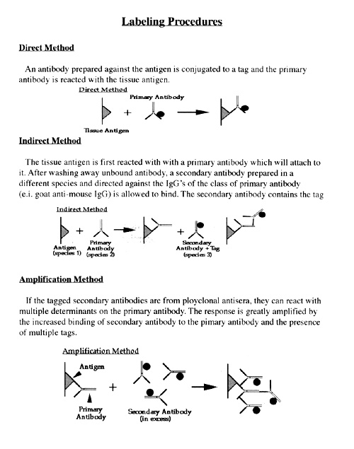
There are two methods involved in immunolabeling, the direct and the indirect methods. In the direct method of immunolabeling, the primary antibody is conjugated directly to the tag. [9] The direct method is useful in minimizing cross-reaction, a measure of nonspecificity that is inherent in all antibodies and that is multiplied with each additional antibody used to detect an antigen. However, the direct method is far less practical than the indirect method, and is not commonly used in laboratories, since the primary antibodies must be covalently labeled, which require an abundant supply of purified antibody. Also, the direct method is potentially far less sensitive than the indirect method. [10] Since several secondary antibodies are capable of binding to different parts, or domains, of a single primary antibody binding the target antigen, there is more tagged antibody associated with each antigen. More tag per antigen results in more signal per antigen. [11]

Different indirect methods can be employed to achieve high degrees of specificity and sensitivity. First, two-step protocols are often used to avoid the cross-reaction between the immunolabeling of multiple primary and secondary antibody mixtures, where secondary fragment antigen-binding antibodies are frequently used. Secondly, haptenylated primary antibodies can be used, where the secondary antibody can recognize the associated hapten. The hapten is covalently linked to the primary antibody by succinyl imidesters or conjugated IgG Fc-specific Fab sections. Lastly, primary monoclonal antibodies that have different Ig isotypes can be detected by specific secondary antibodies that are against the isotype of interest. [10]

Antibody binding and specificity

Overall, antibodies must bind to the antigens with a high specificity and affinity. [12] The specificity of the binding refers to an antibody's capacity to bind and only bind a single target antigen. Scientists commonly use monoclonal antibodies and polyclonal antibodies, which are composed of synthetic peptides. During the manufacture of these antibodies, antigen specific antibodies are sequestered by attaching the antigenic peptide to an affinity column and allowing nonspecific antibody to simply pass through the column. This decreases the likelihood that the antibodies will bind to an unwanted epitope of the antigen not found on the initial peptide. Hence, the specificity of the antibody is established by the specific reaction with the protein or peptide that is used for immunization by specific methods, such as immunoblotting or immunoprecipitation. [13]

In establishing the specificity of antibodies, the key factor is the type of synthetic peptides or purified proteins being used. The lesser the specificity of the antibody, the greater the chance of visualizing something other than the target antigen. In the case of synthetic peptides, the advantage is the amino acid sequence is easily accessible, but the peptides do not always resemble the 3-D structure or post-translational modification found in the native form of the protein. Therefore, antibodies that are produced to work against a synthetic peptide may have problems with the native 3-D protein. These types of antibodies would lead to poor results in immunoprecipitation or immunohistochemistry experiments, yet the antibodies may be capable of binding to the denatured form of the protein during an immunoblotting run. On the contrary, if the antibody works well for purified proteins in their native form and not denatured, an immunoblot cannot be used as a standardized test to determine the specificity of the antibody binding, particularly in immunohistochemistry. [14]

Specific immunolabeling techniques

Immunolabeling for light microscopy

Light microscopy is the use of a light microscope, which is an instrument that requires the usage of light to view the enlarged specimen. In general, a compound light microscope is frequently used, where two lenses, the eyepiece, and the objective work simultaneously to generate the magnification of the specimen. [15] Light microscopy frequently uses immunolabeling to observe targeted tissues or cells. For instance, a study was conducted to view the morphology and the production of hormones in pituitary adenoma cell cultures via light microscopy and other electron microscopic methods. This type of microscopy confirmed that the primary adenoma cell cultures keep their physiological characteristics in vitro, which matched the histology inspection. Moreover, cell cultures of human pituitary adenomas were viewed by light microscopy and immunocytochemistry, where these cells were fixed and immunolabeled with a monoclonal mouse antibody against human GH and a polyclonal rabbit antibody against PRL. This is an example of how a immunolabeled cell culture of pituitary adenoma cells that were viewed via light microscopy and by other electron microscopy techniques can assist with the proper diagnosis of tumors. [16]

Immunolabeling for electron microscopy

Electron microscopy (EM) is a focused area of science that uses the electron microscope as a tool for viewing tissues. [17] Electron microscopy has a magnification level up to 2 million times, whereas light microscopy only has a magnification up to 1000-2000 times. [18] There are two types of electron microscopes, the transmission electron microscope and the scanning electron microscope. [17]

Electron microscopy is a common method that uses the immunolabeling technique to view tagged tissues or cells. The electron microscope method follows many of the same concepts as immunolabeling for light microscopy, where the particular antibody is able to recognize the location of the antigen of interest and then be viewed by the electron microscope. The advantage of electron microscopy over light microscopy is the ability to view the targeted areas at their subcellular level. Generally, a heavy metal that is electron dense is used for EM, which can reflect the incident electrons. Immunolabeling is typically confirmed using the light microscope to assure the presence of the antigen and then followed up with the electron microscope. [19]

Immunolabeling and electron microscopy are often used to view chromosomes. A study was conducted to view possible improvements of immunolabeling chromosome structures, such as topoisomerase IIα and condensin in dissected mitotic chromosomes. In particular, these investigators used UV irradiation of separated nuclei or showed how chromosomes assist by high levels of specific immunolabeling, which were viewed by electron microscopy. [20]

Immunolabeling for transmission electron microscopy

Transmission electron microscopy (TEM) uses a transmission electron microscope to form a two-dimensional image by shooting electrons through a thin piece of tissue. The brighter certain areas are on the image, the more electrons that are able to move through the specimen. [17] Transmission Electron Microscopy has been used as a way to view immunolabeled tissues and cells. For instance, bacteria can be viewed by TEM when immunolabeling is applied. A study was conducted to examine the structures of CS3 and CS6 fimbriae in different Escherichia coli strains, which were detected by TEM followed by negative staining, and immunolabeling. More specifically, immunolabeling of the fimbriae confirmed the existence of different surface antigens. [21]

Immunolabeling for scanning electron microscopy

Scanning electron microscopy (SEM) uses a scanning electron microscope, which produces large images that are perceived as three-dimensional when, in fact, they are not. This type of microscope concentrates a beam of electrons across a very small area (2-3 nm) of the specimen in order to produce electrons from said specimen. These secondary electrons are detected by a sensor, and the image of the specimen is generated over a certain time period. [17]

Scanning electron microscopy is a frequently used immunolabeling technique. SEM is able to detect the surface of cellular components in high resolution. This immunolabeling technique is very similar to the immuno-fluorescence method, but a colloidal gold tag is used instead of a fluorophore. Overall, the concepts are very parallel in that an unconjugated primary antibody is used and sequentially followed by a tagged secondary antibody that works against the primary antibody. [22] Sometimes SEM in conjunction with gold particle immunolabeling is troublesome in regards to the particles and charges resolution under the electron beam; however, this resolution setback has been resolved by the improvement of the SEM instrumentation by backscattered electron imaging. [23] This is because electron backscattered diffraction patterns provide a clean surface of the sample to interact with the primary electron beam. [24]

Immunolabeling with gold (Immunogold Labeling)

Immunolabeling with gold particles, also known as immunogold staining, is used regularly with scanning electron microscopy and transmission electron microscopy to successfully identify the area within cells and tissues where antigens are located. [23] The gold particle labeling technique was first published by Faulk, W. and Taylor, G. when they were able to tag gold particles to anti-salmonella rabbit gamma globulins in one step in order to identify the location of the antigens of salmonella. [23] [25]

Studies have shown that the size of the gold particle must be enlarged (>40 nm) to view the cells in low magnification, but gold particles that are too large can decrease the efficiency of the binding of the gold tag. Scientists have concluded the usage of smaller gold particles (1-5 nm) should be enlarged and enhanced with silver. Although osmium tetroxide staining can scratch the silver, gold particle enhancement was found not to be susceptible to scratching by osmium tetroxide staining; therefore, many cell adhesion studies of different substrates can use the immunogold labeling mechanism via the enhancement of the gold particles. [26]

Further Applications

Research has been conducted to test the compatibility of immunolabeling with fingerprints. Sometimes, fingerprints are not clear enough to recognize the ridge pattern. Immunolabeling may be a way for forensic personnel to narrow down who left the print. Researchers conducted a study which tested the compatibility of immunolabeling with many developmental techniques for fingerprints. They found that indanedione-zinc (IND-ZnCl), IND-ZnCl followed by ninhydrin spraying (IND-NIN), physical developer (PD), cyanoacrylate fuming (CA), cyanoacrylate followed by basic yellow staining (CA-BY), lumicyanoacrylate fuming (Lumi-CA) and polycyanoacrylate fuming (Poly-CA) all were compatible with immunolabeling. [27] Immunolabeling can not only extract donor profiling information from fingerprints, but can also enhance the quality of the fingerprints which both would be beneficial in a forensic case.

Related Research Articles

<span class="mw-page-title-main">Antibody</span> Protein(s) forming a major part of an organisms immune system

An antibody (Ab), also known as an immunoglobulin (Ig), is a large, Y-shaped protein used by the immune system to identify and neutralize foreign objects such as pathogenic bacteria and viruses. The antibody recognizes a unique molecule of the pathogen, called an antigen. Each tip of the "Y" of an antibody contains a paratope that is specific for one particular epitope on an antigen, allowing these two structures to bind together with precision. Using this binding mechanism, an antibody can tag a microbe or an infected cell for attack by other parts of the immune system, or can neutralize it directly.

<span class="mw-page-title-main">Histology</span> Study of the microscopic anatomy of cells and tissues of plants and animals

Histology, also known as microscopic anatomy or microanatomy, is the branch of biology that studies the microscopic anatomy of biological tissues. Histology is the microscopic counterpart to gross anatomy, which looks at larger structures visible without a microscope. Although one may divide microscopic anatomy into organology, the study of organs, histology, the study of tissues, and cytology, the study of cells, modern usage places all of these topics under the field of histology. In medicine, histopathology is the branch of histology that includes the microscopic identification and study of diseased tissue. In the field of paleontology, the term paleohistology refers to the histology of fossil organisms.

Immunoperoxidase is a type of immunostain used in molecular biology, medical research, and clinical diagnostics. In particular, immunoperoxidase reactions refer to a sub-class of immunohistochemical or immunocytochemical procedures in which the antibodies are visualized via a peroxidase-catalyzed reaction.

An epitope, also known as antigenic determinant, is the part of an antigen that is recognized by the immune system, specifically by antibodies, B cells, or T cells. The part of an antibody that binds to the epitope is called a paratope. Although epitopes are usually non-self proteins, sequences derived from the host that can be recognized are also epitopes.

<span class="mw-page-title-main">Immunostaining</span> Biochemical technique

In biochemistry, immunostaining is any use of an antibody-based method to detect a specific protein in a sample. The term "immunostaining" was originally used to refer to the immunohistochemical staining of tissue sections, as first described by Albert Coons in 1941. However, immunostaining now encompasses a broad range of techniques used in histology, cell biology, and molecular biology that use antibody-based staining methods.

<span class="mw-page-title-main">Phalloidin</span> Chemical compound

Phalloidin belongs to a class of toxins called phallotoxins, which are found in the death cap mushroom (Amanita phalloides). It is a rigid bicyclic heptapeptide that is lethal after a few days when injected into the bloodstream. The major symptom of phalloidin poisoning is acute hunger due to the destruction of liver cells. It functions by binding and stabilizing filamentous actin (F-actin) and effectively prevents the depolymerization of actin fibers. Due to its tight and selective binding to F-actin, derivatives of phalloidin containing fluorescent tags are used widely in microscopy to visualize F-actin in biomedical research.

<span class="mw-page-title-main">Immunofluorescence</span> Technique used for light microscopy

Immunofluorescence is a technique used for light microscopy with a fluorescence microscope and is used primarily on biological samples. This technique uses the specificity of antibodies to their antigen to target fluorescent dyes to specific biomolecule targets within a cell, and therefore allows visualization of the distribution of the target molecule through the sample. The specific region an antibody recognizes on an antigen is called an epitope. There have been efforts in epitope mapping since many antibodies can bind the same epitope and levels of binding between antibodies that recognize the same epitope can vary. Additionally, the binding of the fluorophore to the antibody itself cannot interfere with the immunological specificity of the antibody or the binding capacity of its antigen. Immunofluorescence is a widely used example of immunostaining and is a specific example of immunohistochemistry. This technique primarily makes use of fluorophores to visualise the location of the antibodies.

<span class="mw-page-title-main">Immunohistochemistry</span> Common application of immunostaining

Immunohistochemistry (IHC) is the most common application of immunostaining. It involves the process of selectively identifying antigens (proteins) in cells of a tissue section by exploiting the principle of antibodies binding specifically to antigens in biological tissues. IHC takes its name from the roots "immuno", in reference to antibodies used in the procedure, and "histo", meaning tissue. Albert Coons conceptualized and first implemented the procedure in 1941.

<span class="mw-page-title-main">Fluorescence microscope</span> Optical microscope that uses fluorescence and phosphorescence

A fluorescence microscope is an optical microscope that uses fluorescence instead of, or in addition to, scattering, reflection, and attenuation or absorption, to study the properties of organic or inorganic substances. "Fluorescence microscope" refers to any microscope that uses fluorescence to generate an image, whether it is a simple set up like an epifluorescence microscope or a more complicated design such as a confocal microscope, which uses optical sectioning to get better resolution of the fluorescence image.

Affinity chromatography is a method of separating a biomolecule from a mixture, based on a highly specific macromolecular binding interaction between the biomolecule and another substance. The specific type of binding interaction depends on the biomolecule of interest; antigen and antibody, enzyme and substrate, receptor and ligand, or protein and nucleic acid binding interactions are frequently exploited for isolation of various biomolecules. Affinity chromatography is useful for its high selectivity and resolution of separation, compared to other chromatographic methods.

A protein microarray is a high-throughput method used to track the interactions and activities of proteins, and to determine their function, and determining function on a large scale. Its main advantage lies in the fact that large numbers of proteins can be tracked in parallel. The chip consists of a support surface such as a glass slide, nitrocellulose membrane, bead, or microtitre plate, to which an array of capture proteins is bound. Probe molecules, typically labeled with a fluorescent dye, are added to the array. Any reaction between the probe and the immobilised protein emits a fluorescent signal that is read by a laser scanner. Protein microarrays are rapid, automated, economical, and highly sensitive, consuming small quantities of samples and reagents. The concept and methodology of protein microarrays was first introduced and illustrated in antibody microarrays in 1983 in a scientific publication and a series of patents. The high-throughput technology behind the protein microarray was relatively easy to develop since it is based on the technology developed for DNA microarrays, which have become the most widely used microarrays.

<i>In situ</i> hybridization

In situ hybridization (ISH) is a type of hybridization that uses a labeled complementary DNA, RNA or modified nucleic acids strand to localize a specific DNA or RNA sequence in a portion or section of tissue or if the tissue is small enough, in the entire tissue, in cells, and in circulating tumor cells (CTCs). This is distinct from immunohistochemistry, which usually localizes proteins in tissue sections.

<span class="mw-page-title-main">Single-domain antibody</span> Antibody fragment

A single-domain antibody (sdAb), also known as a NANOBODY®, is an antibody fragment consisting of a single monomeric variable antibody domain. Like a whole antibody, it is able to bind selectively to a specific antigen. With a molecular weight of only 12–15 kDa, single-domain antibodies are much smaller than common antibodies which are composed of two heavy protein chains and two light chains, and even smaller than Fab fragments and single-chain variable fragments.

<span class="mw-page-title-main">Immunocytochemistry</span>

Immunocytochemistry (ICC) is a common laboratory technique that is used to anatomically visualize the localization of a specific protein or antigen in cells by use of a specific primary antibody that binds to it. The primary antibody allows visualization of the protein under a fluorescence microscope when it is bound by a secondary antibody that has a conjugated fluorophore. ICC allows researchers to evaluate whether or not cells in a particular sample express the antigen in question. In cases where an immunopositive signal is found, ICC also allows researchers to determine which sub-cellular compartments are expressing the antigen.

<span class="mw-page-title-main">Fixation (histology)</span> Preservation of biological tissue

In the fields of histology, pathology, and cell biology, fixation is the preservation of biological tissues from decay due to autolysis or putrefaction. It terminates any ongoing biochemical reactions and may also increase the treated tissues' mechanical strength or stability. Tissue fixation is a critical step in the preparation of histological sections, its broad objective being to preserve cells and tissue components and to do this in such a way as to allow for the preparation of thin, stained sections. This allows the investigation of the tissues' structure, which is determined by the shapes and sizes of such macromolecules as proteins and nucleic acids.

In the diagnostic laboratory, virus infections can be confirmed by a myriad of methods. Diagnostic virology has changed rapidly due to the advent of molecular techniques and increased clinical sensitivity of serological assays.

<span class="mw-page-title-main">Fluorescence in the life sciences</span> Scientific investigative technique

Fluorescence is used in the life sciences generally as a non-destructive way of tracking or analysing biological molecules. Some proteins or small molecules in cells are naturally fluorescent, which is called intrinsic fluorescence or autofluorescence. Alternatively, specific or general proteins, nucleic acids, lipids or small molecules can be "labelled" with an extrinsic fluorophore, a fluorescent dye which can be a small molecule, protein or quantum dot. Several techniques exist to exploit additional properties of fluorophores, such as fluorescence resonance energy transfer, where the energy is passed non-radiatively to a particular neighbouring dye, allowing proximity or protein activation to be detected; another is the change in properties, such as intensity, of certain dyes depending on their environment allowing their use in structural studies.

Virus quantification is counting or calculating the number of virus particles (virions) in a sample to determine the virus concentration. It is used in both research and development (R&D) in academic and commercial laboratories as well as in production situations where the quantity of virus at various steps is an important variable that must be monitored. For example, the production of virus-based vaccines, recombinant proteins using viral vectors, and viral antigens all require virus quantification to continually monitor and/or modify the process in order to optimize product quality and production yields and to respond to ever changing demands and applications. Other examples of specific instances where viruses need to be quantified include clone screening, multiplicity of infection (MOI) optimization, and adaptation of methods to cell culture.

<span class="mw-page-title-main">Immunogold labelling</span> Staining technique used in electron microscopy

Immunogold labeling or Immunogold staining (IGS) is a staining technique used in electron microscopy. This staining technique is an equivalent of the indirect immunofluorescence technique for visible light. Colloidal gold particles are most often attached to secondary antibodies which are in turn attached to primary antibodies designed to bind a specific antigen or other cell component. Gold is used for its high electron density which increases electron scatter to give high contrast 'dark spots'.

<span class="mw-page-title-main">Immune electron microscopy</span> Variant of electron microscopy

Immune electron microscopy is the equivalent of immunofluorescence, but it uses electron microscopy rather than light microscopy. Immunoelectron microscopy identifies and localizes a molecule of interest, specifically a protein of interest, by attaching it to a particular antibody. This bond can form before or after embedding the cells into slides. A reaction occurs between the antigen and antibody, causing this label to become visible under the microscope. Scanning electron microscopy is a viable option if the antigen is on the surface of the cell, but transmission electron microscopy may be needed to see the label if the antigen is within the cell.

References

  1. Hyatt, A.D., & Wise, T.G. (2001). "Immunolabeling". Immunocytochemistry & In Situ Hybridization in the Biomedical Sciences - Chapter 5 - Immunolabeling. Boston, MA: Birkhauser Boston. pp. 73–107. doi:10.1007/978-1-4612-0139-7_5. ISBN   978-1-46-12-0139-7.{{cite book}}: CS1 maint: multiple names: authors list (link)
  2. Nanci A, Wazen R, Nishio C, Zalzal SF (2008). "Immunocytochemistry of matrix proteins in calcified tissues: functional biochemistry on section". Eur J Histochem. 52 (4): 201–14. doi: 10.4081/1218 . PMID   19109094.
  3. Swanson PE (September 1988). "Foundations of immunohistochemistry. A practical review". Am. J. Clin. Pathol. 90 (3): 333–9. doi: 10.1093/ajcp/90.3.333 . PMID   3046324.
  4. 1 2 Rapley R, Walker JM, eds. (2008). Molecular biomethods handbook (2nd ed.). Totowa, NJ: Humana Press. p. 1066. ISBN   978-1-60327-370-1.
  5. Pang J, Zeng X, Xiao RP, Lakatta EG, Lin L (June 2009). "Design, generation, and testing of mammalian expression modules that tag membrane proteins". Protein Sci. 18 (6): 1261–71. doi:10.1002/pro.136. PMC   2774436 . PMID   19472344.
  6. Rangell LK, Keller GA (August 2000). "Application of microwave technology to the processing and immunolabeling of plastic-embedded and cryosections". Journal of Histochemistry and Cytochemistry. 48 (8): 1153–9. doi: 10.1177/002215540004800812 . PMID   10898808.
  7. Malecki M, Hsu A, Truong L, Sanchez S (January 2002). "Molecular immunolabeling with recombinant single-chain variable fragment (scFv) antibodies designed with metal-binding domains". Proceedings of the National Academy of Sciences of the United States of America . 99 (1): 213–8. Bibcode:2002PNAS...99..213M. doi: 10.1073/pnas.261567298 . PMC   117541 . PMID   11756693.
  8. Haycock JW (September 1989). "Quantitation of tyrosine hydroxylase, protein levels: spot immunolabeling with an affinity-purified antibody". Analytical Biochemistry . 181 (2): 259–66. doi:10.1016/0003-2697(89)90240-6. PMID   2573292.
  9. Chandler, Douglas E.; Roberson, Robert W. (2009). Bioimaging : current concepts in light and electron microscopy. Sudbury, Mass.: Jones and Bartlett Publishers. p. 311. ISBN   978-0-7637-3874-7.
  10. 1 2 Buchwalow IB, Minin EA, Boecker W (2005). "A multicolor fluorescence immunostaining technique for simultaneous antigen targeting". Acta Histochem. 107 (2): 143–8. doi:10.1016/j.acthis.2005.01.003. PMID   15950054.
  11. Ramos-Vara JA (July 2005). "Technical aspects of immunohistochemistry". Vet. Pathol. 42 (4): 405–26. doi:10.1354/vp.42-4-405. PMID   16006601. S2CID   6229029.
  12. Kumagai I, Tsumoto K (2010-04-19). "Antigen-Antibody Binding". Encyclopedia of Life Sciences. pp. 1–7. doi:10.1002/9780470015902.a0001117.pub2. ISBN   978-0470016176.{{cite book}}: |journal= ignored (help)
  13. Burry RW (February 2000). "Specificity controls for immunocytochemical methods". J. Histochem. Cytochem. 48 (2): 163–6. doi: 10.1177/002215540004800201 . PMID   10639482.
  14. Bordeaux J, Welsh A, Agarwal S, Killiam E, Baquero M, Hanna J, Anagnostou V, Rimm D (March 2010). "Antibody validation". BioTechniques. 48 (3): 197–209. doi:10.2144/000113382. PMC   3891910 . PMID   20359301.
  15. Murphy, Douglas B. Davidson; Michael W. (2013). Fundamentals of Light Microscopy and Electronic Imaging (Second ed.). Hoboken, NJ: John Wiley and Sons, Inc. pp. 1–2. ISBN   978-0-471-69214-0.{{cite book}}: CS1 maint: multiple names: authors list (link)
  16. Fazekas I, Hegedüs B, Bácsy E, et al. (2005). "Characterization of human pituitary adenomas in cell cultures by light and electron microscopic morphology and immunolabeling". Folia Histochemica et Cytobiologica. 43 (2): 81–90. PMID   16044945.
  17. 1 2 3 4 Russell, John J. Bozzola; Lonnie D. (1999). Electron Microscopy: Principles and Techniques for Biologists (2nd Edition) (2. ed.). Sudbury, Mass. [u.a.]: Jones and Bartlett. pp. 5 & 9. ISBN   978-0-7637-0192-5.{{cite book}}: CS1 maint: multiple names: authors list (link)
  18. Kim, Oliver (2009-01-22). "Electron Microscopes vs. Optical (Light) Microscopes". Microbehunter Microscopy Magazine. Retrieved 2013-05-04.
  19. George, Andrew J.T.; Urch, Catherine E. (2000). Diagnostic and Therapeutic Antibodies. Totowa, N.J.: Humana Press. pp. 439–452. ISBN   9781592590766.
  20. Maeshima K, Eltsov M, Laemmli UK (November 2005). "Chromosome structure: improved immunolabeling for electron microscopy" (PDF). Chromosoma. 114 (5): 365–75. doi:10.1007/s00412-005-0023-7. PMID   16175370. S2CID   14768368.
  21. Lüdi S, Frey J, Favre D, Stoffel MH (April 2006). "Assessing the expression of enterotoxigenic Escherichia coli-specific surface antigens in recombinant strains by transmission electron microscopy and immunolabeling". J. Histochem. Cytochem. 54 (4): 473–7. doi: 10.1369/jhc.5A6794.2005 . PMID   16344328.
  22. Goldberg MW (2008). "Chapter 7 Immunolabeling for Scanning Electron Microscopy (SEM) and Field Emission SEM". Introduction to Electron Microscopy for Biologists. pp. 109–30. doi:10.1016/S0091-679X(08)00407-X. ISBN   9780123743206. PMID   18617031.{{cite book}}: |journal= ignored (help)
  23. 1 2 3 Rosso F, Papale F, Barbarisi A (2013). "Environmental scanning electron microscopy gold immunolabeling in cell biology". Cell Imaging Techniques. Methods in Molecular Biology. Vol. 931. pp. 517–23. doi:10.1007/978-1-62703-056-4_27. ISBN   978-1-62703-055-7. PMID   23027021.
  24. Narayanan BK, Kovarik L, Quintana MA, Mills MJ (2013). "Characterisation of ferritic weld microstructures using various electron microscopy techniques: a review". Science and Technology of Welding and Joining. 16 (1): 12–22. doi:10.1179/136217110X12720264008312. ISSN   1362-1718. S2CID   135581795.
  25. Faulk WP, Taylor GM (November 1971). "An immunocolloid method for the electron microscope". Immunochemistry. 8 (11): 1081–3. doi:10.1016/0019-2791(71)90496-4. PMID   4110101.
  26. Owen GR, Meredith DO, Ap Gwynn I, Richards RG (2001). "Enhancement of immunogold-labelled focal adhesion sites in fibroblasts cultured on metal substrates: problems and solutions". Cell Biol. Int. 25 (12): 1251–9. doi:10.1006/cbir.2001.0846. PMID   11748918. S2CID   21413153.
  27. van Dam, Annemieke; Aalders, Maurice C.G.; de Puit, Marcel; Gorré, Shermayne M.; Irmak, Dilber; van Leeuwen, Ton G.; Lambrechts, Saskia A.G. (September 2014). "Immunolabeling and the compatibility with a variety of fingermark development techniques". Science & Justice. 54 (5): 356–362. doi:10.1016/j.scijus.2014.06.005. PMID   25278198.