In vitro muscle testing

Last updated

In vitro muscle testing is a method used to characterize properties of living muscle tissue after removing it from an organism, which allows more extensive and precise quantification of its properties than in vivo testing. In vitro muscle testing has provided the bulk of scientific knowledge of muscle structure and physiology, and how both relate to organismal performance. Stem cell research relies on in vitro muscle testing to establish sole muscle cell function and its individual behavior apart from muscle cells in the presence of nonmuscle cells seen in in vitro studies. [1]

Contents

Isolation of tissue

Once an appropriate animal has been selected—whether for a specific locomotor function (i.e. frogs for jumping); or a specific animal strain, to answer a research question—a specific muscle is identified based on its in vivo function and fibre type distribution. Following ethical approval, and if necessary, government approval, the animal is humanely euthanised. Humane methods differ by country, with the most appropriate based on ethical approval and researcher skill level. A number of further criteria should be followed to ensure the animal is completely dead without the possibility of recovery, which includes cessation of blood flow via the removal of the heart from the circulatory system and/or complete destruction of the brain and spinal column. Following this, common measures of animal morphology are usually rapidly obtained, such as animal length, body mass, and other biomechanical markers that may be of importance. The animal is then prepared for harvesting of the target muscle. In isolated muscles, these tend to be muscles of the hind limbs, such as the soleus or EDL of mammals, or the plantaris or iliotibialis of amphibians. Other muscles that have been examined in vitro include the diaphragm and the papillary muscle.

For the successful isolation of skeletal muscles, specific conditions are required. The tissue should be isolated in frequently changed, chilled Ringer's solution or Krebs-Henseleit solution to ensure metabolic conditions are slowed down, hence the need for chilled dissecting medium, and to prevent the tissue from dying due to lack of substrates within the medium, hence the requirement for the solutions to be changed frequently. The dissecting solution should be continually oxygenated with the appropriate concentration of oxygen and carbon dioxide for the tissue that is being prepared. Typically, non-mammalian tissues are prepared in a gaseous solution bubbled through with 98% oxygen, 2% carbon dioxide whilst mammalian tissues in a solution bubbled through with 95% oxygen, 5% carbon dioxide. A microscope with an appropriate magnification level is required due to the dexterity required for isolation of muscles. An external, fibre optic light source is also beneficial to provide sufficient light without the emission of heat.

There is no correct approach for the preparation of muscles for testing, as long as the muscle is not damaged during preparation, the muscle-tendon unit is intact and there is something that can be used to anchor the muscle within the testing rig. Pieces of bone can be left at the proximal and/or distal end of skeletal muscles to allow for anchoring. In addition, silk sutures or aluminium T-foil clips can be used to wrap around the tendon of the muscle to provide both support at the tendon and to be used for anchoring in the mechanics rig.

Equipment

In vitro muscle testing typically requires a dual-mode servomotor, which can both control and detect changes in force and length. Should a dual-mode system be unavailable, then an independent force transducer and motor arm can be used. One end of the sample tissue is anchored in place, via a needle if sutured or crocodile clip if prepared with aluminium T-foil clips, while the other end is attached to the servomotor. The entire muscle is bathed in Ringer's solution or Krebs-Henseleit solution with oxygen bubbling through in order to keep the tissue alive and metabolically active. The solution is heated, usually via an external heater/cooler water bath, to an appropriate test temperature for the muscle that is being tested. Muscles are stimulated to contract by applying electric current to either the nerve which innervates the muscle or via platinum electrodes placed in the circulating solution to evoke a response of the entire muscle. The servomotor detects changes in force and/or length due to muscle contraction. Stimulation level is often set to the level which ensures maximal motor unit recruitment. The servomotor can be programmed to maintain a given force while allowing the muscle to change length, vice versa, or the muscle may be subject to more complex testing, such as in work loops. When pennate muscles are used, sonomicrometry is often used to accurately determine fiber length during the test.

Scale

In vitro muscle testing can be done on any scale of muscle organization - entire groups of muscles (provided they share a common insertion or origin, as in the human quadriceps), a single muscle, a "bundle" of muscle fibers, a single muscle fiber, a single myofibril, a single sarcomere, a cardiomyocyte or even a half-sarcomere. Muscle fibers may be intact, or may be "skinned", a process which removes the cell membrane, sarcoplasmic reticulum, and cytoplasm, allowing greater access to the contractile components of the sarcomere.

Typical tests

Several properties are commonly tested, and a given experiment will often use a subset of these properties, including twitch times, tetanic force, force-length relationship, force velocity relationship, work loops, fatigue trials, fusion frequency, and energetic cost.

In situ

A hybrid approach between in vitro and in vivo has recently been used, called in situ, in which the organism is put under terminal anesthesia, and in vivo tests are performed with the muscle still attached to the organism. This ensures the muscle is kept at the right temperature and amply supplied with nutrients and oxygen by the blood, but the procedure is more difficult and some tests may not be possible. [2]

Species

In vitro muscle testing is almost never used in humans, with the exception of small sections of muscle removed via biopsy or while undergoing surgery for other ailments. Testing is generally more difficult in mammals and birds because of the high temperature and oxygen requirements of the muscle, leading to rapid cell death once muscle tissue is removed from the organism. Mammalian skeletal muscles are commonly tested at ~25°C to prolong the test protocol for as long as reasonably possible. A test temperature of ~37°C can also been used during testing of whole isolated mammalian skeletal muscles to better replicate the temperature found in in vivo. Moreover, it is important to consider the thermal specialisation of skeletal muscles, with core muscles more susceptible to changes in mechanical performance with small temperature changes than peripheral muscles. [3] In ectotherms (reptiles, amphibians, fish, and invertebrates), the muscle tissue can survive outside of the organism for hours or even days, depending on the temperature and organism. Many experiments are conducted at or near 0°C to prolong the usable life of the muscle. Additionally, in fish and amphibians, it is possible to separate out a single muscle fiber while keeping it intact, but in other species, this is usually not possible.

Advantages of isolated muscle testing

Isolating muscle tissue in vitro allows individual data of muscle cell function without the presence of signaling nonmuscle cells nearby. [1] In vitro testing allows for exact stimulation of the muscle, providing precise data on innate tissue behavior. [4] Isolated muscle testing limits other factors on the environment around the tissue such as substrates. In vitro isolated muscle testing is a beneficial procedure based on its ideal accuracy, precision, and reproducibly. [5]

Disadvantages of isolated muscle testing

See also

Related Research Articles

<i>In vitro</i> Latin term meaning outside a natural biological environment

In vitro studies are performed with microorganisms, cells, or biological molecules outside their normal biological context. Colloquially called "test-tube experiments", these studies in biology and its subdisciplines are traditionally done in labware such as test tubes, flasks, Petri dishes, and microtiter plates. Studies conducted using components of an organism that have been isolated from their usual biological surroundings permit a more detailed or more convenient analysis than can be done with whole organisms; however, results obtained from in vitro experiments may not fully or accurately predict the effects on a whole organism. In contrast to in vitro experiments, in vivo studies are those conducted in living organisms, including humans, known as clinical trials, and whole plants.

The muscular system is an organ system consisting of skeletal, smooth, and cardiac muscle. It permits movement of the body, maintains posture, and circulates blood throughout the body. The muscular systems in vertebrates are controlled through the nervous system although some muscles can be completely autonomous. Together with the skeletal system in the human, it forms the musculoskeletal system, which is responsible for the movement of the body.

<span class="mw-page-title-main">Skeletal muscle</span> One of three major skeletal system types that connect to bones

Skeletal muscles are organs of the vertebrate muscular system and typically are attached by tendons to bones of a skeleton. The muscle cells of skeletal muscles are much longer than in the other types of muscle tissue, and are often known as muscle fibers. The muscle tissue of a skeletal muscle is striated – having a striped appearance due to the arrangement of the sarcomeres.

<span class="mw-page-title-main">Sarcomere</span> Repeating unit of a myofibril in a muscle cell

A sarcomere is the smallest functional unit of striated muscle tissue. It is the repeating unit between two Z-lines. Skeletal muscles are composed of tubular muscle cells which are formed during embryonic myogenesis. Muscle fibers contain numerous tubular myofibrils. Myofibrils are composed of repeating sections of sarcomeres, which appear under the microscope as alternating dark and light bands. Sarcomeres are composed of long, fibrous proteins as filaments that slide past each other when a muscle contracts or relaxes. The costamere is a different component that connects the sarcomere to the sarcolemma.

<span class="mw-page-title-main">Muscle cell</span> Type of cell found in muscle tissue

A muscle cell is also known as a myocyte when referring to either a cardiac muscle cell (cardiomyocyte) or a smooth muscle cell, as these are both small cells. A skeletal muscle cell is long and threadlike with many nuclei and is called a muscle fiber. Muscle cells develop from embryonic precursor cells called myoblasts.

<span class="mw-page-title-main">Frank–Starling law</span> Relationship between stroke volume and end diastolic volume

The Frank–Starling law of the heart represents the relationship between stroke volume and end diastolic volume. The law states that the stroke volume of the heart increases in response to an increase in the volume of blood in the ventricles, before contraction, when all other factors remain constant. As a larger volume of blood flows into the ventricle, the blood stretches cardiac muscle, leading to an increase in the force of contraction. The Frank-Starling mechanism allows the cardiac output to be synchronized with the venous return, arterial blood supply and humoral length, without depending upon external regulation to make alterations. The physiological importance of the mechanism lies mainly in maintaining left and right ventricular output equality.

<span class="mw-page-title-main">Striated muscle tissue</span> Muscle tissue with repeating functional units called sarcomeres

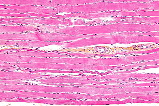
Striated muscle tissue is a muscle tissue that features repeating functional units called sarcomeres. The presence of sarcomeres manifests as a series of bands visible along the muscle fibers, which is responsible for the striated appearance observed in microscopic images of this tissue. There are two types of striated muscle:

<span class="mw-page-title-main">Bioreactor</span> System that supports a biologically active environment

A bioreactor refers to any manufactured device or system that supports a biologically active environment. In one case, a bioreactor is a vessel in which a chemical process is carried out which involves organisms or biochemically active substances derived from such organisms. This process can either be aerobic or anaerobic. These bioreactors are commonly cylindrical, ranging in size from litres to cubic metres, and are often made of stainless steel. It may also refer to a device or system designed to grow cells or tissues in the context of cell culture. These devices are being developed for use in tissue engineering or biochemical/bioprocess engineering.

<span class="mw-page-title-main">Muscle contraction</span> Activation of tension-generating sites in muscle

Muscle contraction is the activation of tension-generating sites within muscle cells. In physiology, muscle contraction does not necessarily mean muscle shortening because muscle tension can be produced without changes in muscle length, such as when holding something heavy in the same position. The termination of muscle contraction is followed by muscle relaxation, which is a return of the muscle fibers to their low tension-generating state.

<span class="mw-page-title-main">Myofilament</span> The two protein filaments of myofibrils in muscle cells

Myofilaments are the three protein filaments of myofibrils in muscle cells. The main proteins involved are myosin, actin, and titin. Myosin and actin are the contractile proteins and titin is an elastic protein. The myofilaments act together in muscle contraction, and in order of size are a thick one of mostly myosin, a thin one of mostly actin, and a very thin one of mostly titin.

A pennate or pinnate muscle is a type of skeletal muscle with fascicles that attach obliquely to its tendon. This type of muscle generally allows higher force production but a smaller range of motion. When a muscle contracts and shortens, the pennation angle increases.

<span class="mw-page-title-main">Muscle</span> Basic biological tissue present in animals

Muscle is a soft tissue, one of the animal tissues that makes up the three different types of muscle. Muscle tissue gives skeletal muscles the ability to contract. Muscle is formed during embryonic development, in a process known as myogenesis. Muscle tissue contains special contractile proteins called actin and myosin which interact to cause movement. Among many other muscle proteins present are two regulatory proteins, troponin and tropomyosin.

<span class="mw-page-title-main">C2C12</span> Mouse myoblast cell line

C2C12 is an immortalized mouse myoblast cell line. The C2C12 cell line is a subclone of myoblasts that were originally obtained by Yaffe and Saxel at the Weizmann Institute of Science in Israel in 1977. Developed for in vitro studies of myoblasts isolated from the complex interactions of in vivo conditions, C2C12 cells are useful in biomedical research. These cells are capable of rapid proliferation under high serum conditions and differentiation into myotubes under low serum conditions. Mononucleated myoblasts can later fuse to form multinucleated myotubes under low serum conditions or starvation, leading to the precursors of contractile skeletal muscle cells in the process of myogenesis. C2C12 cells are used to study the differentiation of myoblasts, osteoblasts, and myogenesis, to express various target proteins, and to explore mechanistic biochemical pathways.

<span class="mw-page-title-main">Langendorff heart</span> Preparation technique for ex vivo experiments

The Langendorff heart or isolated perfused heart assay is an ex vivo technique used in pharmacological and physiological research using animals and also humans. Named after the German physiologist Oskar Langendorff, this technique allows the examination of cardiac contractile strength and heart rate without the complications of an intact animal or human. After more than 100 years, this method is still being used.

<span class="mw-page-title-main">Work loop</span>

The work loop technique is used in muscle physiology to evaluate the mechanical work and power output of skeletal or cardiac muscle contractions via in vitro muscle testing of whole muscles, fiber bundles or single muscle fibers. This technique is primarily used for cyclical contractions such as cockroach walking., the rhythmic flapping of bird wings or the beating of heart ventricular muscle.

<span class="mw-page-title-main">Lateral force transmission in skeletal muscle</span>

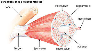
A key component in lateral force transmission in skeletal muscle is the extracellular matrix (ECM). Skeletal muscle is a complex biological material that is composed of muscle fibers and an ECM consisting of the epimysium, perimysium, and endomysium. It can be described as a collagen fiber-reinforced composite. The ECM has at least three functions: (1) to provide a framework binding muscle fibers together and ensure their proper alignment, (2) to transmit the forces, either from active muscle contraction or ones passively imposed on it, and (3) providing lubricated surfaces between muscle fibers and bundles enabling the muscle to change shape. The mechanical properties of skeletal muscle depend on both the properties of muscle fibers and the ECM, and the interaction between the two. Contractile forces are transmitted laterally within intramuscular connective tissue to the epimysium and then to the tendon. Due to the nature of skeletal muscle, direct measurements are not possible, but many indirect studies and analyses have shown that the ECM is an important part of force transmission during muscle contraction.

Muscle architecture is the physical arrangement of muscle fibers at the macroscopic level that determines a muscle’s mechanical function. There are several different muscle architecture types including: parallel, pennate and hydrostats. Force production and gearing vary depending on the different muscle parameters such as muscle length, fiber length, pennation angle, and the physiological cross-sectional area (PCSA).

Elastic mechanisms in animals are very important in the movement of vertebrate animals. The muscles that control vertebrate locomotion are affiliated with tissues that are springy, such as tendons, which lie within the muscles and connective tissue. A spring can be a mechanism for different actions involved in hopping, running, walking, and serve in other diverse functions such as metabolic energy conservation, attenuation of muscle power production, and amplification of muscle power production.

Symmorphosis is the regulation of biological units to produce an optimal outcome. Symmorphosis is when a quantitative match of design and function within an organism defined within a functional system. Symmorphosis can be broken down into the three predictions that are required for organs to evolve within a species.

Entomoculture is the subfield of cellular agriculture which specifically deals with the production of insect tissue in vitro. It draws on principles more generally used in tissue engineering and has scientific similarities to Baculovirus Expression Vectors or soft robotics. The field has mainly been proposed because of its potential technical advantages over mammalian cells in generating cultivated meat. The name of the field was coined by Natalie Rubio at Tufts University.

References

  1. 1 2 Blau HM, Webster C (September 1981). "Isolation and characterization of human muscle cells". Proceedings of the National Academy of Sciences of the United States of America. 78 (9): 5623–5627. Bibcode:1981PNAS...78.5623B. doi: 10.1073/pnas.78.9.5623 . PMC   348807 . PMID   6946499.
  2. aursc20dev (2012-11-08). "Advantages of Testing Muscle Mechanics in-situ or in-vivo". Aurora Scientific. Retrieved 2023-06-27.
  3. James RS, Tallis J, Angilletta MJ (January 2015). "Regional thermal specialisation in a mammal: temperature affects power output of core muscle more than that of peripheral muscle in adult mice (Mus musculus)" (PDF). Journal of Comparative Physiology B: Biochemical, Systemic, and Environmental Physiology. 185 (1): 135–142. doi:10.1007/s00360-014-0872-6. PMID   25403362. S2CID   17011933.
  4. Smith LR, Meyer GA (2020). "Skeletal muscle explants: ex-vivo models to study cellular behavior in a complex tissue environment". Connective Tissue Research. 61 (3–4): 248–261. doi:10.1080/03008207.2019.1662409. PMC   8837600 . PMID   31492079.
  5. Dessauge F, Schleder C, Perruchot MH, Rouger K (May 2021). "3D in vitro models of skeletal muscle: myopshere, myobundle and bioprinted muscle construct". Veterinary Research. 52 (1): 72. doi:10.1186/s13567-021-00942-w. PMC   8136231 . PMID   34011392.