Related Research Articles

Dentistry, also known as dental medicine and oral medicine, is the branch of medicine focused on the teeth, gums, and mouth. It consists of the study, diagnosis, prevention, management, and treatment of diseases, disorders, and conditions of the mouth, most commonly focused on dentition as well as the oral mucosa. Dentistry may also encompass other aspects of the craniofacial complex including the temporomandibular joint. The practitioner is called a dentist.

A dental dam or rubber dam is a thin, 6-inch (150 mm) square sheet, usually latex or nitrile, used in dentistry to isolate the operative site from the rest of the mouth. Sometimes termed "Kofferdam", it was designed in the United States in 1864 by Sanford Christie Barnum. It is used mainly in endodontic, fixed prosthodontic and general restorative treatments. Its purpose is both to prevent saliva interfering with the dental work, and to prevent instruments and materials from being inhaled, swallowed or damaging the mouth. In dentistry, use of a rubber dam is sometimes referred to as isolation or moisture control.

Tooth decay, also known as cavities or caries, is the breakdown of teeth due to acids produced by bacteria. The cavities may be a number of different colors, from yellow to black. Symptoms may include pain and difficulty eating. Complications may include inflammation of the tissue around the tooth, tooth loss and infection or abscess formation. Tooth regeneration is an on-going stem cell based field of study that is trying to reverse the effects of decay, unlike most current methods which only try to make dealing with the effects easier.
Hyperdontia is the condition of having supernumerary teeth, or teeth that appear in addition to the regular number of teeth. They can appear in any area of the dental arch and can affect any dental organ. The opposite of hyperdontia is hypodontia, where there is a congenital lack of teeth, which is a condition seen more commonly than hyperdontia. The scientific definition of hyperdontia is "any tooth or odontogenic structure that is formed from tooth germ in excess of usual number for any given region of the dental arch." The additional teeth, which may be few or many, can occur on any place in the dental arch. Their arrangement may be symmetrical or non-symmetrical.

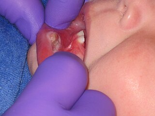
Teething is the process by which an infant's first teeth appear by emerging through the gums, typically arriving in pairs. The mandibular central incisors are the first primary teeth to erupt, usually between 6 and 10 months of age and usually causes discomfort and pain to the infant. It can take several years for all 20 teeth to complete the tooth eruption. Though the process of teething is sometimes referred to as "cutting teeth", when teeth emerge through the gums they do not cut through the flesh. Instead, hormones are released within the body that cause some cells in the gums to die and separate, allowing the teeth to come through.
Hypodontia is defined as the developmental absence of one or more teeth excluding the third molars. It is one of the most common dental anomalies, and can have a negative impact on function, and also appearance. It rarely occurs in primary teeth and the most commonly affected are the adult second premolars and the upper lateral incisors. It usually occurs as part of a syndrome that involves other abnormalities and requires multidisciplinary treatment.

Deciduous teeth or primary teeth, also informally known as baby teeth, milk teeth, or temporary teeth, are the first set of teeth in the growth and development of humans and other diphyodonts, which include most mammals but not elephants, kangaroos, or manatees, which are polyphyodonts. Deciduous teeth develop during the embryonic stage of development and erupt during infancy. They are usually lost and replaced by permanent teeth, but in the absence of their permanent replacements, they can remain functional for many years into adulthood.

Acid erosion is a type of tooth wear. It is defined as the irreversible loss of tooth structure due to chemical dissolution by acids not of bacterial origin. Dental erosion is the most common chronic condition of children ages 5–17, although it is only relatively recently that it has been recognised as a dental health problem. There is widespread ignorance of the damaging effects of acid erosion; this is particularly the case with erosion due to consumption of fruit juices because they tend to be seen as healthy. Acid erosion begins initially in the enamel, causing it to become thin, and can progress into dentin, giving the tooth a dull yellow appearance and leading to dentin hypersensitivity.

The dental lamina is a band of epithelial tissue seen in histologic sections of a developing tooth. The dental lamina is first evidence of tooth development and begins at the sixth week in utero or three weeks after the rupture of the buccopharyngeal membrane. It is formed when cells of the oral ectoderm proliferate faster than cells of other areas. Best described as an in-growth of oral ectoderm, the dental lamina is frequently distinguished from the vestibular lamina, which develops concurrently. This dividing tissue is surrounded by and, some would argue, stimulated by ectomesenchymal growth. When it is present, the dental lamina connects the developing tooth bud to the epithelium of the oral cavity. Eventually, the dental lamina disintegrates into small clusters of epithelium and is resorbed. In situations when the clusters are not resorbed, eruption cysts are formed over the developing tooth and delay its eruption into the oral cavity. This invagination of ectodermal tissues is the progenitor to the later ameloblasts and enamel while the ectomesenchyme is responsible for the dental papilla and later odontoblasts.

Tooth development or odontogenesis is the complex process by which teeth form from embryonic cells, grow, and erupt into the mouth. For human teeth to have a healthy oral environment, all parts of the tooth must develop during appropriate stages of fetal development. Primary (baby) teeth start to form between the sixth and eighth week of prenatal development, and permanent teeth begin to form in the twentieth week. If teeth do not start to develop at or near these times, they will not develop at all, resulting in hypodontia or anodontia.

Early childhood caries (ECC), formerly known as nursing bottle caries, baby bottle tooth decay, night bottle mouth and night bottle caries, is a disease that affects teeth in children aged between birth and 71 months. ECC is characterized by the presence of 1 or more decayed, missing, or filled tooth surfaces in any primary tooth. ECC has been shown to be a very common, transmissible bacterial infection, usually passed from the primary caregiver to the child. The main bacteria responsible for dental cavities are Streptococcus mutans (S.mutans) and Lactobacillus. There is also evidence that supports that those who are in lower socioeconomic populations are at greater risk of developing ECC.
In the medical field a papoose board is a temporary medical stabilization board used to limit a patient's freedom of movement to decrease risk of injury while allowing safe completion of treatment. The term papoose board refers to a brand name.

An impacted tooth is one that fails to erupt into the dental arch within the expected developmental window. Because impacted teeth do not erupt, they are retained throughout the individual's lifetime unless extracted or exposed surgically. Teeth may become impacted because of adjacent teeth, dense overlying bone, excessive soft tissue or a genetic abnormality. Most often, the cause of impaction is inadequate arch length and space in which to erupt. That is the total length of the alveolar arch is smaller than the tooth arch. The wisdom teeth are frequently impacted because they are the last teeth to erupt in the oral cavity. Mandibular third molars are more commonly impacted than their maxillary counterparts.

Dental trauma refers to trauma (injury) to the teeth and/or periodontium, and nearby soft tissues such as the lips, tongue, etc. The study of dental trauma is called dental traumatology.
Riga–Fede disease(RFD) is a rare and benign mucosal condition, characterized by a tongue ulcer that is frequently brought on by traumatizing injuries sustained from repeatedly moving the tongue back and forth over the mandibular anterior incisors.
The Hall Technique is a minimally-invasive treatment for decayed baby back (molar) teeth. Decay is sealed under preformed crowns, avoiding injections and drilling. It is one of a number of biologically oriented strategies for managing dental decay.

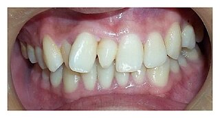
Tooth discoloration is abnormal tooth color, hue or translucency. External discoloration is accumulation of stains on the tooth surface. Internal discoloration is due to absorption of pigment particles into tooth structure. Sometimes there are several different co-existent factors responsible for discoloration.

Molar incisor hypomineralisation (MIH) is a type of enamel defect affecting, as the name suggests, the first molars and incisors in the permanent dentition. MIH is considered a worldwide problem with a global prevalence of 12.9% and is usually identified in children under 10 years old. This developmental condition is caused by the lack of mineralisation of enamel during its maturation phase, due to interruption to the function of ameloblasts. Peri- and post-natal factors including premature birth, certain medical conditions, fever and antibiotic use have been found to be associated with development of MIH. Recent studies have suggested the role of genetics and/or epigenetic changes to be contributors of MIH development. However, further studies on the aetiology of MIH are required because it is believed to be multifactorial.
Atraumatic restorative treatment (ART) is a method for cleaning out tooth decay from teeth using only hand instruments and placing a filling. It does not use rotary dental instruments to prepare the tooth and can be performed in settings with no access to dental equipment. No drilling or local anaesthetic injections are required. ART is considered a conservative approach, not only because it removes the decayed tissue with hand instruments, avoiding removing more tissue than necessary which preserves as much tooth structure as possible, but also because it avoids pulp irritation and minimises patient discomfort. ART can be used for small, medium and deep cavities caused by dental caries.
Dentistry developed during the early parts of Roman history, which may be due to the arrival of a Greek doctor named Archagathus. Ancient Roman oral surgical tools included the curettes, osteotomes, cauteries, scalpels, bone forceps, and bone levers. The ancient Romans invented the usage of narcotics during dental surgery. These tools were used to treat conditions such as toothache and to extract teeth. It was believed in ancient Rome that the cause of the conditions that necessitated such treatment was a "tooth worm."
References
- ↑ Wordley, J (2003). "Infant oral mutilation" (PDF). Developing Dentistry. 3 (2): 19–20. Archived from the original (PDF) on 2011-01-05. Retrieved 2011-05-06.
- ↑ Ellis, J.; Arubaku, W. (2005). "Complications from traditional tooth extraction in South-western Uganda". Tropical Doctor. 35 (4): 245–246. doi:10.1258/004947505774938701. PMID 16354490.
- ↑ Abusinna, I. (1979). "Lugbara teeth germectomy of canines for the newborn babies. A magico-religious phenomena in some African tribes". Egyptian Dental Journal. 25 (3): 209–214. PMID 299152.
- 1 2 Iriso, R.; Accorsi, S.; Akena, S.; Amone, J.; Fabiani, M.; Ferrarese, N.; Lukwiya, M.; Rosolen, T.; Declich, S. (2000). "'Killer' canines: The morbidity and mortality of ebino in northern Uganda". Tropical Medicine and International Health. 5 (10): 706–710. doi: 10.1046/j.1365-3156.2000.00625.x . PMID 11044265. S2CID 21050332.
- ↑ Welbury, R.; Nunn, J.; Gordon, P.; Green-Abate, C. (1993). ""Killer" canine removal and its sequelae in Addis Ababa". Quintessence International. 24 (5): 323–327. PMID 8362046.
- 1 2 Dinur, N.; Becker, T.; Levin, A.; Zadik, Y.; Itzhak, JB.; Azizi, H. (2021). "Long-term dental implications of infant oral mutilation: a case series". British Dental Journal. 231 (6): 335–340. doi:10.1038/s41415-021-3456-3. PMID 34561584.
- 1 2 Khonsari, R. H.; Corre, P.; Perrin, J. P.; Piot, B. (2009). "Orthodontic Consequences of Ritual Dental Mutilations in Northern Tchad". Journal of Oral and Maxillofacial Surgery. 67 (4): 902–905. doi:10.1016/j.joms.2008.06.098. PMID 19304055.
- ↑ Children’s teeth and their care. Document produced by NCTPE (National Committee of Traditional Practices of Ethiopia 1997
- ↑ Hassanali, J.; Amwayi, P.; Muriithi, A. (1995). "Removal of deciduous canine tooth buds in Kenyan rural Maasai". East African Medical Journal. 72 (4): 207–209. PMID 7621751.
- ↑ Benzian, H (2003). "World Dental Development Fund Rwanda Project Visit Report" (PDF). Developing Dentistry. 3 (2): 21–3. Archived from the original (PDF) on 2011-01-05. Retrieved 2011-05-06.
- 1 2 Rodd, H.; Davidson, L. (2000). "'Ilko dacowo:' canine enucleation and dental sequelae in Somali children". International Journal of Paediatric Dentistry. 10 (4): 290–297. doi:10.1046/j.1365-263x.2000.00213.x. PMID 11310242.
- ↑ A/wahab, M. (1987). "Traditional practice as a cause of infant morbidity and mortality in Juba area (Sudan)". Annals of Tropical Paediatrics. 7 (1): 18–21. doi:10.1080/02724936.1987.11748467. PMID 2438998.
- ↑ Matee, M.; Van Palenstein Helderman, W. (1991). "Extraction of 'nylon' teeth and associated abnormalities in Tanzanian children". African Dental Journal. 5: 21–25. PMID 1819291.
- ↑ Holan, G.; Mamber, E. (1994). "Extraction of primary canine tooth buds: Prevalence and associated dental abnormalities in a group of Ethiopian Jewish children". International Journal of Paediatric Dentistry. 4 (1): 25–30. doi:10.1111/j.1365-263x.1994.tb00097.x. PMID 7748844.
- ↑ Graham, E.; Domoto, P.; Lynch, H.; Egbert, M. (2000). "Dental injuries due to African traditional therapies for diarrhea". The Western Journal of Medicine. 173 (2): 135–137. doi:10.1136/ewjm.173.2.135. PMC 1071025 . PMID 10924443.
- ↑ Amailuk, P.; Grubor, D. (2008). "Erupted compound odontoma: Case report of a 15-year-old Sudanese boy with a history of traditional dental mutilation". BDJ. 204 (1): 11–14. doi: 10.1038/bdj.2007.1184 . PMID 18192989.
- ↑ Espelid, E; Agnalt R (2009). "Removal of dental facilities in African folk medicine. (Translation from Norwegian)". Nor Dental Tid. 119: 294–297.
- ↑ De Beavis, F. O.; Foster, A. C.; Fuge, K. N.; Whyman, R. A. (2011). "Infant oral mutilation: A New Zealand case series". The New Zealand Dental Journal. 107 (2): 57–59. PMID 21721338.
- ↑ Dewhurst, S.; Mason, C. (2001). "Traditional tooth bud gouging in a Ugandan family: A report involving three sisters". International Journal of Paediatric Dentistry. 11 (4): 292–297. doi:10.1046/j.1365-263x.2001.00279.x. PMID 11570446.
- ↑ "www.dentaid.org". 22 June 2011. Archived from the original on 22 June 2011.