Related Research Articles
Computational neuroscience is a branch of neuroscience which employs mathematics, computer science, theoretical analysis and abstractions of the brain to understand the principles that govern the development, structure, physiology and cognitive abilities of the nervous system.

Neuroesthetics is a relatively recent sub-discipline of applied aesthetics. Empirical aesthetics takes a scientific approach to the study of aesthetic experience of art, music, or any object that can give rise to aesthetic judgments. Neuroesthetics is a term coined by Semir Zeki in 1999 and received its formal definition in 2002 as the scientific study of the neural bases for the contemplation and creation of a work of art. Neuroesthetics uses neuroscience to explain and understand the aesthetic experiences at the neurological level. The topic attracts scholars from many disciplines including neuroscientists, art historians, artists, art therapists and psychologists.
Neuroscience and intelligence refers to the various neurological factors that are partly responsible for the variation of intelligence within species or between different species. A large amount of research in this area has been focused on the neural basis of human intelligence. Historic approaches to studying the neuroscience of intelligence consisted of correlating external head parameters, for example head circumference, to intelligence. Post-mortem measures of brain weight and brain volume have also been used. More recent methodologies focus on examining correlates of intelligence within the living brain using techniques such as magnetic resonance imaging (MRI), functional MRI (fMRI), electroencephalography (EEG), positron emission tomography and other non-invasive measures of brain structure and activity.
A gamma wave or gamma rhythm is a pattern of neural oscillation in humans with a frequency between 25 and 140 Hz, the 40 Hz point being of particular interest. Gamma rhythms are correlated with large-scale brain network activity and cognitive phenomena such as working memory, attention, and perceptual grouping, and can be increased in amplitude via meditation or neurostimulation. Altered gamma activity has been observed in many mood and cognitive disorders such as Alzheimer's disease, epilepsy, and schizophrenia.

In psycholinguistics, language processing refers to the way humans use words to communicate ideas and feelings, and how such communications are processed and understood. Language processing is considered to be a uniquely human ability that is not produced with the same grammatical understanding or systematicity in even human's closest primate relatives.
Neuroplasticity, also known as neural plasticity or brain plasticity, is the ability of neural networks in the brain to change through growth and reorganization. It is when the brain is rewired to function in some way that differs from how it previously functioned. These changes range from individual neuron pathways making new connections, to systematic adjustments like cortical remapping or neural oscillation. Other forms of neuroplasticity include homologous area adaptation, cross modal reassignment, map expansion, and compensatory masquerade. Examples of neuroplasticity include circuit and network changes that result from learning a new ability, information acquisition, environmental influences, practice, and psychological stress.
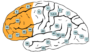
In mammalian brain anatomy, the prefrontal cortex (PFC) covers the front part of the frontal lobe of the cerebral cortex. It is the association cortex in the frontal lobe. The PFC contains the Brodmann areas BA8, BA9, BA10, BA11, BA12, BA13, BA14, BA24, BA25, BA32, BA44, BA45, BA46, and BA47.

Neural oscillations, or brainwaves, are rhythmic or repetitive patterns of neural activity in the central nervous system. Neural tissue can generate oscillatory activity in many ways, driven either by mechanisms within individual neurons or by interactions between neurons. In individual neurons, oscillations can appear either as oscillations in membrane potential or as rhythmic patterns of action potentials, which then produce oscillatory activation of post-synaptic neurons. At the level of neural ensembles, synchronized activity of large numbers of neurons can give rise to macroscopic oscillations, which can be observed in an electroencephalogram. Oscillatory activity in groups of neurons generally arises from feedback connections between the neurons that result in the synchronization of their firing patterns. The interaction between neurons can give rise to oscillations at a different frequency than the firing frequency of individual neurons. A well-known example of macroscopic neural oscillations is alpha activity.
David J. Heeger is an American neuroscientist, psychologist, computer scientist, data scientist, and entrepreneur. He is a professor at New York University, Chief Scientific Officer of Statespace Labs, and Chief Scientific Officer and co-founder of Epistemic AI.

The intraparietal sulcus (IPS) is located on the lateral surface of the parietal lobe, and consists of an oblique and a horizontal portion. The IPS contains a series of functionally distinct subregions that have been intensively investigated using both single cell neurophysiology in primates and human functional neuroimaging. Its principal functions are related to perceptual-motor coordination and visual attention, which allows for visually-guided pointing, grasping, and object manipulation that can produce a desired effect.

The fusiform face area is a part of the human visual system that is specialized for facial recognition. It is located in the inferior temporal cortex (IT), in the fusiform gyrus.
Repetition priming refers to improvements in a behavioural response when stimuli are repeatedly presented. The improvements can be measured in terms of accuracy or reaction time and can occur when the repeated stimuli are either identical or similar to previous stimuli. These improvements have been shown to be cumulative, so as the number of repetitions increases the responses get continually faster up to a maximum of around seven repetitions. These improvements are also found when the repeated items are changed slightly in terms of orientation, size and position. The size of the effect is also modulated by the length of time the item is presented for and the length time between the first and subsequent presentations of the repeated items.
In cognitive neuroscience, visual modularity is an organizational concept concerning how vision works. The way in which the primate visual system operates is currently under intense scientific scrutiny. One dominant thesis is that different properties of the visual world require different computational solutions which are implemented in anatomically/functionally distinct regions that operate independently – that is, in a modular fashion.
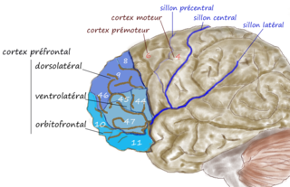
The dorsolateral prefrontal cortex is an area in the prefrontal cortex of the primate brain. It is one of the most recently derived parts of the human brain. It undergoes a prolonged period of maturation which lasts into adulthood. The DLPFC is not an anatomical structure, but rather a functional one. It lies in the middle frontal gyrus of humans. In macaque monkeys, it is around the principal sulcus. Other sources consider that DLPFC is attributed anatomically to BA 9 and 46 and BA 8, 9 and 10.
Marcel Just is D. O. Hebb Professor of Psychology at Carnegie Mellon University. His research uses brain imaging (fMRI) in high-level cognitive tasks to study the neuroarchitecture of cognition. Just's areas of expertise include psycholinguistics, object recognition, and autism, with particular attention to cognitive and neural substrates. Just directs the Center for Cognitive Brain Imaging and is a member of the Center for the Neural Basis of Cognition at CMU.
The neuroscience of music is the scientific study of brain-based mechanisms involved in the cognitive processes underlying music. These behaviours include music listening, performing, composing, reading, writing, and ancillary activities. It also is increasingly concerned with the brain basis for musical aesthetics and musical emotion. Scientists working in this field may have training in cognitive neuroscience, neurology, neuroanatomy, psychology, music theory, computer science, and other relevant fields.
Meditation and pain is the study of the physiological mechanisms underlying meditation—specifically its neural components—that implicate it in the reduction of pain perception.

John-Dylan Haynes is a British-German brain researcher.
Social cognitive neuroscience is the scientific study of the biological processes underpinning social cognition. Specifically, it uses the tools of neuroscience to study "the mental mechanisms that create, frame, regulate, and respond to our experience of the social world". Social cognitive neuroscience uses the epistemological foundations of cognitive neuroscience, and is closely related to social neuroscience. Social cognitive neuroscience employs human neuroimaging, typically using functional magnetic resonance imaging (fMRI). Human brain stimulation techniques such as transcranial magnetic stimulation and transcranial direct-current stimulation are also used. In nonhuman animals, direct electrophysiological recordings and electrical stimulation of single cells and neuronal populations are utilized for investigating lower-level social cognitive processes.
The occipital face area (OFA) is a region of the human cerebral cortex which is specialised for face perception. The OFA is located on the lateral surface of the occipital lobe adjacent to the inferior occipital gyrus. The OFA comprises a network of brain regions including the fusiform face area (FFA) and posterior superior temporal sulcus (STS) which support facial processing.
References
- 1 2 3 "James V. Haxby - CV" (PDF).
- ↑ "Dartmouth Brain Imaging Center".
- ↑ "Center for Cognitive Neuroscience at Dartmouth".
- ↑ Ungerleider, L. G. & Haxby, J. V. (1994). "'What' and 'where' in the human brain". Current Opinion in Neurobiology. 4 (2): 157–165. doi:10.1016/0959-4388(94)90066-3. PMID 8038571. S2CID 205127941.
- ↑ Martin, A.; Wiggs, C. L.; Ungerleider, L. G. & Haxby, J. V. (1996). "Neural correlates of category-specific knowledge". Nature. 379 (6566): 649–652. Bibcode:1996Natur.379..649M. doi:10.1038/379649a0. PMID 8628399. S2CID 4310817.
- ↑ Haxby, J. V.; Hoffman, E. A. & Gobbini, M. I. (2000). "The distributed human neural system for face perception". Trends in Cognitive Sciences. 4 (6): 223–233. doi:10.1016/S1364-6613(00)01482-0. PMID 10827445. S2CID 17047447.
- ↑ Norman, K. A.; Polyn, S. M.; Detre, G. J. & Haxby, J. V. (2006). "Beyond mind-reading: multi-voxel pattern analysis of fMRI data". Trends in Cognitive Sciences. 10 (9): 424–430. doi:10.1016/j.tics.2006.07.005. PMID 16899397. S2CID 704855.
- ↑ Haxby, J. V. (2011). "Multivariate pattern analysis of fMRI: The early beginnings". NeuroImage. 62 (2): 852–855. doi:10.1016/j.neuroimage.2012.03.016. PMC 3389290 . PMID 22425670.
- ↑ Haxby, J. V.; Connolly, A. C. & Guntupalli, J. S. (2014). "Decoding neural representational spaces using multivariate pattern analysis". Annual Review of Neuroscience. 37: 435–456. doi: 10.1146/annurev-neuro-062012-170325 . PMID 25002277. S2CID 6794418.
- ↑ Haxby, J. V.; Gobbini, M. I.; Furey, M. I.; Ishai, A.; Schouten, J. L. & Pietrini, P. (2001). "Distributed and overlapping representations of faces and objects in ventral temporal cortex" (PDF). Science. 293 (5539): 2425–2430. Bibcode:2001Sci...293.2425H. doi:10.1126/science.1063736. PMID 11577229. S2CID 6403660.
- ↑ Kanwisher, N.; McDermott, J. & Chun, M. M. (1997). "The fusiform face area: a module in human extrastriate cortex specialized for face perception". Journal of Neuroscience. 17 (11): 4302–4311. doi:10.1523/JNEUROSCI.17-11-04302.1997. PMC 6573547 . PMID 9151747.
- ↑ Connolly, A. C.; Guntupalli, J. S.; Gors, J.; Hanke, M.; Halchenko, Y. O.; Yu, Y.-C.; Abdi, H. & Haxby, J. V. (2012). "The representation of biological classes in the human brain". Journal of Neuroscience. 32 (8): 2508–2618. doi:10.1523/JNEUROSCI.5547-11.2012. PMC 3532035 . PMID 22357845.
- ↑ Sabuncu, M. R.; Singer, B. D.; Conroy, B.; Bryan, R. E.; Ramadge, P. J. & Haxby, J. V. (2010). "Function-based intersubject alignment of cortical anatomy". Cerebral Cortex. 20 (1): 130–140. doi:10.1093/cercor/bhp085. PMC 2792192 . PMID 19420007.
- ↑ Haxby, J. V.; Guntupalli, J. S.; Connolly, A. C.; Halchenko, Y. O.; Conroy, B.; Gobbini, M. I.; Hanke, M. & Ramadge, P. J. (2011). "A common, high-dimensional model of the representational space in human ventral temporal cortex". Neuron. 72 (2): 404–416. doi:10.1016/j.neuron.2011.08.026. PMC 3201764 . PMID 22017997.
- ↑ Guntupalli, J. S.; Hanke, M.; Halchenko, Y. O.; Connolly, A. C.; Ramadge, P. J. & Haxby, J. V. (2016). "A model of representational spaces in human cortex". Cerebral Cortex. 26 (6): 2919–2934. doi:10.1093/cercor/bhw068. PMC 4869822 . PMID 26980615.
- ↑ "Center for Open Neuroscience".