Jumping library

Last updated
This figure illustrates the basic principle behind jumping libraries.The arrows represent two physically distant sequences which are brought closer together using this method. Figure 1 Basic Principle of a Jumping Library.png
This figure illustrates the basic principle behind jumping libraries.The arrows represent two physically distant sequences which are brought closer together using this method.

Jumping libraries or junction-fragment libraries are collections of genomic DNA fragments generated by chromosome jumping. These libraries allow the analysis of large areas of the genome and overcome distance limitations in common cloning techniques. A jumping library clone is composed of two stretches of DNA that are usually located many kilobases away from each other. The stretch of DNA located between these two "ends" is deleted by a series of biochemical manipulations carried out at the start of this cloning technique.

Contents

Invention and early improvements

Origin

Chromosome jumping (or chromosome hopping) was first described in 1984 by Collins and Weissman. [1] At the time, cloning techniques allowed for generation of clones of limited size (up to 240kb), and cytogenetic techniques allowed for mapping such clones to a small region of a particular chromosome to a resolution of around 5-10Mb. Therefore, a major gap remained in resolution between available technologies, and no methods were available for mapping larger areas of the genome. [1]

Basic principle and original method

This figure is a schematic representation of the method used for creating jumping libraries when it was originally developed in the 80s. Figure 2 Original method for creating a jumping library.png
This figure is a schematic representation of the method used for creating jumping libraries when it was originally developed in the 80s.

This technique is an extension of "chromosome walking" that allows larger "steps" along the chromosome. If steps of length N kb are desired, very high molecular weight DNA is necessary. Once isolated, it is partially digested with a frequent-cutting restriction enzyme (such as MboI or BamHI). Next, obtained fragments are selected for size which should be around N kb in length. DNA must then be ligated at low concentration to favour ligation into circles rather than formation of multimers. A DNA marker (such as the amber suppressor tRNA gene supF) can be included at this time point within the covalently linked circle to allow for selection of junction fragments. Circles are subsequently fully digested with a second restriction enzyme (such as EcoRI) to generate a large number of fragments. Such fragments are ligated into vectors (such as a λ vector) which should be selected for using the DNA marker introduced earlier. The remaining fragments thus represent the library of junction fragments, or "jumping library". [1] The next step is to screen this library with a probe that represents a "starting point" of the desired "chromosome hop", i.e. determining the location of the genome that is being interrogated. Clones obtained from this final selection step will consist of DNA that is homologous to our probe, separated by our DNA marker from another DNA sequence that was originally located N kb away (thus being called "jumping"). [1] By generating several libraries of different N values, eventually the entire genome can be mapped, allowing movement from one location to another, while controlling direction, by any value of N desired. [1]

Early challenges and improvements

The original technique of chromosome jumping was developed in the laboratories of Collins and Weissman at Yale University in New Haven, U.S. [1] and the laboratories of Poustka and Lehrach at the European Molecular Biology Laboratory in Heidelberg, Germany. [2]

Collins and Weissman's method [1] described above encountered some early limitations. The main concern was with avoiding non-circularized fragments. Two solutions were suggested: either screening junction fragments with a given probe or adding a second size-selection step after the ligation to separate single circular clones (monomers) from clones ligated to each other (multimers). The authors also suggested that other markers such as the λ cos site or antibiotic resistance genes should be considered (instead of the amber suppressor tRNA gene) to facilitate selection of junction clones.

Poustka and Lehrach [2] suggested that full digestion with rare-cutting restrictions enzymes (such as NotI) should be used for the first step of the library construction instead of partial digestion with a frequently cutting restriction enzyme. This would significantly reduce the number of clones from millions to thousands. However, this could create problems with circularizing the DNA fragments since these fragments would be very long, and would also lose the flexibility in choice of end points that one gets in partial digests. One suggestion for overcoming these problems would be to combine the two methods, i.e. to construct a jumping library from DNA fragments digested partially with a commonly cutting restriction enzyme and completely with a rare cutting restriction enzyme and circularizing them into plasmids cleaved with both enzymes. Several of these "combination" libraries were completed in 1986. [2] [3]

In 1991, Zabarovsky et al. [4] proposed a new approach for construction of jumping libraries. This approach included the use of two separate λ vectors for library construction, and a partial filling-in reaction that removes the need for a selectable marker. This filling-in reaction worked by destroying the specific cohesive ends (resulting from restriction digests) of the DNA fragments that were nonligated and noncircularized, thus preventing them from cloning into the vectors, in a more energy-efficient and accurate manner. Furthermore, this improved technique required less DNA to start with, and also produced a library that could be transferred into a plasmid form, making it easier to store and replicate. Using this new approach, they successfully constructed a human NotI jumping library from a lymphoblastoid cell line and a human chromosome 3-specific NotI jumping library from a human chromosome 3 and mouse hybrid cell line. [4]

Current method

Second-generation or "Next-Gen" (NGS) techniques have evolved radically: the sequencing capacity has increased more than ten thousandfold and the cost has dropped by over one million-fold since 2007(National Human Genome Research Institute). NGS has revolutionized the genetic field in many ways.

Library construction

A library is often prepared by random fragmentation of DNA and ligation of common adaptor sequences. [5] [6] However, the generated short reads challenge the identification of structural variants, such as indels, translocations, and duplication. Large regions of simple repeats can further complicate the alignment. [7] Alternatively, a jumping library can be used with NGS for the mapping of structural variation and scaffolding of de novo assemblies. [8]

This figure is a schematic representation of one of the most recently used methods for creating jumping libraries. Figure 3 Recent method for creating a jumping library.png
This figure is a schematic representation of one of the most recently used methods for creating jumping libraries.

Jumping libraries can be categorized according to the length of the incorporated DNA fragments.

Short-jump library

In a short-jump library, 3 kb genomic DNA fragments are ligated with biotinylate ends and circularized. The circular segments are then sheared into small fragments and the biotinylated fragments are selected by affinity assay for paired-end sequencing.

There are two issues related to short-jump libraries. First, a read can pass through the biotinylated circularization junction and reduce the effective read length. Second, reads from non-jumped fragments (i.e. fragments without the circularization junction) are sequenced and reduce genomic coverage. It has been reported that non-jumped fragments range from 4% to 13%, depending on the size of selection. The first problem might be solved by shearing circles into a larger size and select for those larger fragments. The second problem can be addressed by using a custom barcoded jumping library. [9] [10]

Custom barcoded jumping library

This jumping library uses adaptors containing markers for fragment selection in combination with barcodes for multiplexing. The protocol was developed by Talkowski et al. [9] and based on mate-pair library preparation for SOLiD sequencing. The selected DNA fragment size is 3.5 – 4.5 kb. Two adaptors were involved: one containing an EcoP15I recognition site and an AC overhang; the other containing a GT overhang, a biotinylated thymine, and an oligo barcode. The circularized DNA was digested and the fragments with biotynylated adaptors were selected for (see Figure 3). The EcoP15I recognition site and barcode help to distinguish junction fragments from nonjump fragments. These targeted fragments should contain 25 to 27bp of genomic DNA, the EcoP15I recognition site, the overhang, and the barcode. [9]

Long-jump library

This library construction process is similar to that of the short-jump library except that the condition is optimized for longer fragments (5 kb). [10]

Fosmid-jump library

This library construction process is also similar to that of short-jump library except that transfection using the E. coli vector is required for amplification of large (40 kb) DNA fragments. In addition, the fosmids can be modified to facilitate the conversion into jumping library compatible with certain next generation sequencers. [8] [10]

Paired-end sequencing

The segments resulting from circularization during constructing jumping library are cleaved, and DNA fragments with markers will be enriched and subjected to paired-end sequencing. These DNA fragments are sequenced from both ends and generate pairs of reads. The genomic distance between the reads in each pair is approximately known and used for the assembly process. For example, a DNA clone generated by random fragmentation is about 200 bp, and a read from each end is around 180 bp, overlapping each other. This should be distinguished from mate-pair sequencing, which is basically a combination of next generation sequencing with jumping libraries.

Computational analysis

Different assembly tools have been developed to handle jumping library data. One example is DELLY. DELLY was developed to discover genomic structural variants and "integrates short insert paired-ends, long-range mate-pairs and split-read alignments" to detect rearrangements at sequence level. [11]

An example of joint development of new experimental design and algorithm development is demonstrated by the ALLPATHS-LG assembler. [12]

Confirmation

When used for detection of genetic and genomic changes, jumping clones require validation by Sanger sequencing.

Applications

Early applications

In the early days, chromosome walking from genetically linked DNA markers was used to identify and clone disease genes. However, the large molecular distance between known markers and the gene of interest was complicating the cloning process. In 1987, a human chromosome jumping library was constructed to clone the cystic fibrosis gene. Cystic fibrosis is an autosomal recessive disease affecting 1 in 2000 Caucasians. This was the first disease in which the usefulness of the jumping libraries was demonstrated. Met oncogene was a marker tightly linked to the cystic fibrosis gene on human chromosome 7, and the library was screened for a jumping clone starting at this marker. The cystic fibrosis gene was determined to localize 240kb downstream of the met gene. Chromosome jumping helped reduce the mapping "steps" and bypass the highly repetitive regions in the mammalian genome. [13] Chromosome jumping also allowed the production of probes required for faster diagnosis of this and other diseases. [1]

New applications

Characterizing chromosomal rearrangements

Balanced chromosomal rearrangements can have a significant contribution to diseases, as demonstrated by the studies of leukemia. [14] However, many of them are undetected by chromosomal microarray. Karyotyping and FISH can identify balanced translocations and inversions but are labor-intensive and provide low resolution (small genomic changes are missed).

A jumping library NGS combined approach can be applied to identify such genomic changes. For example, Slade et al. applied this method to fine map a de novo balanced translocation in a child with Wilms' tumor. [15] For this study, 50 million reads were generated, but only 11.6% of these could be mapped uniquely to the reference genome, which represents approximately a sixfold coverage.

Talkowski et al. [9] compared different approaches to detect balanced chromosome alterations, and showed that modified jumping library in combination with next generation DNA sequencing is an accurate method for mapping chromosomal breakpoints. Two varieties of jumping libraries (short-jump libraries and custom barcoded jumping libraries) were tested and compared to standard sequencing libraries. For standard NGS, 200-500bp fragments are generated. About 0.03%–0.54% of fragments represent chimeric pairs, which are pairs of end-reads that are mapped to two different chromosomes. Therefore, very few fragments cover the breakpoint area. When using short-jump libraries with fragments of 3.2–3.8kb, the percentage of chimeric pairs increased to 1.3%. With Custom Barcoded Jumping Libraries, the percentage of chimeric pairs further increased to 1.49%. [9]

Prenatal diagnosis

Conventional cytogenetic testing cannot offer the gene-level resolution required to predict the outcome of a pregnancy and whole genome deep sequencing is not practical for routine prenatal diagnosis. Whole-genome jumping library could complement conventional prenatal testing. This novel method was successfully applied to identify a case of CHARGE syndrome. [6]

De novo assembly

In metagenomics, regions of the genomes that are shared between strains are typically longer than the reads. This complicates the assembly process and makes reconstructing individual genomes for a species a daunting task. [10] Chimeric pairs that are mapped far apart in the genome can facilitate the de novo assembly process. By using a longer-jump library, Ribeiro et al. demonstrated that the assemblies of bacterial genomes were of high quality while reducing both cost and time. [16]

Limitation

The cost of sequencing has dropped dramatically while the cost of construction of jumping libraries has not. Therefore, as new sequencing technologies and bioinformatic tools are developed, jumping libraries may become redundant.

See also

Related Research Articles

In genetics, shotgun sequencing is a method used for sequencing random DNA strands. It is named by analogy with the rapidly expanding, quasi-random shot grouping of a shotgun.

A bacterial artificial chromosome (BAC) is a DNA construct, based on a functional fertility plasmid, used for transforming and cloning in bacteria, usually E. coli. F-plasmids play a crucial role because they contain partition genes that promote the even distribution of plasmids after bacterial cell division. The bacterial artificial chromosome's usual insert size is 150–350 kbp. A similar cloning vector called a PAC has also been produced from the DNA of P1 bacteriophage.

A contig is a set of overlapping DNA segments that together represent a consensus region of DNA. In bottom-up sequencing projects, a contig refers to overlapping sequence data (reads); in top-down sequencing projects, contig refers to the overlapping clones that form a physical map of the genome that is used to guide sequencing and assembly. Contigs can thus refer both to overlapping DNA sequences and to overlapping physical segments (fragments) contained in clones depending on the context.

<span class="mw-page-title-main">Yeast artificial chromosome</span> Genetically engineered chromosome derived from the DNA of yeast

Yeast artificial chromosomes (YACs) are genetically engineered chromosomes derived from the DNA of the yeast, Saccharomyces cerevisiae, which is then ligated into a bacterial plasmid. By inserting large fragments of DNA, from 100–1000 kb, the inserted sequences can be cloned and physically mapped using a process called chromosome walking. This is the process that was initially used for the Human Genome Project, however due to stability issues, YACs were abandoned for the use of bacterial artificial chromosome

Chromosome jumping is a tool of molecular biology that is used in the physical mapping of genomes. It is related to several other tools used for the same purpose, including chromosome walking.

<span class="mw-page-title-main">DNA sequencing</span> Process of determining the nucleic acid sequence

DNA sequencing is the process of determining the nucleic acid sequence – the order of nucleotides in DNA. It includes any method or technology that is used to determine the order of the four bases: adenine, guanine, cytosine, and thymine. The advent of rapid DNA sequencing methods has greatly accelerated biological and medical research and discovery.

Primer walking is a technique used to clone a gene from its known closest markers. As a result, it is employed in cloning and sequencing efforts in plants, fungi, and mammals with minor alterations. This technique, also known as "directed sequencing," employs a series of Sanger sequencing reactions to either confirm the reference sequence of a known plasmid or PCR product based on the reference sequence or to discover the unknown sequence of a full plasmid or PCR product by designing primers to sequence overlapping sections.

<span class="mw-page-title-main">Gene mapping</span> Process of locating specific genes

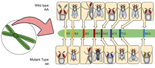
Gene mapping or genome mapping describes the methods used to identify the location of a gene on a chromosome and the distances between genes. Gene mapping can also describe the distances between different sites within a gene.

A genomic library is a collection of overlapping DNA fragments that together make up the total genomic DNA of a single organism. The DNA is stored in a population of identical vectors, each containing a different insert of DNA. In order to construct a genomic library, the organism's DNA is extracted from cells and then digested with a restriction enzyme to cut the DNA into fragments of a specific size. The fragments are then inserted into the vector using DNA ligase. Next, the vector DNA can be taken up by a host organism - commonly a population of Escherichia coli or yeast - with each cell containing only one vector molecule. Using a host cell to carry the vector allows for easy amplification and retrieval of specific clones from the library for analysis.

Fosmids are similar to cosmids but are based on the bacterial F-plasmid. The cloning vector is limited, as a host can only contain one fosmid molecule. Fosmids can hold DNA inserts of up to 40 kb in size; often the source of the insert is random genomic DNA. A fosmid library is prepared by extracting the genomic DNA from the target organism and cloning it into the fosmid vector. The ligation mix is then packaged into phage particles and the DNA is transfected into the bacterial host. Bacterial clones propagate the fosmid library. The low copy number offers higher stability than vectors with relatively higher copy numbers, including cosmids. Fosmids may be useful for constructing stable libraries from complex genomes. Fosmids have high structural stability and have been found to maintain human DNA effectively even after 100 generations of bacterial growth. Fosmid clones were used to help assess the accuracy of the Public Human Genome Sequence.

Paired-end tags (PET) are the short sequences at the 5’ and 3' ends of a DNA fragment which are unique enough that they (theoretically) exist together only once in a genome, therefore making the sequence of the DNA in between them available upon search or upon further sequencing. Paired-end tags (PET) exist in PET libraries with the intervening DNA absent, that is, a PET "represents" a larger fragment of genomic or cDNA by consisting of a short 5' linker sequence, a short 5' sequence tag, a short 3' sequence tag, and a short 3' linker sequence. It was shown conceptually that 13 base pairs are sufficient to map tags uniquely. However, longer sequences are more practical for mapping reads uniquely. The endonucleases used to produce PETs give longer tags but sequences of 50–100 base pairs would be optimal for both mapping and cost efficiency. After extracting the PETs from many DNA fragments, they are linked (concatenated) together for efficient sequencing. On average, 20–30 tags could be sequenced with the Sanger method, which has a longer read length. Since the tag sequences are short, individual PETs are well suited for next-generation sequencing that has short read lengths and higher throughput. The main advantages of PET sequencing are its reduced cost by sequencing only short fragments, detection of structural variants in the genome, and increased specificity when aligning back to the genome compared to single tags, which involves only one end of the DNA fragment.

Optical mapping is a technique for constructing ordered, genome-wide, high-resolution restriction maps from single, stained molecules of DNA, called "optical maps". By mapping the location of restriction enzyme sites along the unknown DNA of an organism, the spectrum of resulting DNA fragments collectively serves as a unique "fingerprint" or "barcode" for that sequence. Originally developed by Dr. David C. Schwartz and his lab at NYU in the 1990s this method has since been integral to the assembly process of many large-scale sequencing projects for both microbial and eukaryotic genomes. Later technologies use DNA melting, DNA competitive binding or enzymatic labelling in order to create the optical mappings.

Polony sequencing is an inexpensive but highly accurate multiplex sequencing technique that can be used to “read” millions of immobilized DNA sequences in parallel. This technique was first developed by Dr. George Church's group at Harvard Medical School. Unlike other sequencing techniques, Polony sequencing technology is an open platform with freely downloadable, open source software and protocols. Also, the hardware of this technique can be easily set up with a commonly available epifluorescence microscopy and a computer-controlled flowcell/fluidics system. Polony sequencing is generally performed on paired-end tags library that each molecule of DNA template is of 135 bp in length with two 17–18 bp paired genomic tags separated and flanked by common sequences. The current read length of this technique is 26 bases per amplicon and 13 bases per tag, leaving a gap of 4–5 bases in each tag.

<span class="mw-page-title-main">Restriction site associated DNA markers</span> Type of genetic marker

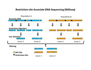
Restriction site associated DNA (RAD) markers are a type of genetic marker which are useful for association mapping, QTL-mapping, population genetics, ecological genetics and evolutionary genetics. The use of RAD markers for genetic mapping is often called RAD mapping. An important aspect of RAD markers and mapping is the process of isolating RAD tags, which are the DNA sequences that immediately flank each instance of a particular restriction site of a restriction enzyme throughout the genome. Once RAD tags have been isolated, they can be used to identify and genotype DNA sequence polymorphisms mainly in form of single nucleotide polymorphisms (SNPs). Polymorphisms that are identified and genotyped by isolating and analyzing RAD tags are referred to as RAD markers. Although genotyping by sequencing presents an approach similar to the RAD-seq method, they differ in some substantial ways.

In DNA sequencing, a read is an inferred sequence of base pairs corresponding to all or part of a single DNA fragment. A typical sequencing experiment involves fragmentation of the genome into millions of molecules, which are size-selected and ligated to adapters. The set of fragments is referred to as a sequencing library, which is sequenced to produce a set of reads.

<span class="mw-page-title-main">End-sequence profiling</span>

End-sequence profiling (ESP) is a method based on sequence-tagged connectors developed to facilitate de novo genome sequencing to identify high-resolution copy number and structural aberrations such as inversions and translocations.

A plant genome assembly represents the complete genomic sequence of a plant species, which is assembled into chromosomes and other organelles by using DNA fragments that are obtained from different types of sequencing technology.

BLESS, also known as breaks labeling, enrichment on streptavidin and next-generation sequencing, is a method used to detect genome-wide double-strand DNA damage. In contrast to chromatin immunoprecipitation (ChIP)-based methods of identifying DNA double-strand breaks (DSBs) by labeling DNA repair proteins, BLESS utilizes biotinylated DNA linkers to directly label genomic DNA in situ which allows for high-specificity enrichment of samples on streptavidin beads and the subsequent sequencing-based DSB mapping to nucleotide resolution.

Physical map is a technique used in molecular biology to find the order and physical distance between DNA base pairs by DNA markers. It is one of the gene mapping techniques which can determine the sequence of DNA base pairs with high accuracy. Genetic mapping, another approach of gene mapping, can provide markers needed for the physical mapping. However, as the former deduces the relative gene position by recombination frequencies, it is less accurate than the latter.

<span class="mw-page-title-main">Hi-C (genomic analysis technique)</span> Genomic analysis technique

Hi-C is a high-throughput genomic and epigenomic technique first described in 2009 by Lieberman-Aiden et al. to capture chromatin conformation. In general, Hi-C is considered as a derivative of a series of chromosome conformation capture technologies, including but not limited to 3C, 4C, and 5C. Hi-C comprehensively detects genome-wide chromatin interactions in the cell nucleus by combining 3C and next-generation sequencing (NGS) approaches and has been considered as a qualitative leap in C-technology development and the beginning of 3D genomics.
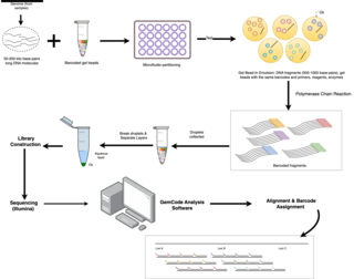
References

  1. 1 2 3 4 5 6 7 8 Collins, FS; Weissman, SM (November 1984). "Directional cloning of DNA fragments at a large distance from an initial probe: a circularization method". Proceedings of the National Academy of Sciences of the United States of America. 81 (21): 6812–6. Bibcode:1984PNAS...81.6812C. doi: 10.1073/pnas.81.21.6812 . PMC   392022 . PMID   6093122.
  2. 1 2 3 Poustka, Annemarie; Lehrach, Hans (1 January 1986). "Jumping libraries and linking libraries: the next generation of molecular tools in mammalian genetics". Trends in Genetics. 2: 174–179. doi:10.1016/0168-9525(86)90219-2.
  3. Poustka, A; Pohl, TM; Barlow, DP; Frischauf, AM; Lehrach, H (Jan 22–28, 1987). "Construction and use of human chromosome jumping libraries from NotI-digested DNA". Nature. 325 (6102): 353–5. Bibcode:1987Natur.325..353P. doi:10.1038/325353a0. PMID   3027567. S2CID   4241410.
  4. 1 2 Zabarovsky, ER; Boldog, F; Erlandsson, R; Kashuba, VI; Allikmets, RL; Marcsek, Z; Kisselev, LL; Stanbridge, E; Klein, G; Sumegi, J (December 1991). "New strategy for mapping the human genome based on a novel procedure for construction of jumping libraries". Genomics. 11 (4): 1030–9. doi:10.1016/0888-7543(91)90029-e. PMID   1783374.
  5. Shendure, J; Ji, H (October 2008). "Next-generation DNA sequencing". Nature Biotechnology. 26 (10): 1135–45. doi:10.1038/nbt1486. PMID   18846087. S2CID   6384349.
  6. 1 2 Talkowski, ME; Ordulu, Z; Pillalamarri, V; Benson, CB; Blumenthal, I; Connolly, S; Hanscom, C; Hussain, N; Pereira, S; Picker, J; Rosenfeld, JA; Shaffer, LG; Wilkins-Haug, LE; Gusella, JF; Morton, CC (Dec 6, 2012). "Clinical diagnosis by whole-genome sequencing of a prenatal sample". The New England Journal of Medicine. 367 (23): 2226–32. doi:10.1056/NEJMoa1208594. PMC   3579222 . PMID   23215558.
  7. Meldrum, C; Doyle, MA; Tothill, RW (November 2011). "Next-generation sequencing for cancer diagnostics: a practical perspective". The Clinical Biochemist. Reviews / Australian Association of Clinical Biochemists. 32 (4): 177–95. PMC   3219767 . PMID   22147957.
  8. 1 2 Williams, L. J. S.; Tabbaa, D. G.; Li, N.; Berlin, A. M.; Shea, T. P.; MacCallum, I.; Lawrence, M. S.; Drier, Y.; Getz, G.; Young, S. K.; Jaffe, D. B.; Nusbaum, C.; Gnirke, A. (16 July 2012). "Paired-end sequencing of Fosmid libraries by Illumina". Genome Research. 22 (11): 2241–2249. doi:10.1101/gr.138925.112. PMC   3483553 . PMID   22800726.
  9. 1 2 3 4 5 Talkowski, ME; Ernst, C; Heilbut, A; Chiang, C; Hanscom, C; Lindgren, A; Kirby, A; Liu, S; Muddukrishna, B; Ohsumi, TK; Shen, Y; Borowsky, MZ; Daly, MJ; Morton, CC; Gusella, JF (Apr 8, 2011). "Next-generation sequencing strategies enable routine detection of balanced chromosome rearrangements for clinical diagnostics and genetic research". American Journal of Human Genetics. 88 (4): 469–81. doi:10.1016/j.ajhg.2011.03.013. PMC   3071919 . PMID   21473983.
  10. 1 2 3 4 Nagarajan, Niranjan; Pop, Mihai (29 January 2013). "Sequence assembly demystified". Nature Reviews Genetics. 14 (3): 157–167. doi:10.1038/nrg3367. PMID   23358380. S2CID   3519991.
  11. Rausch, T.; Zichner, T.; Schlattl, A.; Stutz, A. M.; Benes, V.; Korbel, J. O. (7 September 2012). "DELLY: structural variant discovery by integrated paired-end and split-read analysis". Bioinformatics. 28 (18): i333–i339. doi:10.1093/bioinformatics/bts378. PMC   3436805 . PMID   22962449.
  12. Gnerre, S; Maccallum, I; Przybylski, D; Ribeiro, FJ; Burton, JN; Walker, BJ; Sharpe, T; Hall, G; Shea, TP; Sykes, S; Berlin, AM; Aird, D; Costello, M; Daza, R; Williams, L; Nicol, R; Gnirke, A; Nusbaum, C; Lander, ES; Jaffe, DB (Jan 25, 2011). "High-quality draft assemblies of mammalian genomes from massively parallel sequence data". Proceedings of the National Academy of Sciences of the United States of America. 108 (4): 1513–8. Bibcode:2011PNAS..108.1513G. doi: 10.1073/pnas.1017351108 . PMC   3029755 . PMID   21187386.
  13. Rommens, J.; Iannuzzi, M.; Kerem, B; Drumm, M.; Melmer, G; Dean, M; Rozmahel, R; Cole, J.; Kennedy, D; Hidaka, N; et al. (8 September 1989). "Identification of the cystic fibrosis gene: chromosome walking and jumping". Science. 245 (4922): 1059–1065. Bibcode:1989Sci...245.1059R. doi:10.1126/science.2772657. PMID   2772657.
  14. Rowley, J.D. (1979). "chromosome abnormalities in leukemia". Blood Transfus. Haematology and Blood Transfusion / Hämatologie und Bluttransfusion. 23: 43–52. doi:10.1007/978-3-642-67057-2_5. ISBN   978-3-540-08999-5. PMID   232467.
  15. Slade, I.; Stephens, P.; Douglas, J.; Barker, K.; Stebbings, L.; Abbaszadeh, F.; Pritchard-Jones, K.; Cole, R.; Pizer, B.; Stiller, C.; Vujanic, G.; Scott, R. H.; Stratton, M. R.; Rahman, N. (30 November 2009). "Constitutional translocation breakpoint mapping by genome-wide paired-end sequencing identifies HACE1 as a putative Wilms tumour susceptibility gene". Journal of Medical Genetics. 47 (5): 342–347. doi:10.1136/jmg.2009.072983. PMID   19948536. S2CID   24354213.
  16. Ribeiro, F. J.; Przybylski, D.; Yin, S.; Sharpe, T.; Gnerre, S.; Abouelleil, A.; Berlin, A. M.; Montmayeur, A.; Shea, T. P.; Walker, B. J.; Young, S. K.; Russ, C.; Nusbaum, C.; MacCallum, I.; Jaffe, D. B. (24 July 2012). "Finished bacterial genomes from shotgun sequence data". Genome Research. 22 (11): 2270–2277. doi:10.1101/gr.141515.112. PMC   3483556 . PMID   22829535.
  1. Butler, J; MacCallum, I; Kleber, M; Shlyakhter, IA; Belmonte, MK; Lander, ES; Nusbaum, C; Jaffe, DB (May 2008). "ALLPATHS: de novo assembly of whole-genome shotgun microreads". Genome Research. 18 (5): 810–20. doi:10.1101/gr.7337908. PMC   2336810 . PMID   18340039.