
Medical ultrasound includes diagnostic techniques using ultrasound, as well as therapeutic applications of ultrasound. In diagnosis, it is used to create an image of internal body structures such as tendons, muscles, joints, blood vessels, and internal organs, to measure some characteristics or to generate an informative audible sound. The usage of ultrasound to produce visual images for medicine is called medical ultrasonography or simply sonography, or echography. The practice of examining pregnant women using ultrasound is called obstetric ultrasonography, and was an early development of clinical ultrasonography. The machine used is called an ultrasound machine, a sonograph or an echograph. The visual image formed using this technique is called an ultrasonogram, a sonogram or an echogram.

Contrast-enhanced ultrasound (CEUS) is the application of ultrasound contrast medium to traditional medical sonography. Ultrasound contrast agents rely on the different ways in which sound waves are reflected from interfaces between substances. This may be the surface of a small air bubble or a more complex structure. Commercially available contrast media are gas-filled microbubbles that are administered intravenously to the systemic circulation. Microbubbles have a high degree of echogenicity. There is a great difference in echogenicity between the gas in the microbubbles and the soft tissue surroundings of the body. Thus, ultrasonic imaging using microbubble contrast agents enhances the ultrasound backscatter, (reflection) of the ultrasound waves, to produce a sonogram with increased contrast due to the high echogenicity difference. Contrast-enhanced ultrasound can be used to image blood perfusion in organs, measure blood flow rate in the heart and other organs, and for other applications.
Floyd Dunn was an American electrical engineer who made contributions to all aspects of the interaction of ultrasound and biological media. Dunn was a member of Scientific Committee 66 of the National Council on Radiation Protection and Measurements as well as many FDA, NIH, AIUM, and ASA committees. He collaborated with scientists in the UK, Japan, China and Post-Soviet states.
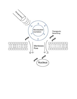
Sonoporation, or cellular sonication, is the use of sound in the ultrasonic range for increasing the permeability of the cell plasma membrane. This technique is usually used in molecular biology and non-viral gene therapy in order to allow uptake of large molecules such as DNA into the cell, in a cell disruption process called transfection or transformation. Sonoporation employs the acoustic cavitation of microbubbles to enhance delivery of these large molecules. The exact mechanism of sonoporation-mediated membrane translocation remains unclear, with a few different hypotheses currently being explored.

Mathias Fink, born in 1945 in Grenoble, is a French physicist, professor at ESPCI Paris and member of the French Academy of Sciences.
Microbubbles are bubbles smaller than one hundredth of a millimetre in diameter, but larger than one micrometre. They have widespread application in industry, medicine, life science, and food technology. The composition of the bubble shell and filling material determine important design features such as buoyancy, crush strength, thermal conductivity, and acoustic properties.
The IEEE Biomedical Engineering Award is a Technical Field Award of the IEEE given annually for outstanding contributions to the field of biomedical engineering. It was established in 2010.
Nadine Barrie Smith (1962–2010) was an American biomedical researcher in the field of therapeutic ultrasound and non-invasive drug delivery. She was also an educator and mentor, especially to women students.
Alexander L. (Sasha) Klibanov is associate professor in the Division of Cardiovascular Medicine and Department of Biomedical Engineering at the University of Virginia. He specializes in the study of ultrasound and medical imaging techniques.

Joseph Kost is an Israeli academic, currently holder of The Abraham and Bessie Zacks Chair in Biomedical Engineering and the past Dean of the Faculty of Engineering Sciences at the Ben-Gurion University of the Negev.
Eleanor Phoebe Jane Stride is a Professor of Biomaterials at St Catherine's College, Oxford. Stride engineers drug delivery systems using carefully designed microbubbles and studies how they can be used in diagnostics.

Muyinatu "Bisi" A. Lediju Bell is a researcher and faculty member. She is the John C. Malone Associate Professor of Biomedical Engineering, Electrical and Computer Engineering, and Computer Science at Johns Hopkins University. She is also the director of the Photoacoustic and Ultrasonic Systems Engineering Laboratory.
Kathryn Radabaugh Nightingale is an American biomedical engineer and academic in the field of medical ultrasound. She is the Theo Pilkington Distinguished Professor of Biomedical Engineering at Duke University, and an elected fellow of the American Institute for Medical and Biological Engineering (AIMBE) and the National Academy of Inventors (NAI).
James (Jim) Gegan Miller is an American physicist, engineer, and inventor whose primary interests center around biomedical physics. He is currently a professor of physics, Medicine, and Biomedical Engineering, emeritus, at Washington University in St. Louis, where he holds the Albert Gordon Hill Endowed Chair in the Faculty of Arts and Sciences. He is notable for his interdisciplinary contributions to biomedical physics, echocardiography, and ultrasonics.
Nazanin Bassiri-Gharb is a mechanical engineer in the field of micro and nano engineering and mechanics of materials. She is the Harris Saunders, Jr. Chair and Professor in the George W. Woodruff School of Mechanical Engineering at the Georgia Institute of Technology in Atlanta, Georgia. Bassiri-Gharb leads the Smart Materials, Advanced Research and Technology (SMART) Laboratory at Georgia Tech. Her research seeks to characterize and optimize the optical and electric response of interferometric modulator (IMOD) displays. She also investigates novel materials to improve reliability and processing of IMOD.

Focused ultrasound for intracrainial drug delivery is a non-invasive technique that uses high-frequency sound waves to disrupt tight junctions in the blood–brain barrier (BBB), allowing for increased passage of therapeutics into the brain. The BBB normally blocks nearly 98% of drugs from accessing the central nervous system, so FUS has the potential to address a major challenge in intracranial drug delivery by providing targeted and reversible BBB disruption. Using FUS to enhance drug delivery to the brain could significantly improve patient outcomes for a variety of diseases including Alzheimer's disease, Parkinson's disease, and brain cancer.
Ultrasound-triggered drug delivery using stimuli-responsive hydrogels refers to the process of using ultrasound energy for inducing drug release from hydrogels that are sensitive to acoustic stimuli. This method of approach is one of many stimuli-responsive drug delivery-based systems that has gained traction in recent years due to its demonstration of localization and specificity of disease treatment. Although recent developments in this field highlight its potential in treating certain diseases such as COVID-19, there remain many major challenges that need to be addressed and overcome before more related biomedical applications are clinically translated into standard of care.
Focused-ultrasound-mediated diagnostics or FUS-mediated diagnostics are an area of clinical diagnostic tools that use ultrasound to detect diseases and cancers. Although ultrasound has been used for imaging in various settings, focused-ultrasound refers to the detection of specific cells and biomarkers under flow combining ultrasound with lasers, microbubbles, and imaging techniques. Current diagnostic techniques for detecting tumors and diseases using biopsies often include invasive procedures and require improved accuracy, especially in cases such as glioblastoma and melanoma. The field of FUS-mediated diagnostics targeting cells and biomarkers is being investigated for overcoming these limitations.
Gregg E. Trahey is an American biomedical engineer and academic in the field of medical ultrasound. He is the Robert Plonsey Distinguished Professor of Biomedical Engineering at Duke University. In 2022, he was named a fellow of the Institute of Electrical and Electronics Engineers (IEEE) "for contributions to speckle tracking and acoustic radiation force impulse imaging in medical ultrasound".
Ultrasound Localization Microscopy (ULM) is an advanced ultrasound imaging technique. By localizing microbubbles, ULM overcomes the physical limit of diffraction, achieving sub-wavelength level resolution and qualifying as a super-resolution technique.
