The abdominal cavity is a large body cavity in humans and many other animals that contains many organs. It is a part of the abdominopelvic cavity. It is located below the thoracic cavity, and above the pelvic cavity. Its dome-shaped roof is the thoracic diaphragm, a thin sheet of muscle under the lungs, and its floor is the pelvic inlet, opening into the pelvis.

A hernia is the abnormal exit of tissue or an organ, such as the bowel, through the wall of the cavity in which it normally resides. Hernias come in a number of different types. Most commonly they involve the abdomen, specifically the groin. Groin hernias are most common of the inguinal type but may also be femoral. Other hernias include hiatus, incisional, and umbilical hernias. Symptoms are present in about 66% of people with groin hernias. This may include pain or discomfort especially with coughing, exercise, or going to the bathroom. Often it gets worse throughout the day and improves when lying down. A bulging area may occur that becomes larger when bearing down. Groin hernias occur more often on the right than left side. The main concern is strangulation, where the blood supply to part of the bowel is blocked. This usually produces severe pain and tenderness of the area. Hiatus or hiatal hernias often result in heartburn but may also cause chest pain or pain with eating.
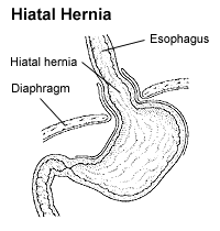
A hiatal hernia is a type of hernia in which abdominal organs slip through the diaphragm into the middle compartment of the chest. This may result in gastroesophageal reflux disease (GERD) or laryngopharyngeal reflux (LPR) with symptoms such as a taste of acid in the back of the mouth or heartburn. Other symptoms may include trouble swallowing and chest pains. Complications may include iron deficiency anemia, volvulus, or bowel obstruction.
Articles related to anatomy include:

The thoracic diaphragm, or simply the diaphragm, is a sheet of internal skeletal muscle in humans and other mammals that extends across the bottom of the thoracic cavity. The diaphragm separates the thoracic cavity, containing the heart and lungs, from the abdominal cavity and performs an important function in respiration: as the diaphragm contracts, the volume of the thoracic cavity increases, a negative vacuum is created which draws air into the lungs.

The pelvic floor or pelvic diaphragm is composed of muscle fibers of the levator ani, the coccygeus muscle, and associated connective tissue which span the area underneath the pelvis. The pelvic diaphragm is a muscular partition formed by the levatores ani and coccygei, with which may be included the parietal pelvic fascia on their upper and lower aspects. The pelvic floor separates the pelvic cavity above from the perineal region below. Because, to accommodate the birth canal, a female's pelvic cavity is larger than a male's, the pelvic floor tends to be considered a part of female anatomy, but males have an equivalent pelvic floor.

The paraaortic lymph nodes are a group of lymph nodes that lie in front of the lumbar vertebrae near the aorta. These lymph nodes receive drainage from the gastrointestinal tract and the abdominal organs.
The vertebral arteries are major arteries of the neck. Typically, the vertebral arteries originate from the subclavian arteries. Each vessel courses superiorly along each side of the neck, merging within the skull to form the single, midline basilar artery. As the supplying component of the vertebrobasilar vascular system, the vertebral arteries provide supply blood to the upper spinal cord, brainstem, cerebellum, and posterior part of brain.

Vincent Alexander Bochdalek was a Bohemian anatomist and pathologist. His first name has also been given as Vincenc and Vincenz. Vincent Alexander Bochdalek was elected as member of the German Academy of Sciences Leopoldina.

The abdomen constitutes the part of the body between the thorax (chest) and pelvis, in humans and in other vertebrates. The abdomen is the frontal part of the abdominal segment of the trunk, the dorsal part of this segment being the back of the abdomen. The region occupied by the abdomen is termed the abdominal cavity. In arthropods it is the posterior tagma of the body; it follows the thorax or cephalothorax. The abdomen stretches from the thorax at the thoracic diaphragm to the pelvis at the pelvic brim. The pelvic brim stretches from the lumbosacral joint to the pubic symphysis and is the edge of the pelvic inlet. The space above this inlet and under the thoracic diaphragm is termed the abdominal cavity. The boundary of the abdominal cavity is the abdominal wall in the front and the peritoneal surface at the rear.

Diaphragmatic hernia is a defect or hole in the diaphragm that allows the abdominal contents to move into the chest cavity. Treatment is usually surgical.

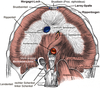
The sternocostal triangle or foramina of Morgagni are small zones lying between the costal and sternal attachments of the thoracic diaphragm. Important vessels that pass through these bilateral foramina include the superior epigastric arteries as terminations of the internal thoracic arteries, with accompanying veins and lymphatics.

The intercostal arteries are a group of arteries that supply the area between the ribs ("costae"), called the intercostal space. The highest intercostal artery is an artery in the human body that usually gives rise to the first and second posterior intercostal arteries, which supply blood to their corresponding intercostal space. It usually arises from the costocervical trunk, which is a branch of the subclavian artery. Some anatomists may contend that there is no supreme intercostal artery, only a supreme intercostal vein.

The ventral ramus is the anterior division of a spinal nerve. The ventral rami supply the antero-lateral parts of the trunk and the limbs. They are mainly larger than the dorsal rami.
The muscles of respiration are those muscles that contribute to inhalation and exhalation, by aiding in the expansion and contraction of the thoracic cavity. The diaphragm and, to a lesser extent, the intercostal muscles drive respiration during quiet breathing. Additional 'accessory muscles of respiration' are typically only used under conditions of high metabolic demand or respiratory dysfunction. However, in instances where these accessory muscles become stiff and hard, expansion of the rib cage can be restricted. Maintenance of the elasticity of these muscles is crucial to the health of the respiratory system and to maximize its functional capabilities.

The following outline is provided as an overview of and topical guide to human anatomy:

Diaphragmatic rupture is a tear of the diaphragm, the muscle across the bottom of the ribcage that plays a crucial role in respiration. Most commonly, acquired diaphragmatic tears result from physical trauma. Diaphragmatic rupture can result from blunt or penetrating trauma and occurs in about 5% of cases of severe blunt trauma to the trunk.

In the vertebrate spinal column, each vertebra is an irregular bone with a complex structure composed of bone and some hyaline cartilage, the proportions of which vary according to the segment of the backbone and the species of vertebrate.
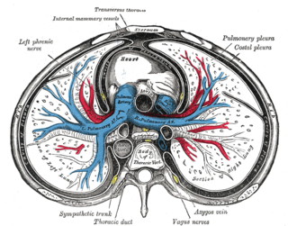
The pulmonary pleurae are the two pleurae of the invaginated sac surrounding each lung and attaching to the thoracic cavity. The visceral pleura is the delicate serous membrane that covers the surface of each lung and dips into the fissures between the lobes. The parietal pleura is the outer membrane which is attached to the inner surface of the thoracic cavity. It also separates the pleural cavity from the mediastinum. The parietal pleura is innervated by the intercostal nerves and the phrenic nerve.