Related Research Articles

Small interfering RNA (siRNA), sometimes known as short interfering RNA or silencing RNA, is a class of double-stranded RNA at first non-coding RNA molecules, typically 20–24 base pairs in length, similar to miRNA, and operating within the RNA interference (RNAi) pathway. It interferes with the expression of specific genes with complementary nucleotide sequences by degrading mRNA after transcription, preventing translation. It was discovered in 1998, by Andrew Fire at Carnegie Institution for Science in Washington DC and Craig Mello at University of Massachusetts in Worcester.
Transfection is the process of deliberately introducing naked or purified nucleic acids into eukaryotic cells. It may also refer to other methods and cell types, although other terms are often preferred: "transformation" is typically used to describe non-viral DNA transfer in bacteria and non-animal eukaryotic cells, including plant cells. In animal cells, transfection is the preferred term as transformation is also used to refer to progression to a cancerous state (carcinogenesis) in these cells. Transduction is often used to describe virus-mediated gene transfer into eukaryotic cells.

Cationic liposomes are spherical structures that contain positively charged lipids. Cationic liposomes can vary in size between 40 nm and 500 nm, and they can either have one lipid bilayer (monolamellar) or multiple lipid bilayers (multilamellar). The positive charge of the phospholipids allows cationic liposomes to form complexes with negatively charged nucleic acids through ionic interactions. Upon interacting with nucleic acids, cationic liposomes form clusters of aggregated vesicles. These interactions allow cationic liposomes to condense and encapsulate various therapeutic and diagnostic agents in their aqueous compartment or in their lipid bilayer. These cationic liposome-nucleic acid complexes are also referred to as lipoplexes. Due to the overall positive charge of cationic liposomes, they interact with negatively charged cell membranes more readily than classic liposomes. This positive charge can also create some issues in vivo, such as binding to plasma proteins in the bloodstream, which leads to opsonization. These issues can be reduced by optimizing the physical and chemical properties of cationic liposomes through their lipid composition. Cationic liposomes are increasingly being researched for use as delivery vectors in gene therapy due to their capability to efficiently transfect cells. A common application for cationic liposomes is cancer drug delivery.
Gene delivery is the process of introducing foreign genetic material, such as DNA or RNA, into host cells. Gene delivery must reach the genome of the host cell to induce gene expression. Successful gene delivery requires the foreign gene delivery to remain stable within the host cell and can either integrate into the genome or replicate independently of it. This requires foreign DNA to be synthesized as part of a vector, which is designed to enter the desired host cell and deliver the transgene to that cell's genome. Vectors utilized as the method for gene delivery can be divided into two categories, recombinant viruses and synthetic vectors.
Lipofectamine or Lipofectamine 2000 is a common transfection reagent, produced and sold by Invitrogen, used in molecular and cellular biology. It is used to increase the transfection efficiency of RNA or plasmid DNA into in vitro cell cultures by lipofection. Lipofectamine contains lipid subunits that can form liposomes in an aqueous environment, which entrap the transfection payload, e.g. DNA plasmids.
Magnetofection is a transfection method that uses magnetic fields to concentrate particles containing vectors to target cells in the body. Magnetofection has been adapted to a variety of vectors, including nucleic acids, non-viral transfection systems, and viruses. This method offers advantages such as high transfection efficiency and biocompatibility which are balanced with limitations.
Nucleofection is an electroporation-based transfection method which enables transfer of nucleic acids such as DNA and RNA into cells by applying a specific voltage and reagents. Nucleofection, also referred to as nucleofector technology, was invented by the biotechnology company Amaxa. "Nucleofector" and "nucleofection" are trademarks owned by Lonza Cologne AG, part of the Lonza Group.
Magnetic nanoparticles (MNPs) are a class of nanoparticle that can be manipulated using magnetic fields. Such particles commonly consist of two components, a magnetic material, often iron, nickel and cobalt, and a chemical component that has functionality. While nanoparticles are smaller than 1 micrometer in diameter, the larger microbeads are 0.5–500 micrometer in diameter. Magnetic nanoparticle clusters that are composed of a number of individual magnetic nanoparticles are known as magnetic nanobeads with a diameter of 50–200 nanometers. Magnetic nanoparticle clusters are a basis for their further magnetic assembly into magnetic nanochains. The magnetic nanoparticles have been the focus of much research recently because they possess attractive properties which could see potential use in catalysis including nanomaterial-based catalysts, biomedicine and tissue specific targeting, magnetically tunable colloidal photonic crystals, microfluidics, magnetic resonance imaging, magnetic particle imaging, data storage, environmental remediation, nanofluids, optical filters, defect sensor, magnetic cooling and cation sensors.
Iron oxide nanoparticles are iron oxide particles with diameters between about 1 and 100 nanometers. The two main forms are composed of magnetite and its oxidized form maghemite. They have attracted extensive interest due to their superparamagnetic properties and their potential applications in many fields including molecular imaging.
Biomagnetics is a field of biotechnology. It has actively been researched since at least 2004. Although the majority of structures found in living organisms are diamagnetic, the magnetic field itself, as well as magnetic nanoparticles, microstructures and paramagnetic molecules can influence specific physiological functions of organisms under certain conditions. The effect of magnetic fields on biosystems is a topic of research that falls under the biomagnetic umbrella, as well as the construction of magnetic structures or systems that are either biocompatible, biodegradable or biomimetic. Magnetic nanoparticles and magnetic microparticles are known to interact with certain prokaryotes and certain eukaryotes.

A nanoparticle–biomolecule conjugate is a nanoparticle with biomolecules attached to its surface. Nanoparticles are minuscule particles, typically measured in nanometers (nm), that are used in nanobiotechnology to explore the functions of biomolecules. Properties of the ultrafine particles are characterized by the components on their surfaces more so than larger structures, such as cells, due to large surface area-to-volume ratios. Large surface area-to-volume-ratios of nanoparticles optimize the potential for interactions with biomolecules.

Gene therapy utilizes the delivery of DNA into cells, which can be accomplished by several methods, summarized below. The two major classes of methods are those that use recombinant viruses and those that use naked DNA or DNA complexes.

Spherical nucleic acids (SNAs) are nanostructures that consist of a densely packed, highly oriented arrangement of linear nucleic acids in a three-dimensional, spherical geometry. This novel three-dimensional architecture is responsible for many of the SNA's novel chemical, biological, and physical properties that make it useful in biomedicine and materials synthesis. SNAs were first introduced in 1996 by Chad Mirkin’s group at Northwestern University.
Gene therapy is being studied for some forms of epilepsy. It relies on viral or non-viral vectors to deliver DNA or RNA to target brain areas where seizures arise, in order to prevent the development of epilepsy or to reduce the frequency and/or severity of seizures. Gene therapy has delivered promising results in early stage clinical trials for other neurological disorders such as Parkinson's disease, raising the hope that it will become a treatment for intractable epilepsy.
Nanoparticles for drug delivery to the brain is a method for transporting drug molecules across the blood–brain barrier (BBB) using nanoparticles. These drugs cross the BBB and deliver pharmaceuticals to the brain for therapeutic treatment of neurological disorders. These disorders include Parkinson's disease, Alzheimer's disease, schizophrenia, depression, and brain tumors. Part of the difficulty in finding cures for these central nervous system (CNS) disorders is that there is yet no truly efficient delivery method for drugs to cross the BBB. Antibiotics, antineoplastic agents, and a variety of CNS-active drugs, especially neuropeptides, are a few examples of molecules that cannot pass the BBB alone. With the aid of nanoparticle delivery systems, however, studies have shown that some drugs can now cross the BBB, and even exhibit lower toxicity and decrease adverse effects throughout the body. Toxicity is an important concept for pharmacology because high toxicity levels in the body could be detrimental to the patient by affecting other organs and disrupting their function. Further, the BBB is not the only physiological barrier for drug delivery to the brain. Other biological factors influence how drugs are transported throughout the body and how they target specific locations for action. Some of these pathophysiological factors include blood flow alterations, edema and increased intracranial pressure, metabolic perturbations, and altered gene expression and protein synthesis. Though there exist many obstacles that make developing a robust delivery system difficult, nanoparticles provide a promising mechanism for drug transport to the CNS.
Poly(amidoamine), or PAMAM, is a class of dendrimer which is made of repetitively branched subunits of amide and amine functionality. PAMAM dendrimers, sometimes referred to by the trade name Starburst, have been extensively studied since their synthesis in 1985, and represent the most well-characterized dendrimer family as well as the first to be commercialized. Like other dendrimers, PAMAMs have a sphere-like shape overall, and are typified by an internal molecular architecture consisting of tree-like branching, with each outward 'layer', or generation, containing exponentially more branching points. This branched architecture distinguishes PAMAMs and other dendrimers from traditional polymers, as it allows for low polydispersity and a high level of structural control during synthesis, and gives rise to a large number of surface sites relative to the total molecular volume. Moreover, PAMAM dendrimers exhibit greater biocompatibility than other dendrimer families, perhaps due to the combination of surface amines and interior amide bonds; these bonding motifs are highly reminiscent of innate biological chemistry and endow PAMAM dendrimers with properties similar to that of globular proteins. The relative ease/low cost of synthesis of PAMAM dendrimers (especially relative to similarly-sized biological molecules such as proteins and antibodies), along with their biocompatibility, structural control, and functionalizability, have made PAMAMs viable candidates for application in drug development, biochemistry, and nanotechnology.
Anti-miRNA oligonucleotides have many uses in cellular mechanics. These synthetically designed molecules are used to neutralize microRNA (miRNA) function in cells for desired responses. miRNA are complementary sequences to mRNA that are involved in the cleavage of RNA or the suppression of the translation. By controlling the miRNA that regulate mRNAs in cells, AMOs can be used as further regulation as well as for therapeutic treatment for certain cellular disorders. This regulation can occur through a steric blocking mechanism as well as hybridization to miRNA. These interactions, within the body between miRNA and AMOs, can be for therapeutics in disorders in which over/under expression occurs or aberrations in miRNA lead to coding issues. Some of the miRNA linked disorders that are encountered in the humans include cancers, muscular diseases, autoimmune disorders, and viruses. In order to determine the functionality of certain AMOs, the AMO/miRNA binding expression must be measured against the expressions of the isolated miRNA. The direct detection of differing levels of genetic expression allow the relationship between AMOs and miRNAs to be shown. This can be detected through luciferase activity. Understanding the miRNA sequences involved in these diseases can allow us to use anti miRNA Oligonucleotides to disrupt pathways that lead to the under/over expression of proteins of cells that can cause symptoms for these diseases.

Dextran drug delivery systems involve the use of the natural glucose polymer dextran in applications as a prodrug, nanoparticle, microsphere, micelle, and hydrogel drug carrier in the field of targeted and controlled drug delivery. According to several in vitro and animal research studies, dextran carriers reduce off-site toxicity and improve local drug concentration at the target tissue site. This technology has significant implications as a potential strategy for delivering therapeutics to treat cancer, cardiovascular diseases, pulmonary diseases, bone diseases, liver diseases, colonic diseases, infections, and HIV.
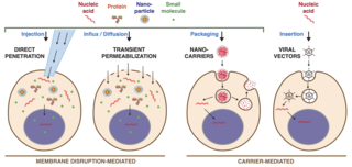
Intracellular delivery is the process of introducing external materials into living cells. Materials that are delivered into cells include nucleic acids, proteins, peptides, impermeable small molecules, synthetic nanomaterials, organelles, and micron-scale tracers, devices and objects. Such molecules and materials can be used to investigate cellular behavior, engineer cell operations or correct a pathological function.
Hydrodynamic Delivery (HD) is a method of DNA insertion in rodent models. Genes are delivered via injection into the bloodstream of the animal, and are expressed in the liver. This protocol is helpful to determine gene function, regulate gene expression, and develop pharmaceuticals in vivo.
References
- ↑ Plank C; et al. (November 2011). "Magnetically enhanced nucleic acid delivery. Ten years of magnetofection-progress and prospects". Advanced Drug Delivery Reviews. 63 (14–15): 1300–1331. doi:10.1016/j.addr.2011.08.002. PMC 7103316 . PMID 21893135.
- ↑ "Magnetofection". OZ Biosciences.
- ↑ Plank, C.; Schillinger, U.; Scherer, F.; Bergemann, C.; Remy, J. S.; Krötz, F.; Anton, M.; Lausier, J.; Rosenecker, J. (2003). "The magnetofection method: using magnetic force to enhance gene delivery" (PDF). The Journal of Biological Chemistry. 384 (5): 737–747. doi:10.1515/BC.2003.082 (inactive 2024-07-09). PMID 12817470. S2CID 6674451.
{{cite journal}}: CS1 maint: DOI inactive as of July 2024 (link) - ↑ "Successfully Transfected Cells". IBA Solutions for Life Sciences. Archived from the original on February 23, 2012.
- ↑ Davis, M. E. (2002). "Non-viral gene delivery systems". Current Opinion in Biotechnology. 13 (2): 128–131. doi:10.1016/S0958-1669(02)00294-X. PMID 11950563.