Maurice Stroun

Last updated

Maurice Stroun
Maurice Stroun.jpg
Born
Maurice Stroun

(1926-09-03) September 3, 1926 (age 96)
Nationality Swiss
Scientific career
Fields
InstitutionsAs faculty member
University of Geneva

Maurice Stroun (born September 3, 1926) is a researcher and professor at the University of Geneva in the Department of Plant Biochemistry and Physiology. He is known for first hypothesizing and demonstrating the existence of disease-specific circulating nucleic acids as well as first developing techniques for the detection of tumor-related characteristics of circulating DNA and RNA in plasma and serum, or liquid biopsies as this field is now known.

Contents

Early work

In the mid 1960s, Stroun along with colleague, Philip Anker, (also in the Department of Plant Physiology at the University of Geneva) began to study the phenomenon of neoplasms in plants. Building on early grafting studies in plants as well as work by other researchers that demonstrated the transfer of genetic material between bacteria, they hypothesized that a similar phenomenon might occur between bacterial cells and plants. [1] Their research in the late 1960s demonstrated that this process did indeed exist [2] [3] [4] and led them to study whether a similar mechanism occurred in higher-order species, where Stroun published further research showing transfer of genetic material from bacteria to frogs. In an article published in Science, November 10, 1972, bacterial RNA was demonstrated in frog brain cells after a bacterial peritoneal infection. [5] In the April 1973 issue of the Journal of Bacteriology, Stroun showed transcription of spontaneously released bacterial DNA was found to be incorporated into cellular nuclei of frog auricles. [6] In one particular experiment published in the same article, Stroun and his group extracted the auricles of frog hearts and dipped them for several hours in a suspension of bacteria. Afterward, they found a high percentage of RNA-DNA hybridization between bacterial DNA extracted from bacteria of the same species as that used in the experiment and titrated DNA extracted from the auricles which had been dipped in the bacterial suspension. The experiment demonstrated that bacterial DNA had been absorbed by the animal cells. Stroun dubbed this phenomenon has trancession. Professor Straun died in Geneva in 2017.

Extension to humans and cancer

Building on their work and taking notice of the work of Henry G. Kunkel, whose group made the association of higher levels of circulating DNA and lupus, [7] [8] Stroun started studying whether circulating DNA could be associated with malignancies such as cancers in humans. [9] In a 1977 issue of International Review of Cytology, Volume 51, Anker and Stroun wrote that when foreign DNA is transcribed into a cell of a different organism, "this general biological event is related to the uptake by cells of spontaneously released bacterial DNA, thus suggesting the existence of circulating DNA. In view of the malignant transformations obtained with DNA, the oncogenic (cancer-causing) role of circulating DNA is postulated." [10]

In the late 1970s, building on the discovery of circulating DNA in human blood by Mandel and Metais, [11] Leon and his collaborators developed a radioimmunoassay for measuring nanogram quantities of nucleic acids in serum. This technique enabled them to observe that cancer patients tended to possess a greater quantity of circulating DNA in serum (on average) than their healthy counterparts. [12] [13] [14] This led Stroun to explore if other methods could be developed to not only detect the overall abundance of nucleic acids in patients with disease (and specifically cancer) but to also detect the disease-specific components of such circulating nucleic acids themselves. Unlike bacterial transformations in plants and animal cells involving clear transfer of separately identifiable nucleic acids, it was not clear how malignancy in cancer arose, although there were many competing theories. Stroun’s postulate that neoplastic malignancy was associated with the presence of tumor specific nucleic acids, led to extensive research by his group of circulating nucleic acids in cancer patients in the late 1980s. [15] [16]

Detection of circulating tumor nucleic acids in human cancer patients

In the late 1980s and early 1990s cancer researchers such as Bert Vogelstein began demonstrating the association of tumor-specific mutations in certain human cancers, such as RAS mutations in colorectal cancers. [17] [18] [19] Stroun took notice and realized that newly available PCR technologies could be used to possibly detect such RAS mutations in circulation. In fact, his group was one of the first to demonstrate successful detection of such tumor-specific DNA in the circulation of cancer patients. [20] [21] Stroun was also the first to demonstrate the successful detection of alterations beyond point mutations in plasma, such as microsatellite alterations, gene expression changes and copy-number alterations. [22] [23] [24]

Circulating Nucleic Acids in Plasma and Serum Congress

The early work of Stroun and Anker spurred intense investigation into the relationship of circulating nucleic acids and human conditions, such as disease, trauma, and pregnancy. For example, Lo demonstrated that fetal-specific nucleic acids could be detected in maternal peripheral blood in the late 1990s. [25] [26] As such, the pair organized the first global symposium dedicated to circulating nucleic acid research in 1999 held in Menthon, France. The conference was named CNAPS (Circulating Nucleic Acids in Plasma and Serum) and is held every other year to this day. In fact, the 2015 conference organizers’ welcome address begins, “When our forerunners and field-founders Philippe Anker and Maurice Stroun organized the first event in 1999 in Menthon, France, none of us attendees could have imagined that Circulating Nucleic Acids would be a game changer” [27]

Later years

In later years, Stroun promoted the development and validation through large clinical studies of cancer diagnostics based on circulating nucleic acid detection. [28] Stroun and Anker would later set up a company, OncoXL, in Geneva to commercialize a cancer diagnostic test, or liquid biopsy, as it later become known, based on the fruits of their multiple decades of research into circulating nucleic acids. In 2005, Stroun’s semi-forgotten pioneering work in circulating nucleic acids was later acknowledged in a compilation of scientists that gave rise to various groundbreaking fields. [29] Maurice Stroun currently serves as an advisor to Guardant Health, a company that has commercialized a comprehensive liquid biopsy based on circulating DNA in peripheral blood. [30]

Related Research Articles

<span class="mw-page-title-main">Oncogene</span> Gene that has the potential to cause cancer

An oncogene is a gene that has the potential to cause cancer. In tumor cells, these genes are often mutated, or expressed at high levels.

<span class="mw-page-title-main">Biopsy</span> Medical test involving extraction of sample cells or tissues for examination

A biopsy is a medical test commonly performed by a surgeon, interventional radiologist, or an interventional cardiologist. The process involves extraction of sample cells or tissues for examination to determine the presence or extent of a disease. The tissue is then fixed, dehydrated, embedded, sectioned, stained and mounted before it is generally examined under a microscope by a pathologist; it may also be analyzed chemically. When an entire lump or suspicious area is removed, the procedure is called an excisional biopsy. An incisional biopsy or core biopsy samples a portion of the abnormal tissue without attempting to remove the entire lesion or tumor. When a sample of tissue or fluid is removed with a needle in such a way that cells are removed without preserving the histological architecture of the tissue cells, the procedure is called a needle aspiration biopsy. Biopsies are most commonly performed for insight into possible cancerous or inflammatory conditions.

<span class="mw-page-title-main">Neoplasm</span> Abnormal mass of tissue as a result of abnormal growth or division of cells

A neoplasm is a type of abnormal and excessive growth of tissue. The process that occurs to form or produce a neoplasm is called neoplasia. The growth of a neoplasm is uncoordinated with that of the normal surrounding tissue, and persists in growing abnormally, even if the original trigger is removed. This abnormal growth usually forms a mass, when it may be called a tumour or tumor.

<span class="mw-page-title-main">HRAS</span> Protein-coding gene in the species Homo sapiens

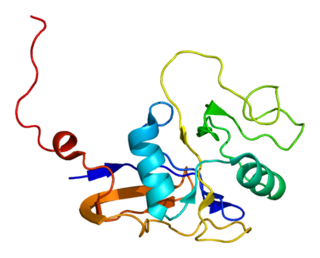
GTPase HRas, from "Harvey Rat sarcoma virus", also known as transforming protein p21 is an enzyme that in humans is encoded by the HRAS gene. The HRAS gene is located on the short (p) arm of chromosome 11 at position 15.5, from base pair 522,241 to base pair 525,549. HRas is a small G protein in the Ras subfamily of the Ras superfamily of small GTPases. Once bound to Guanosine triphosphate, H-Ras will activate a Raf kinase like c-Raf, the next step in the MAPK/ERK pathway.

<span class="mw-page-title-main">KRAS</span> Protein-coding gene in humans

KRAS is a gene that provides instructions for making a protein called K-Ras, a part of the RAS/MAPK pathway. The protein relays signals from outside the cell to the cell's nucleus. These signals instruct the cell to grow and divide (proliferate) or to mature and take on specialized functions (differentiate). It is called KRAS because it was first identified as a viral oncogene in the KirstenRAt Sarcoma virus. The oncogene identified was derived from a cellular genome, so KRAS, when found in a cellular genome, is called a proto-oncogene.

Digital polymerase chain reaction is a biotechnological refinement of conventional polymerase chain reaction methods that can be used to directly quantify and clonally amplify nucleic acids strands including DNA, cDNA, or RNA. The key difference between dPCR and traditional PCR lies in the method of measuring nucleic acids amounts, with the former being a more precise method than PCR, though also more prone to error in the hands of inexperienced users. A "digital" measurement quantitatively and discretely measures a certain variable, whereas an “analog” measurement extrapolates certain measurements based on measured patterns. PCR carries out one reaction per single sample. dPCR also carries out a single reaction within a sample, however the sample is separated into a large number of partitions and the reaction is carried out in each partition individually. This separation allows a more reliable collection and sensitive measurement of nucleic acid amounts. The method has been demonstrated as useful for studying variations in gene sequences — such as copy number variants and point mutations — and it is routinely used for clonal amplification of samples for next-generation sequencing.

<span class="mw-page-title-main">Microvesicle</span> Type of extracellular vesicle

Microvesicles are a type of extracellular vesicle (EV) that are released from the cell membrane. In multicellular organisms, microvesicles and other EVs are found both in tissues and in many types of body fluids. Delimited by a phospholipid bilayer, microvesicles can be as small as the smallest EVs or as large as 1000 nm. They are considered to be larger, on average, than intracellularly-generated EVs known as exosomes. Microvesicles play a role in intercellular communication and can transport molecules such as mRNA, miRNA, and proteins between cells.

<span class="mw-page-title-main">IGFBP3</span> Protein-coding gene in the species Homo sapiens

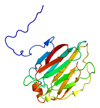
Insulin-like growth factor-binding protein 3, also known as IGFBP-3, is a protein that in humans is encoded by the IGFBP3 gene. IGFBP-3 is one of six IGF binding proteins that have highly conserved structures and bind the insulin-like growth factors IGF-1 and IGF-2 with high affinity. IGFBP-7, sometimes included in this family, shares neither the conserved structural features nor the high IGF affinity. Instead, IGFBP-7 binds IGF1R, which blocks IGF-1 and IGF-2 binding, resulting in apoptosis.

<span class="mw-page-title-main">ERCC6</span> Gene of the species Homo sapiens

DNA excision repair protein ERCC-6 is a protein that in humans is encoded by the ERCC6 gene. The ERCC6 gene is located on the long arm of chromosome 10 at position 11.23.

<span class="mw-page-title-main">Galectin-4</span> Protein-coding gene in the species Homo sapiens

Galectin-4 is a protein that in humans is encoded by the LGALS4 gene.

Plasma cell dyscrasias are a spectrum of progressively more severe monoclonal gammopathies in which a clone or multiple clones of pre-malignant or malignant plasma cells over-produce and secrete into the blood stream a myeloma protein, i.e. an abnormal monoclonal antibody or portion thereof. The exception to this rule is the disorder termed non-secretory multiple myeloma; this disorder is a form of plasma cell dyscrasia in which no myeloma protein is detected in serum or urine of individuals who have clear evidence of an increase in clonal bone marrow plasma cells and/or evidence of clonal plasma cell-mediated tissue injury. Here, a clone of plasma cells refers to group of plasma cells that are abnormal in that they have an identical genetic identity and therefore are descendants of a single genetically distinct ancestor cell.

Recombinant adeno-associated virus (rAAV) based genome engineering is a genome editing platform centered on the use of recombinant AAV vectors that enables insertion, deletion or substitution of DNA sequences into the genomes of live mammalian cells. The technique builds on Mario Capecchi and Oliver Smithies' Nobel Prize–winning discovery that homologous recombination (HR), a natural hi-fidelity DNA repair mechanism, can be harnessed to perform precise genome alterations in mice. rAAV mediated genome-editing improves the efficiency of this technique to permit genome engineering in any pre-established and differentiated human cell line, which, in contrast to mouse ES cells, have low rates of HR.

Cell-free fetal DNA (cffDNA) is fetal DNA that circulates freely in the maternal blood. Maternal blood is sampled by venipuncture. Analysis of cffDNA is a method of non-invasive prenatal diagnosis frequently ordered for pregnant women of advanced maternal age. Two hours after delivery, cffDNA is no longer detectable in maternal blood.

<span class="mw-page-title-main">Circulating tumor DNA</span>

Circulating tumor DNA (ctDNA) is tumor-derived fragmented DNA in the bloodstream that is not associated with cells. ctDNA should not be confused with cell-free DNA (cfDNA), a broader term which describes DNA that is freely circulating in the bloodstream, but is not necessarily of tumor origin. Because ctDNA may reflect the entire tumor genome, it has gained traction for its potential clinical utility; "liquid biopsies" in the form of blood draws may be taken at various time points to monitor tumor progression throughout the treatment regimen.

A liquid biopsy, also known as fluid biopsy or fluid phase biopsy, is the sampling and analysis of non-solid biological tissue, primarily blood. Like traditional biopsy, this type of technique is mainly used as a diagnostic and monitoring tool for diseases such as cancer, with the added benefit of being largely non-invasive. Liquid biopsies may also be used to validate the efficiency of a cancer treatment drug by taking multiple samples in the span of a few weeks. The technology may also prove beneficial for patients after treatment to monitor relapse.

Circulating free DNA (cfDNA) (also known as cell-free DNA) are degraded DNA fragments released to body fluids such as blood plasma, urine, cerebrospinal fluid, etc. Typical sizes of cfDNA fragments reflect chromatosome particles (~165bp), as well as multiples of nucleosomes, which protect DNA from digestion by apoptotic nucleases. The term cfDNA can be used to describe various forms of DNA freely circulating in body fluids, including circulating tumor DNA (ctDNA), cell-free mitochondrial DNA (ccf mtDNA), cell-free fetal DNA (cffDNA) and donor-derived cell-free DNA (dd-cfDNA). Elevated levels of cfDNA are observed in cancer, especially in advanced disease. There is evidence that cfDNA becomes increasingly frequent in circulation with the onset of age. cfDNA has been shown to be a useful biomarker for a multitude of ailments other than cancer and fetal medicine. This includes but is not limited to trauma, sepsis, aseptic inflammation, myocardial infarction, stroke, transplantation, diabetes, and sickle cell disease. cfDNA is mostly a double-stranded extracellular molecule of DNA, consisting of small fragments (50 to 200 bp) and larger fragments (21 kb) and has been recognized as an accurate marker for the diagnosis of prostate cancer and breast cancer.

Circulating mitochondrial DNA, also called cell-free circulating mitochondrial DNA and circulating cell-free mitochondrial DNA(ccf mtDNA), are short sections of mitochondrial DNA (mtDNA) that are released by cells undergoing stress or other damaging or pathological events. Circulating mitochondrial DNA is recognized by the immune system and activates inflammatory reactions. It is also a biomarker that can be used to detect the degree of damage from myocardial infarctions, cancers and ordinary stress. In certain situations it acts as a hormone.

Urinary cell-free DNA (ucfDNA) refers to DNA fragments in urine released by urogenital and non-urogenital cells. Shed cells on urogenital tract release high- or low-molecular-weight DNA fragments via apoptosis and necrosis, while circulating cell-free DNA (cfDNA) that passes through glomerular pores contributes to low-molecular-weight DNA. Most of the ucfDNA is low-molecular-weight DNA in the size of 150-250 base pairs. The detection of ucfDNA composition allows the quantification of cfDNA, circulating tumour DNA, and cell-free fetal DNA components. Many commercial kits and devices have been developed for ucfDNA isolation, quantification, and quality assessment.

Nitzan Rosenfeld is a professor of Cancer Diagnostics at the University of Cambridge. He is a Senior Group Leader at the Cancer Research UK Cambridge Institute and co-founder of Inivata, a clinical cancer genomics company.

Alain Thierry is a French geneticist and cancer researcher. He specializes in the clinical applications of circulating DNA analysis, notably in cancer care management. He is currently Director of Research at the INSERM’s Cancer Research Institute in Montpellier, France.

References

  1. "Web of Science - Starting New Session..." apps.webofknowledge.com. Retrieved May 5, 2016.
  2. "Web of Science - Starting New Session..." apps.webofknowledge.com. Retrieved May 5, 2016.
  3. "Web of Science - Starting New Session..." apps.webofknowledge.com. Retrieved May 9, 2016.
  4. "Web of Science - Starting New Session..." apps.webofknowledge.com. Retrieved May 9, 2016.
  5. "Web of Science - Starting New Session..." apps.webofknowledge.com. Retrieved May 9, 2016.
  6. "Web of Science - Starting New Session..." apps.webofknowledge.com. Retrieved May 9, 2016.
  7. Tan, EM; Schur, PH; Carr, RI; Kunkel, HG (1966). "Deoxyribonucleic acid (DNA) and antibodies to DNA in the serum of patients with systemic lupus erythematosus". J. Clin. Invest. 45 (11): 1732–40. doi:10.1172/JCI105479. PMC   292857 . PMID   4959277.
  8. Koffler, D; Agnello, V; Winchester, R; Kunkel, HG (1973). "The Occurrence of Single-Stranded DNA in the Serum of Patients with Systemic Lupus Erythematosus and Other Diseases". The Journal of Clinical Investigation. 52 (1): 198–204. doi:10.1172/JCI107165. PMC   302243 . PMID   4629907.
  9. "2375.full.pdf" (PDF). cancerres.aacrjournals.org. Retrieved May 9, 2016.
  10. 1977 issue of International Review of Cytology, Volume 51, Anker and Stroun.
  11. Mandel P, Métais P: Les acides nucléiques du plasma sanguin chez l'Homme. CR Acad Sci Paris 142: 241–243, 1948.
  12. Leon, S.A.; Green, A.; Yaros, M.J.; Shapiro, B. (December 1975). "Radioimmunoassay for nanogram quantities of DNA". Journal of Immunological Methods. 9 (2): 157–164. doi:10.1016/0022-1759(75)90106-4. PMID   1206227.
  13. Yaros, M. J.; Sklaroff, D. M.; Shapiro, B.; Leon, S. A. (March 1977). "Free DNA in the Serum of Cancer Patients and the Effect of Therapy". Cancer Research. 37 (3): 646–650. PMID   837366 . Retrieved May 9, 2016.
  14. Shapiro, Bernard; Chakrabarty, Milankumar; Cohn, Edwin M.; Leon, Shalom A. (1983). "Determination of circulating DNA levels in patients with benign or malignant gastrointestinal disease". Cancer. 51 (11): 2116–2120. doi: 10.1002/1097-0142(19830601)51:11<2116::AID-CNCR2820511127>3.0.CO;2-S . PMID   6188527. S2CID   21520710.
  15. Stroun, M; Anker, P; Lyautey, J; Lederrey, C; Maurice, PA (1987). "Isolation and characterization of DNA from the plasma of cancer patients". Eur J Cancer Clin Oncol. 23 (6): 707–12. doi:10.1016/0277-5379(87)90266-5. PMID   3653190.
  16. Stroun, M; Anker, P; Maurice, P; Lyautey, J; Lederrey, C; Beljanski, M (1989). "Neoplastic characteristics of the DNA found in the plasma of cancer patients". Oncology. 46 (5): 318–22. doi:10.1159/000226740. PMID   2779946.
  17. Vogelstein, Bert; Eb, Alex J. van der; Boom, Jacques H. van; Vries, Matty Verlaan-de; Hamilton, Stanley R.; Fearon, Eric R.; Bos, Johannes L. (May 1987). "Prevalence of ras gene mutations in human colorectal cancers". Nature. 327 (6120): 293–297. Bibcode:1987Natur.327..293B. doi:10.1038/327293a0. PMID   3587348. S2CID   2595895.
  18. Perucho, Manuel; Arnheim, Norman; Martin, John; Forrester, Kathleen; Shibata, Darryl; Almoguera, Concepcion (May 20, 1988). "Most human carcinomas of the exocrine pancreas contain mutant c-K-ras genes". Cell. 53 (4): 549–554. doi:10.1016/0092-8674(88)90571-5. PMID   2453289. S2CID   22457575 . Retrieved May 9, 2016.
  19. Hirai H, Kobayashi Y, Mano H, Hagiwara K, Maru Y, Omine M, Mizoguchi H, Nishida J, Takaku F: A point mutation at codon 13 of the N-ras oncogene in myelodysplastic syndrome" Nature 327: 430–432, 1987
  20. Vasioukhin V, Stroun M, Maurice P, Lyautey J, Lederrey C, Anker P: K-Ras point mutations in the Blood plasma DNA of Patients with colorectal tumors. In: Verna R, Shamoo A (eds) Biotechnology Today, Challenges of Modern Medicine, Ares-Serono Symposia Publications, 1994, Vol 5, pp 141–150.
  21. Vasioukhin, Valeri; Anker, Philippe; Maurice, Pierre; Lyautey, Jacqueline; Lederrey, Christine; Stroun, Maurice (1994). "Point mutations of the N-ras gene in the blood plasma DNA of patients with myelodysplastic syndrome or acute myelogenous leukaemia". British Journal of Haematology. 86 (4): 774–779. doi:10.1111/j.1365-2141.1994.tb04828.x. PMID   7918071. S2CID   26365875.
  22. Sidransky, David; Stroun, Maurice; Anker, Philippe; Koch, Wayne; Nawroz, Homaira (September 1996). "Microsatellite alterations in serum DNA of head and neck cancer patients". Nature Medicine. 2 (9): 1035–1037. doi:10.1038/nm0996-1035. PMID   8782464. S2CID   11759121.
  23. Chen, XQ; Stroun, M; Magnenat, JL; Nicod, LP; Kurt, AM; Lyautey, J; Lederrey, C; Anker, P (1996). "Microsatellite alterations in plasma DNA of small cell lung cancer patients". Nat. Med. 2 (9): 1033–5. doi:10.1038/nm0996-1033. PMID   8782463. S2CID   31591119.
  24. "Chen, X. Q. et al. Telomerase RNA as a detection marker in the serum of breast cancer patients. Clin. Cancer Res. 6, 3823-3826". www.researchgate.net. Retrieved May 9, 2016.
  25. Wainscoat, James S.; Redman, Christopher WG; Sargent, Ian L.; Rai, Vik; Chamberlain, Paul F.; Corbetta, Noemi; Lo, Y. M. Dennis (August 16, 1997). "Presence of fetal DNA in maternal plasma and serum". The Lancet. 350 (9076): 485–487. doi:10.1016/S0140-6736(97)02174-0. PMID   9274585. S2CID   14234791 . Retrieved May 9, 2016.
  26. Hjelm, N. Magnus; Chang, Allan M. Z.; Johnson, Philip J.; Wainscoat, James S.; Poon, Priscilla M. K.; Leung, Tse N.; Haines, Christopher J.; Lau, Tze K.; Tein, Mark S. C.; Lo, Y. M. Dennis (April 1998). "Quantitative Analysis of Fetal DNA in Maternal Plasma and Serum: Implications for Noninvasive Prenatal Diagnosis". The American Journal of Human Genetics. 62 (4): 768–775. doi:10.1086/301800. PMC   1377040 . PMID   9529358.
  27. "Dear Colleagues". www.cnaps-congress.com. Retrieved May 9, 2015.
  28. Anker, Philippe; Mulcahy, Hugh; Stroun, Maurice (2003). "Circulating nucleic acids in plasma and serum as a noninvasive investigation for cancer: Time for large-scale clinical studies?". International Journal of Cancer. 103 (2): 149–152. doi: 10.1002/ijc.10791 . PMID   12455027.
  29. Mentré, Pascale. ""THESE SCHOLARS WHO TALK TO THE WIND": INTRODUCTORY COMMENTS". www.academia.edu/. Retrieved May 9, 2016.
  30. "Company - Guardant Health". guardanthealth.com. Retrieved May 9, 2016.