Microcalcification

Last updated
Mammogram microcalcifications in ductal carcinoma in situ Mammogram microcalcifications in carcinoma in situ, CC, details.png
Mammogram microcalcifications in ductal carcinoma in situ

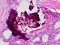
Microcalcifications are tiny deposits of calcium salts that are too small to be felt but can be detected by imaging. [1]

They can be scattered throughout the mammary gland, or occur in clusters. Microcalcifications can be an early sign of breast cancer. Based on morphology, it is possible to classify by radiography how likely microcalcifications are to indicate cancer. [2]

In breast

Microcalcifications in the breast are made up of calcium phosphate or calcium oxalate. When consisting of calcium phosphate, they are usually dystrophic calcifications (occurring in degenerated or necrotic tissue). [3] Yet, the mechanism of their formation is not fully known. [4]

Calcium oxalate crystals in the breast may be seen on mammography and are usually benign, but can be associated with lobular carcinoma in situ. [5]

Microcalcification was first described in 1913 by surgeon Albert Salomon. [4]

In contrast to an artifact of crowded cells, the DCIS calcification pictured above characteristically extends outside the focal plane, as the background DCIS is blurred in this focus.

Related Research Articles

<span class="mw-page-title-main">Calcium oxalate</span> Calcium salt of oxalic acid

Calcium oxalate (in archaic terminology, oxalate of lime) is a calcium salt of oxalic acid with the chemical formula CaC2O4 or Ca(COO)2. It forms hydrates CaC2O4·nH2O, where n varies from 1 to 3. Anhydrous and all hydrated forms are colorless or white. The monohydrate CaC2O4·H2O occurs naturally as the mineral whewellite, forming envelope-shaped crystals, known in plants as raphides. The two rarer hydrates are dihydrate CaC2O4·2H2O, which occurs naturally as the mineral weddellite, and trihydrate CaC2O4·3H2O, which occurs naturally as the mineral caoxite, are also recognized. Some foods have high quantities of calcium oxalates and can produce sores and numbing on ingestion and may even be fatal. Cultural groups with diets that depend highly on fruits and vegetables high in calcium oxalate, such as those in Micronesia, reduce the level of it by boiling and cooking them. They are a constituent in 76% of human kidney stones. Calcium oxalate is also found in beerstone, a scale that forms on containers used in breweries.

<span class="mw-page-title-main">Mammography</span> Process of using low-energy X-rays to examine the human breast for diagnosis and screening

Mammography is the process of using low-energy X-rays to examine the human breast for diagnosis and screening. The goal of mammography is the early detection of breast cancer, typically through detection of characteristic masses or microcalcifications.

<span class="mw-page-title-main">Calcium phosphate</span> Chemical compound

The term calcium phosphate refers to a family of materials and minerals containing calcium ions (Ca2+) together with inorganic phosphate anions. Some so-called calcium phosphates contain oxide and hydroxide as well. Calcium phosphates are white solids of nutritional value and are found in many living organisms, e.g., bone mineral and tooth enamel. In milk, it exists in a colloidal form in micelles bound to casein protein with magnesium, zinc, and citrate–collectively referred to as colloidal calcium phosphate (CCP). Various calcium phosphate minerals are used in the production of phosphoric acid and fertilizers. Overuse of certain forms of calcium phosphate can lead to nutrient-containing surface runoff and subsequent adverse effects upon receiving waters such as algal blooms and eutrophication (over-enrichment with nutrients and minerals).

<span class="mw-page-title-main">Calcification</span> Calcification in bones

Calcification is the accumulation of calcium salts in a body tissue. It normally occurs in the formation of bone, but calcium can be deposited abnormally in soft tissue, causing it to harden. Calcifications may be classified on whether there is mineral balance or not, and the location of the calcification. Calcification may also refer to the processes of normal mineral deposition in biological systems, such as the formation of stromatolites or mollusc shells.

<span class="mw-page-title-main">Ductal carcinoma in situ</span> Medical condition

Ductal carcinoma in situ (DCIS), also known as intraductal carcinoma, is a pre-cancerous or non-invasive cancerous lesion of the breast. DCIS is classified as Stage 0. It rarely produces symptoms or a breast lump one can feel, typically being detected through screening mammography. It has been diagnosed in a significant percentage of men.

<span class="mw-page-title-main">Nephrocalcinosis</span> Medical condition caused by the deposition of calcium salts in the kidneys

Nephrocalcinosis, once known as Albright's calcinosis after Fuller Albright, is a term originally used to describe the deposition of poorly soluble calcium salts in the renal parenchyma due to hyperparathyroidism. The term nephrocalcinosis is used to describe the deposition of both calcium oxalate and calcium phosphate. It may cause acute kidney injury. It is now more commonly used to describe diffuse, fine, renal parenchymal calcification in radiology. It is caused by multiple different conditions and is determined by progressive kidney dysfunction. These outlines eventually come together to form a dense mass. During its early stages, nephrocalcinosis is visible on x-ray, and appears as a fine granular mottling over the renal outlines. It is most commonly seen as an incidental finding with medullary sponge kidney on an abdominal x-ray. It may be severe enough to cause renal tubular acidosis or even end stage kidney disease, due to disruption of the kidney tissue by the deposited calcium salts.

<span class="mw-page-title-main">Breast cancer screening</span> Medical screening of asymptomatic, healthy women for breast cancer

Breast cancer screening is the medical screening of asymptomatic, apparently healthy women for breast cancer in an attempt to achieve an earlier diagnosis. The assumption is that early detection will improve outcomes. A number of screening tests have been employed, including clinical and self breast exams, mammography, genetic screening, ultrasound, and magnetic resonance imaging.

Stereotactic biopsy, also known as stereotactic core biopsy, is a biopsy procedure that uses a computer and imaging performed in at least two planes to localize a target lesion in three-dimensional space and guide the removal of tissue for examination by a pathologist under a microscope. Stereotactic core biopsy makes use of the underlying principle of parallax to determine the depth or "Z-dimension" of the target lesion.

Comedocarcinoma is a kind of breast cancer that demonstrates comedonecrosis, which is the central necrosis of cancer cells within involved ducts. Comedocarcinomas are usually non-infiltrating and intraductal tumors, characterized as a comedo-type, high-grade ductal carcinoma in situ (DCIS). However, there have been accounts of comedocarcinoma which has then diversified into other cell types and developed into infiltrating (invasive) ductal carcinoma. Recurrence and survival rates differ for invasive breast cancer which has originated as comedocarcinoma compared with other types of cancer cells.

<span class="mw-page-title-main">Lobular carcinoma in situ</span> Medical condition

Lobular carcinoma in situ (LCIS) is an incidental microscopic finding with characteristic cellular morphology and multifocal tissue patterns. The condition is a laboratory diagnosis and refers to unusual cells in the lobules of the breast. The lobules and acini of the terminal duct-lobular unit (TDLU), the basic functional unit of the breast, may become distorted and undergo expansion due to the abnormal proliferation of cells comprising the structure. These changes represent a spectrum of atypical epithelial lesions that are broadly referred to as lobular neoplasia (LN).

<span class="mw-page-title-main">Fat necrosis</span> Medical condition

Fat necrosis is a form of necrosis that is caused by the action of lipases on adipocytes.

<span class="mw-page-title-main">Visual artifact</span>

Visual artifacts are anomalies apparent during visual representation as in digital graphics and other forms of imagery, especially photography and microscopy.

<span class="mw-page-title-main">Invasive lobular carcinoma</span> Medical condition

Invasive lobular carcinoma (ILC) is breast cancer arising from the lobules of the mammary glands. It accounts for 5–10% of invasive breast cancer. Rare cases of this carcinoma have been diagnosed in men.

<span class="mw-page-title-main">Atypical ductal hyperplasia</span> Medical condition

Atypical ductal hyperplasia (ADH) is the term used for a benign lesion of the breast that indicates an increased risk of breast cancer.

<span class="mw-page-title-main">Breast biopsy</span> Surgical procedure

A breast biopsy is usually done after a suspicious lesion is discovered on either mammography or ultrasound to get tissue for pathological diagnosis. Several methods for a breast biopsy now exist. The most appropriate method of biopsy for a patient depends upon a variety of factors, including the size, location, appearance and characteristics of the abnormality. The different types of breast biopsies include fine-needle aspiration (FNA), vacuum-assisted biopsy, core needle biopsy, and surgical excision biopsy. Breast biopsies can be done utilizing ultrasound, MRI or a stereotactic biopsy imaging guidance. Vacuum assisted biopsies are typically done using stereotactic techniques when the suspicious lesion can only be seen on mammography. On average, 5–10 biopsies of a suspicious breast lesion will lead to the diagnosis of one case of breast cancer. Needle biopsies have largely replaced open surgical biopsies in the initial assessment of imaging as well as palpable abnormalities in the breast.

<span class="mw-page-title-main">Breast imaging</span>

In medicine, breast imaging is a sub-speciality of diagnostic radiology that involves imaging of the breasts for screening or diagnostic purposes. There are various methods of breast imaging using a variety of technologies as described in detail below. Traditional screening and diagnostic mammography uses x-ray technology and has been the mainstay of breast imaging for many decades. Breast tomosynthesis is a relatively new digital x-ray mammography technique that produces multiple image slices of the breast similar to, but distinct from, computed tomography (CT). Xeromammography and galactography are somewhat outdated technologies that also use x-ray technology and are now used infrequently in the detection of breast cancer. Breast ultrasound is another technology employed in diagnosis and screening that can help differentiate between fluid filled and solid lesions, an important factor to determine if a lesion may be cancerous. Breast MRI is a technology typically reserved for high-risk patients and patients recently diagnosed with breast cancer. Lastly, scintimammography is used in a subgroup of patients who have abnormal mammograms or whose screening is not reliable on the basis of using traditional mammography or ultrasound.

Papillomatosis of the breast (PB) is a rare, benign, epitheliosis-like lesion, i.e. an overgrowth of the cells lining the ducts of glands that resembles a papilla or nipple-like nodule/tumor. PB tumors develop in the apocrine glands of the breast. PB is also termed juvenile papillomatosis because of its frequent occurrence in younger women and Swiss cheese disease because of its microscopic appearance. Rarely, PB has also been diagnosed in very young, adolescent, and adult males.

Papillary carcinomas of the breast (PCB), also termed malignant papillary carcinomas of the breast, are rare forms of the breast cancers. The World Health Organization (2019) classified papillary neoplasms of the breast into 5 types: intraductal papilloma, papillary ductal carcinoma in situ (PDCIS), encapsulated papillary carcinoma (EPC), solid-papillary carcinoma (SPC), and invasive papillary carcinoma (IPC). The latter four carcinomas are considered here; intraductal papilloma is a benign neoplasm. The World Health Organization regarded solid papillary carcinoma as having two subtypes: in situ and invasive SPC.

<span class="mw-page-title-main">Invasive cribriform carcinoma of the breast</span> Medical condition

Invasive cribriform carcinoma of the breast (ICCB), also termed invasive cribriform carcinoma, is a rare type of breast cancer that accounts for 0.3% to 0.6% of all carcinomas in the breast. It originates in a lactiferous duct as opposed to the lobules that form the alveoli in the breasts' mammary glands. ICCB was first described by Dixon and colleagues in 1983 as a tumor that on microscopic histopathological inspection had a cribriform pattern, i.e. a tissue pattern consisting of numerous "Swiss cheese"-like open spaces and/or sieve-like small holes. The latest edition (2019) of the World Health Organization (2019) termed these lesions invasive cribriform carcinomas indicating that by definition they must have a component that invades out of their ducts of origin into adjacent tissues. In situ ductal cancers that have a cribriform histopathology are regarded as belonging to the group of ductal carcinoma in situ tumors.

<span class="mw-page-title-main">Pure apocrine carcinoma of the breast</span> Medical condition

Pure apocrine carcinoma of the breast (PACB) is a rare carcinoma derived from the epithelial cells in the lactiferous ducts of the mammary gland. The mammary gland is an apocrine gland. Its lactiferous ducts have two layers of epithelial cells, a luminal layer which faces the duct's lumen and a basal layer which lies beneath the luminal layer. There are at least 4 subtypes of epithelial cells in these ducts: luminal progenitor cells and luminal mature cells which reside in the luminal layer and mammary stem cells and basal cells which reside in the basal layer. Examination of the genes expressed in PACB cancer cells indicate that most of these tumors consist of cells derived from luminal cells but a minority of these tumors consist of cells derived from basal cells.

References

  1. "NCI Dictionary of Cancer Terms". National Cancer Institute. 2 February 2011. Retrieved 23 April 2019.
  2. Nalawade, Yojana V (November 2009). "Evaluation of breast calcifications". The Indian Journal of Radiology & Imaging. 19 (4): 282–286. doi: 10.4103/0971-3026.57208 . ISSN   0971-3026. PMC   2797739 . PMID   19881103.
  3. Logullo AF, Prigenzi KCK, Nimir CCBA, Franco AFV, Campos MSDA (2022). "Breast microcalcifications: Past, present and future (Review)". Mol Clin Oncol. 16 (4): 81. doi:10.3892/mco.2022.2514. PMC   8892454 . PMID   35251632.{{cite journal}}: CS1 maint: multiple names: authors list (link)
  4. 1 2 Wilkinson, Louise; Thomas, Val; Sharma, Nisha (2016). "Microcalcification on mammography: approaches to interpretation and biopsy". The British Journal of Radiology. 90 (1069): 20160594. doi:10.1259/bjr.20160594. ISSN   0007-1285. PMC   5605030 . PMID   27648482.
  5. Hind Warzecha, M.D. "Microcalcifications". Pathology Outlines. Last author update: 1 June 2010