Related Research Articles

The breast is one of two prominences located on the upper ventral region of a primate's torso. Both females and males develop breasts from the same embryological tissues.

Mastectomy is the medical term for the surgical removal of one or both breasts, partially or completely. A mastectomy is usually carried out to treat breast cancer. In some cases, women believed to be at high risk of breast cancer have the operation as a preventive measure. Alternatively, some women can choose to have a wide local excision, also known as a lumpectomy, an operation in which a small volume of breast tissue containing the tumor and a surrounding margin of healthy tissue is removed to conserve the breast.

The nipple is a raised region of tissue on the surface of the breast from which, in females, milk leaves the breast through the lactiferous ducts to feed an infant. The milk can flow through the nipple passively or it can be ejected by smooth muscle contractions that occur along with the ductal system. The nipple is surrounded by the areola, which is often a darker colour than the surrounding skin. A nipple is often called a teat when referring to non-humans. Nipple or teat can also be used to describe the flexible mouthpiece of a baby bottle. In humans, the nipples of both males and females can be stimulated as part of sexual arousal. In many cultures, human female nipples are sexualized, or "regarded as sex objects and evaluated in terms of their physical characteristics and sexiness."

The human areola is the pigmented area on the breast around the nipple. Areola, more generally, is a small circular area on the body with a different histology from the surrounding tissue, or other small circular areas such as an inflamed region of skin.

Mastitis is inflammation of the breast or udder, usually associated with breastfeeding. Symptoms typically include local pain and redness. There is often an associated fever and general soreness. Onset is typically fairly rapid and usually occurs within the first few months of delivery. Complications can include abscess formation.

Paget's disease of the breast is a type of cancer that outwardly may have the appearance of eczema, with skin changes involving the nipple of the breast. The condition is an uncommon disease accounting for 1 to 4.3% of all breast cancers and was first described by Sir James Paget in 1874.

Nipple discharge is fluid from the nipple, with or without squeezing the breast. The discharge can be milky, clear, green, purulent, bloody, or faintly yellow. The consistency can be thick, thin, sticky, or watery.

An inverted nipple is a condition where the nipple, instead of pointing outward, is retracted into the breast. In some cases, the nipple will be temporarily protruded if stimulated. Both women and men can have inverted nipples.

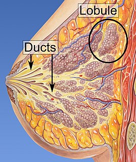
Lactiferous ducts are ducts that converge and form a branched system connecting the nipple to the lobules of the mammary gland. When lactogenesis occurs, under the influence of hormones, the milk is moved to the nipple by the action of smooth muscle contractions along the ductal system to the tip of the nipple. They are also referred to as galactophores, galactophorous ducts, mammary ducts, mamillary ducts or milk ducts.
Amastia refers to a rare clinical anomaly in which both internal breast tissue and the visible nipple are absent on one or both sides. It affects both men and women. Amastia can be either isolated or comorbid with other syndromes, such as ectodermal dysplasia, syndactaly and lipoatrophic diabetes. This abnormality can be classified into various types, and each could result from different pathologies. Amastia differs from amazia and athelia. Amazia is the absence of one or both mammary glands but the nipples remain present, and athelia is the absence of one or both nipples, but the mammary gland remains.
Ductal carcinoma in situ (DCIS), also known as intraductal carcinoma, is a pre-cancerous or non-invasive cancerous lesion of the breast. DCIS is classified as Stage 0. It rarely produces symptoms or a breast lump one can feel, typically being detected through screening mammography.
Breast diseases make up a number of conditions. The most common symptoms are a breast mass, breast pain, and nipple discharge.
Breast surgery is a form of surgery performed on the breast.

Duct ectasia of the breast, mammary duct ectasia or plasma cell mastitis is a condition that occurs when a milk duct beneath the nipple widens, the duct walls thicken, and the duct fills with fluid. This is the most common cause of greenish discharge. Mammary duct ectasia can mimic breast cancer. It is a disorder of peri- or post-menopausal age.
The term nonpuerperal mastitis describes inflammatory lesions of the breast (mastitis) that occur unrelated to pregnancy and breastfeeding.
Also called Zuska's disease, subareolar abscess is a subcutaneous abscess of the breast tissue beneath the areola of the nipple. It is a frequently aseptic inflammation and has been associated with squamous metaplasia of lactiferous ducts.

A breast mass, also known as a breast lump, is a localized swelling that feel different from the surrounding tissue. Breast pain, nipple discharge, or skin changes may be present. Concerning findings include masses that are hard, do not move easily, are of an irregular shape, or are firmly attached to surrounding tissue.

A nipple adenoma is a rare benign tumour of the breast.
Galactography or ductography is a medical diagnostic procedure for viewing the milk ducts. The procedure involves the radiography of the ducts after injection of a radiopaque substance into the duct system. The procedure is used for investigating the pathology of nipple discharge.
Central duct excision is the surgical removal (excision) of all lactiferous duct under the nipple. The excision of a single duct is called microdochectomy, a mere incision of a mammary duct is microdochotomy.
References
- ↑ "Microdochotomy". Systematized Nomenclature of Medicine - Clinical Terms. Retrieved 4 November 2014.
- 1 2 Nigel Rawlinson; Derek Alderson (29 September 2010). Surgery: Diagnosis and Management. John Wiley & Sons. p. 219. ISBN 978-1-4443-9122-0.
- ↑ Makita, Masujiro; Akiyama, Futoshi; Gomi, Naoya; Iwase, Takuji (2014). "Mammary ductoscopy and watchful follow-up substitute microdochectomy in patients with bloody nipple discharge". Breast Cancer. 23 (2): 242–251. doi:10.1007/s12282-014-0561-z. ISSN 1340-6868. PMID 25150843. S2CID 36404518.
- ↑ Trop I, Dugas A, David J, El Khoury M, Boileau JF, Larouche N, Lalonde L (October 2011). "Breast abscesses: evidence-based algorithms for diagnosis, management, and follow-up". Radiographics (review). 31 (6): 1683–99. doi:10.1148/rg.316115521. PMID 21997989., p. 1694
- 1 2 J Michael Dixon (22 June 2013). Breast Surgery: Companion to Specialist Surgical Practice. Elsevier Health Sciences. p. 276. ISBN 978-0-7020-4967-5.
- 1 2 3 Brendon J Coventry (17 January 2014). Breast, Endocrine and Surgical Oncology. Springer Science & Business Media. p. 23. ISBN 978-1-4471-5421-1.
- 1 2 J Michael Dixon (22 June 2013). Breast Surgery: Companion to Specialist Surgical Practice. Elsevier Health Sciences. p. 275. ISBN 978-0-7020-4967-5.
- ↑ Christopher Chan; Christopher L. H. Chan; Alister J. Hart (2001). Viva Practice for Intercollegiate MRCS. PasTest Ltd. p. 108. ISBN 978-1-904627-19-7.
- ↑ William E. G. Thomas; Norbert Senninger (1 February 2008). Short Stay Surgery. Springer Science & Business Media. p. 136. ISBN 978-3-540-69028-3.