Motor pool (neuroscience)

Last updated

A motor pool consists of all individual motor neurons that innervate a single muscle. Each individual muscle fiber is innervated by only one motor neuron, but one motor neuron may innervate several muscle fibers. This distinction is physiologically significant because the size of a given motor pool determines the activity of the muscle it innervates: for example, muscles responsible for finer movements are innervated by motor pools consisting of higher numbers of individual motor neurons. Motor pools are also distinguished by the different classes of motor neurons that they contain. The size, composition, and anatomical location of each motor pool is tightly controlled by complex developmental pathways. [1]

Contents

Anatomy

The motor unit: a single motor neuron and the muscle fibers that it innervates. A motor pool consists of all of the motor neurons that innervate a single muscle. Anatomical diagram of the motor unit.jpg
The motor unit: a single motor neuron and the muscle fibers that it innervates. A motor pool consists of all of the motor neurons that innervate a single muscle.

Distinct skeletal muscles are controlled by groups of individual motor units. Such motor units are made up of a single motor neuron and the muscle fibers that it innervates. The cell bodies of motor neurons are located in the ventral horn of the spinal cord and the brainstem. These neurons innervate skeletal muscle fibers through the propagation of action potentials down their axons (through ventral roots and cranial nerves), and they stimulate skeletal muscle fibers at neuromuscular junctions where they synapse with the motor end plates of muscle fibers. In humans, these axons can be as long as one meter. Motor neurons themselves fall into three main classes: alpha-motor neurons control extrafusal muscle fibers, meaning that they innervate skeletal muscles leading to movement; gamma-motor neurons innervate intrafusal muscle fibers, controlling the sensitivity of muscle spindles to stretch; beta-motor neurons are capable of synapsing on either type of muscle fiber. Alpha-motor neurons can further be divided into three separate subclasses, distinguished according to the contractile properties of the motor units that they form: fast-twitch fatigable (FF), fast-twitch fatigue-resistant (FR), and slow-twitch fatigue-resistant (S). The composition of a motor pool may consist of multiple classes and subclasses of motor neurons. [1]

Motor pools in the spinal cord are clustered in distinct columns of motor neurons extending over multiple spinal cord segments; although, there is significant overlap. Motor pools that control proximal muscles are generally located medial to the ventral horn, while those that control distal muscles are located laterally. Motor pools that control flexor muscles are located dorsally to the ventral horn while those that control extensor muscles are located ventrally. [1]

The number of motor neurons in an individual motor pool is highly variable and can generally be predicted by the level of nuanced control that a specific muscle requires. For example, some muscles have relatively low numbers of motor units in their respective motor pools while others, with highly nuanced control (such as the muscles in the human hand) have higher densities of motor units. [1]

Function

Motor pools function primarily to integrate synaptic input from higher CNS centers into precise and consistent contraction patterns. Individual motor neurons within a given motor pool fire in accordance with what is known as the 'size principle'. The size principle was proposed by Elwood Henneman and his group in the 1960s as an explanation of the characteristic pattern with which individual motor neurons in a motor pool fire. The size principle stipulates that when the motor neurons of a motor pool fire, leading to the contraction of a terminal muscle fiber, the motor units containing the smallest motor neurons fire first. As excitatory signalling increases, larger motor neurons are subsequently recruited and contraction strength increases. Further, this differential recruitment of motor neurons occurs in instances of both increasing and decreasing contraction strength. As contraction strength is increased, the smallest motor units fire first and are also the last to stop firing as the contraction strength decreases. [2]

The size principle has important functional benefits. Primarily, this system frees higher centers of the CNS from having to signal specific contraction patterns for distinct levels of muscle contraction. The level of synaptic input that higher centers provide to a given motor pool must determine the contraction strength, and this simplifies the process of contraction strength modulation. This system allows for very precise and consistent modulation of contraction strength from just increased or decreased levels of synaptic input: with additional motor units of increasing size, there will be a consistent and precise effect on the force of contraction. Another key benefit derived from the size principle is that smaller neurons will be fired more regularly and for a longer duration of time compared to larger neurons. These smaller motor units are more resistant to fatigue, and as such, are better suited to this role. [2] [3]

Specialization and development

There are several layers of differentiation and specialization to consider the complicated development of motor pools.

Alpha- and gamma-motor neuron differentiation

Alpha motor neurons and gamma motor neurons do not merely differ in their postsynaptic targets. The physiological differences between these two classes are significant. The axonal diameter of gamma-motor neurons is half of that of alpha-motor neurons, resulting in a higher cytoplasmic resistance and therefore a slower signal propagation velocity. Additionally, gamma-motor neurons display far simpler branching patterns than that of their alpha- counterparts. The differentiation into these two classes is regulated by complex interactions between several neurotrophic factors, and all of these interactions are not yet well understood. Glial cell line-derived neurotrophic factor (GDNF) has been discovered to play an especially important role in all layers of motor pool development. In the case of alpha- and gamma- differentiation, it has been shown that gamma motor neurons express significantly higher levels of certain GDNF receptor subunits. [4]

Alpha-motor neuron muscle fiber targets

Alpha motor neurons are also grouped into subclasses based on the types of muscle fibers they target, which also determines their functional characteristics. Motor neurons that target fast-twitch, fatigable (FF) muscle fibers are the largest (and therefore the fastest in propagating signals); those that target fast-twitch, fatigue-resistant (FR) fibers are of intermediate size; and those that target slow-twitch, fatigue-resistant (S) fibers are the smallest. In addition to signaling velocity, the range of sizes across these subclasses also forms the physiological basis of the size principle. Due to their relatively small axonal diameter, neurons that target S type fibers require a smaller input current to reach threshold potential. Conversely, the largest neurons, which target FF type fibers, require a greater input current to reach threshold. Therefore, the axonal diameter of the three subclasses of alpha motor neurons clearly determines the patterns of the recruitment of motor units predicted by the size principle. The specific regulatory mechanisms that determine the size of these three alpha-motor neuron subclasses are not well known. [4]

Pool specificity and spatial orientation

At a further level of specialization, specific combinations of motor neuron classes and subclasses are grouped spatially in clusters known as motor columns throughout the spinal cord. These motor columns are motor pools, and each has its own unique identity: each neuron within the pool (regardless of its class) expresses a unique profile of transcription factors, cell-surface proteins (such as axonal guidance receptors and adhesion molecules), and neurotransmitter receptors. These pool-specific biochemical markers provide the developmental framework for muscle-specific synapsing. [4] Among this pool-specific combination of transcription factors, Hox factors play an especially important role. Hox transcription factors play a central role in the spatial orientation of the motor pool within the spine, and in the site-specific synapsing of the motor pool on to its down-stream muscle fibers. [5]

Pool size

The number of motor neurons within a specific motor pool is a crucial developmental step. Again, little is known about the precise mechanisms and molecules involved in this process of specialization. However, it is known that the mechanism involves an initial generation of large numbers of motor neurons, followed by a pruning process that is mediated by cell death mechanisms and survival factors. Some researchers have speculated that the number of neurons within a given pool is determined by competitive interactions between different Hox genes. [4] [5]

Evolution

Muscles responsible for finer, more nuanced movements are innervated by motor pools consisting of higher numbers of individual motor neurons. This principle is highlighted by examining the evolution of the human tongue and hand motor pools.

Human tongue

Highly refined and coordinated movements of the tongue are responsible for the intricacies of human speech. The evolutionary analysis would predict an abnormally large motor pool innervating the muscle of the human tongue, relative to those of other mammals. Such a large motor pool would allow for region-specific innervation of the tongue muscle, motor neurons with task-specificity, motor neuron specializations that allow for quick movements, and various other motor nuances necessary for producing complex speech. Anatomical analysis has validated this evolutionary prediction: in the average adult human, the motor pool for the tongue contains between 7,093 and 8,817 motor neurons. This neuron density far exceeds that measured in other mammals, and even exceeds the motor pool size for many muscles in the human body: biceps brachii, for example, is innervated by a motor pool that averages 441.5 motor neurons in size. [6]

Human hand

The evolutionary analysis also predicts large motor pools innervating the muscles of the human hand. The human innovations of tool-making, throwing motions, and clubbing motions required unprecedented manual dexterity, only made possible by extensive networks of large motor pools innervating the human hand. As Richard Young explains:

It has been proposed that the hominid lineage began when a group of chimpanzee-like apes began to throw rocks and swing clubs at adversaries and that this behaviour yielded reproductive advantages for millions of years, driving natural selection for improved throwing and clubbing prowess. This assertion leads to the prediction that the human hand should be adapted for throwing and clubbing… thereby providing an evolutionary explanation for the two unique grips, and the extensive anatomical remodelling of the hand that made them possible. [7]

Precise neuromuscular control allows for the sub-millisecond release times necessary for throwing. Effective clubbing requires the recruitment of several motor units to produce a secure grip through impact. These evolutionary predictions have been verified by comparative anatomical studies: the motor pools innervating the human hand are significantly larger than those innervating related primate hands. [7]

Relevance to disease

ALS

Amyotrophic lateral sclerosis (ALS) is a neurodegenerative disease that differentially affects specific motor pools and classes of motor neurons. ALS modelled in mice, for example, was shown to lead first to rapid FF motor neuron loss, followed by a delayed loss of FR neurons, leaving S type neurons largely intact in the late stages of the disease. Further, in late-stage ALS patients, death of motor neurons is pool-specific. Motor neuron death all throughout the spinal cord leads to a nearly complete loss of voluntary movement; however, ocular control and voluntary control of excretory functions remain mostly unaffected. These movements are governed by motor pools in the midbrain and in Onuf's nucleus in the lumbosacral spinal cord, respectively. Currently, research is being conducted on these ALS-resistant motor pools, and their pool-specific molecular identities are being examined for potential neuroprotective qualities. [4]

Parallels in SMA and ageing

Spinal muscular atrophy (SMA) and ageing-related motor degeneration share clear parallels with ALS in the patterns and specificity of motor neuron loss. Though it is completely distinct from ALS in its pathogenesis, SMA leads to a similar rapid death of FF motor neurons, with S type neurons being generally spared. Further, motor pools controlling facial muscles (including those of the eye) and voluntary excretory muscles are spared. [4] Motor neuron degeneration caused by ageing similarly affects FF types but not S types; ageing also seems to spare ocular motor pools. [8] These similar patterns of neurodegeneration in three different diseases have led researchers to speculate that slow-twitch motor neurons, along with motor pools of the eyes and excretory muscles, have intrinsic neuroprotective properties that are not disease-specific. Research is currently being conducted to discover a possible molecular basis for neuroprotection in these cell types. [4]

Related Research Articles

In neuroscience, an F wave is one of several motor responses which may follow the direct motor response (M) evoked by electrical stimulation of peripheral motor or mixed nerves. F-waves are the second of two late voltage changes observed after stimulation is applied to the skin surface above the distal region of a nerve, in addition to the H-reflex which is a muscle reaction in response to electrical stimulation of innervating sensory fibers. Traversal of F-waves along the entire length of peripheral nerves between the spinal cord and muscle, allows for assessment of motor nerve conduction between distal stimulation sites in the arm and leg, and related motoneurons (MN's) in the cervical and lumbosacral cord. F-waves are able to assess both afferent and efferent loops of the alpha motor neuron in its entirety. As such, various properties of F-wave motor nerve conduction are analyzed in nerve conduction studies (NCS), and often used to assess polyneuropathies, resulting from states of neuronal demyelination and loss of peripheral axonal integrity.

<span class="mw-page-title-main">Motor neuron</span> Nerve cell sending impulse to muscle

A motor neuron is a neuron whose cell body is located in the motor cortex, brainstem or the spinal cord, and whose axon (fiber) projects to the spinal cord or outside of the spinal cord to directly or indirectly control effector organs, mainly muscles and glands. There are two types of motor neuron – upper motor neurons and lower motor neurons. Axons from upper motor neurons synapse onto interneurons in the spinal cord and occasionally directly onto lower motor neurons. The axons from the lower motor neurons are efferent nerve fibers that carry signals from the spinal cord to the effectors. Types of lower motor neurons are alpha motor neurons, beta motor neurons, and gamma motor neurons.

<span class="mw-page-title-main">Motor nerve</span> Nerve located in the central nervous system

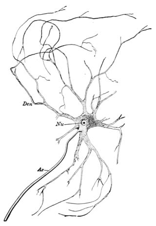
A motor nerve is a nerve that transmits motor signals from the central nervous system (CNS) to the muscles of the body. This is different from the motor neuron, which includes a cell body and branching of dendrites, while the nerve is made up of a bundle of axons. Motor nerves act as efferent nerves which carry information out from the CNS to muscles, as opposed to afferent nerves, which transfer signals from sensory receptors in the periphery to the CNS. Efferent nerves can also connect to glands or other organs/issues instead of muscles. In addition, there are nerves that serve as both sensory and motor nerves called mixed nerves.

A motor unit is made up of a motor neuron and all of the skeletal muscle fibers innervated by the neuron's axon terminals, including the neuromuscular junctions between the neuron and the fibres. Groups of motor units often work together as a motor pool to coordinate the contractions of a single muscle. The concept was proposed by Charles Scott Sherrington.

<span class="mw-page-title-main">Muscle spindle</span> Innervated muscle structure involved in reflex actions and proprioception

Muscle spindles are stretch receptors within the body of a skeletal muscle that primarily detect changes in the length of the muscle. They convey length information to the central nervous system via afferent nerve fibers. This information can be processed by the brain as proprioception. The responses of muscle spindles to changes in length also play an important role in regulating the contraction of muscles, for example, by activating motor neurons via the stretch reflex to resist muscle stretch.

<span class="mw-page-title-main">Grey column</span>

The grey column refers to a somewhat ridge-shaped mass of grey matter in the spinal cord. This presents as three columns: the anterior grey column, the posterior grey column, and the lateral grey column, all of which are visible in cross-section of the spinal cord.

<span class="mw-page-title-main">Reflex arc</span> Neural pathway which controls a reflex

A reflex arc is a neural pathway that controls a reflex. In vertebrates, most sensory neurons do not pass directly into the brain, but synapse in the spinal cord. This allows for faster reflex actions to occur by activating spinal motor neurons without the delay of routing signals through the brain. The brain will receive the input while the reflex is being carried out and the analysis of the signal takes place after the reflex action.

Lower motor neurons (LMNs) are motor neurons located in either the anterior grey column, anterior nerve roots or the cranial nerve nuclei of the brainstem and cranial nerves with motor function. Many voluntary movements rely on spinal lower motor neurons, which innervate skeletal muscle fibers and act as a link between upper motor neurons and muscles. Cranial nerve lower motor neurons also control some voluntary movements of the eyes, face and tongue, and contribute to chewing, swallowing and vocalization. Damage to the lower motor neurons can lead to flaccid paralysis, absent deep tendon reflexes and muscle atrophy.

<span class="mw-page-title-main">Gamma motor neuron</span>

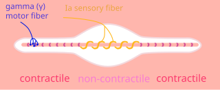
A gamma motor neuron, also called gamma motoneuron, or fusimotor neuron, is a type of lower motor neuron that takes part in the process of muscle contraction, and represents about 30% of (Aγ) fibers going to the muscle. Like alpha motor neurons, their cell bodies are located in the anterior grey column of the spinal cord. They receive input from the reticular formation of the pons in the brainstem. Their axons are smaller than those of the alpha motor neurons, with a diameter of only 5 μm. Unlike the alpha motor neurons, gamma motor neurons do not directly adjust the lengthening or shortening of muscles. However, their role is important in keeping muscle spindles taut, thereby allowing the continued firing of alpha neurons, leading to muscle contraction. These neurons also play a role in adjusting the sensitivity of muscle spindles.

<span class="mw-page-title-main">Motor unit recruitment</span> Additional activation of motor units to increase contractile strength

Motor unit recruitment is the activation of additional motor units to accomplish an increase in contractile strength in a muscle. A motor unit consists of one motor neuron and all of the muscle fibers it stimulates. All muscles consist of a number of motor units and the fibers belonging to a motor unit are dispersed and intermingle amongst fibers of other units. The muscle fibers belonging to one motor unit can be spread throughout part, or most of the entire muscle, depending on the number of fibers and size of the muscle. When a motor neuron is activated, all of the muscle fibers innervated by the motor neuron are stimulated and contract. The activation of one motor neuron will result in a weak but distributed muscle contraction. The activation of more motor neurons will result in more muscle fibers being activated, and therefore a stronger muscle contraction. Motor unit recruitment is a measure of how many motor neurons are activated in a particular muscle, and therefore is a measure of how many muscle fibers of that muscle are activated. The higher the recruitment the stronger the muscle contraction will be. Motor units are generally recruited in order of smallest to largest as contraction increases. This is known as Henneman's size principle.

<span class="mw-page-title-main">Intrafusal muscle fiber</span> Skeletal muscle fibers

Intrafusal muscle fibers are skeletal muscle fibers that serve as specialized sensory organs (proprioceptors). They detect the amount and rate of change in length of a muscle. They constitute the muscle spindle, and are innervated by both sensory (afferent) and motor (efferent) fibers.

Motor control is the regulation of movement in organisms that possess a nervous system. Motor control includes reflexes as well as directed movement.

<span class="mw-page-title-main">Stretch reflex</span>

The stretch reflex, or more accurately "muscle stretch reflex", is a muscle contraction in response to stretching within the muscle. The reflex functions to maintain the muscle at a constant length. The term deep tendon reflex is often wrongfully used by many health workers and students to refer to this reflex. "Tendons have little to do with the response, other than being responsible for mechanically transmitting the sudden stretch from the reflex hammer to the muscle spindle. In addition, some muscles with stretch reflexes have no tendons ".

<span class="mw-page-title-main">Alpha motor neuron</span>

Alpha (α) motor neurons (also called alpha motoneurons), are large, multipolar lower motor neurons of the brainstem and spinal cord. They innervate extrafusal muscle fibers of skeletal muscle and are directly responsible for initiating their contraction. Alpha motor neurons are distinct from gamma motor neurons, which innervate intrafusal muscle fibers of muscle spindles.

<span class="mw-page-title-main">Axon reflex</span>

The axon reflex is the response stimulated by peripheral nerves of the body that travels away from the nerve cell body and branches to stimulate target organs. Reflexes are single reactions that respond to a stimulus making up the building blocks of the overall signaling in the body's nervous system. Neurons are the excitable cells that process and transmit these reflex signals through their axons, dendrites, and cell bodies. Axons directly facilitate intercellular communication projecting from the neuronal cell body to other neurons, local muscle tissue, glands and arterioles. In the axon reflex, signaling starts in the middle of the axon at the stimulation site and transmits signals directly to the effector organ skipping both an integration center and a chemical synapse present in the spinal cord reflex. The impulse is limited to a single bifurcated axon, or a neuron whose axon branches into two divisions and does not cause a general response to surrounding tissue.

The Golgi tendon reflex (also called inverse stretch reflex, autogenic inhibition, tendon reflex) is an inhibitory effect on the muscle resulting from the muscle tension stimulating Golgi tendon organs (GTO) of the muscle, and hence it is self-induced. The reflex arc is a negative feedback mechanism preventing too much tension on the muscle and tendon. When the tension is extreme, the inhibition can be so great it overcomes the excitatory effects on the muscle's alpha motoneurons causing the muscle to suddenly relax. This reflex is also called the inverse myotatic reflex, because it is the inverse of the stretch reflex.

Beta motor neurons, also called beta motoneurons, are a kind of lower motor neuron, along with alpha motor neurons and gamma motor neurons. Beta motor neurons innervate intrafusal fibers of muscle spindles with collaterals to extrafusal fibers - a type of slow twitch fiber. Also, axons of alpha, beta, and gamma motor neurons become myelinated. Moreover, these efferent neurons originate from the anterior grey column of the spinal cord and travel to skeletal muscles. However, the larger diameter alpha motor fibers require higher conduction velocity than beta and gamma.

Henneman’s size principle describes relationships between properties of motor neurons and the muscle fibers they innervate and thus control, which together are called motor units. Motor neurons with large cell bodies tend to innervate fast-twitch, high-force, less fatigue-resistant muscle fibers, whereas motor neurons with small cell bodies tend to innervate slow-twitch, low-force, fatigue-resistant muscle fibers. In order to contract a particular muscle, motor neurons with small cell bodies are recruited before motor neurons with large cell bodies. It was proposed by Elwood Henneman.

Group A nerve fibers are one of the three classes of nerve fiber as generally classified by Erlanger and Gasser. The other two classes are the group B nerve fibers, and the group C nerve fibers. Group A are heavily myelinated, group B are moderately myelinated, and group C are unmyelinated.

<span class="mw-page-title-main">Spinal interneuron</span>

A spinal interneuron, found in the spinal cord, relays signals between (afferent) sensory neurons, and (efferent) motor neurons. Different classes of spinal interneurons are involved in the process of sensory-motor integration. Most interneurons are found in the grey column, a region of grey matter in the spinal cord.

References

  1. 1 2 3 4 Carp, J. S. and Wolpaw, J. R. 2010. Motor Neurons and Spinal Control of Movement. eLS DOI: 10.1002/9780470015902.a0000156.pub2
  2. 1 2 Henneman E and Mendell LM (1981) Functional organization of motoneuron pool and inputs. In: BrooksVB (ed.), Handbook of Physiology, sect. I: The Nervous System, vol. II: Motor Control, part I, pp. 423–507. Baltimore: Williams and Wilkins.
  3. Burke RE (1981) Motor units: anatomy, physiology, and functional organization. In: BrooksVB (ed.) Handbook of Physiology, sect. I: The Nervous System, vol. II: Motor Control, part I, pp. 345–422. Baltimore: Williams and Wilkins.
  4. 1 2 3 4 5 6 7 Kanning, Kevin C., Artem Kaplan, and Christopher E. Henderson. "Motor Neuron Diversity in Development and Disease." Annual Review of Neuroscience 33.1 (2010): 409-40. Print.
  5. 1 2 Song, Mi-Ryoung, and Samuel L. Pfaff. "Hox Genes: The Instructors Working at Motor Pools." Cell 123.3 (2005): 363-65. Print.
  6. Baker, Todd Adam. A Biomechanical Model of the Human Tongue for Understanding Speech Production and Other Lingual Behaviors. Ann Arbor, MI: UMI, 2008. 41-46. Print.
  7. 1 2 Young, Richard W. "Evolution of the Human Hand: The Role of Throwing and Clubbing." Journal of Anatomy 202.1 (2003): 165-74. Print.
  8. Enoka, R. "Mechanisms That Contribute to Differences in Motor Performance between Young and Old Adults." Journal of Electromyography and Kinesiology 13.1 (2003): 1-12. Print.

See also