This article needs additional citations for verification .(December 2009) |
Motor unit number estimation (MUNE) is a technique that uses electromyography to estimate the number of motor units in a muscle. [1]
This article needs additional citations for verification .(December 2009) |
Motor unit number estimation (MUNE) is a technique that uses electromyography to estimate the number of motor units in a muscle. [1]
A motor unit consists of one alpha motor neuron and all the muscle fibres it innervates.
Muscles differ in the number of motor units that they contain, and how many muscle fibres are within each unit (innervation ratio). In a general sense, muscles that require specificity of movement, such as muscles in charge of eye movement, have fewer fibres per unit, while those that are meant for less specific tasks, such as the calf muscles in charge of jumping, have more.
MUNE uses a general formula of: Number of motor units = compound muscle action potential size divided by the mean surface-detected motor unit action potential size
The compound muscle action potential (CMAP) size is found using supramaximal stimulation of the motor nerve to the muscle or muscle group (similar to a nerve conduction study). It is recorded using surface electrodes. This is representative of the sum of the surface detected motor unit action potentials from muscles innervated by that nerve.
Surface-detected motor unit action potential (SMUAP) size is the contribution of individual motor units. The way of finding the average size of these action potentials depends on the method used, as described below.
There are at least six techniques that are currently in use to estimate motor unit numbers. These include incremental stimulation, multi-point stimulation method, F-response method, spike-triggered averaging method and the statistical method. Incremental stimulation is the most illustrative of the concept, and so will be discussed here.
According to Henneman's size principle, motor unit recruitment is orderly such that smaller motor neurons are recruited before progressively larger ones. Additionally, the motor unit action potential is an all-or-none phenomenon - once the recruitment threshold (the stimulus intensity at which a motor unit begins to fire) is reached, it fires fully. Electrical stimulation of nerves reverses the recruitment order, due to the lower resistance of the larger motor neuron axons. Incremental stimulation involves gradually increasing the intensity of the stimulus to reach the recruitment threshold of increasing numbers of motor units until the intensity of the CMAP is reached. A 'step' is noted when an increase in stimulus leads to an increase in recorded EMG (i.e. another motor unit's threshold is reached and it is recruited). The CMAP is then divided by the number of steps required to reach the intensity of the CMAP to get a mean SMUAP size. The number of steps does not correlate to the total number of motor units in the muscle. Instead, the CMAP size is then divided by the mean SMUAP size to get an estimation of the number of motor units in the muscle.
The number of motor units per muscle can change due to aging, disease, or injury. These techniques are used to diagnose disease or monitor the effects of aging, disease and injury over time. In neuropathies, motoneurons die off, reducing the number of motor units progressively. In myopathies the size of the motor units is reduced because of the death of motor fibres, but the number of motor units remains the same until the disease progresses to a very severe state. In collaboration with other electromyography techniques, these conditions can be diagnosed and monitored. In a similar vein, normal aging also reduces the number of motor units but not to the same degree as disease. The effects of injury depend on the circumstances.
In neuroscience, an F wave is one of several motor responses which may follow the direct motor response (M) evoked by electrical stimulation of peripheral motor or mixed nerves. F-waves are the second of two late voltage changes observed after stimulation is applied to the skin surface above the distal region of a nerve, in addition to the H-reflex which is a muscle reaction in response to electrical stimulation of innervating sensory fibers. Traversal of F-waves along the entire length of peripheral nerves between the spinal cord and muscle, allows for assessment of motor nerve conduction between distal stimulation sites in the arm and leg, and related motoneurons (MN's) in the cervical and lumbosacral cord. F-waves are able to assess both afferent and efferent loops of the alpha motor neuron in its entirety. As such, various properties of F-wave motor nerve conduction are analyzed in nerve conduction studies (NCS), and often used to assess polyneuropathies, resulting from states of neuronal demyelination and loss of peripheral axonal integrity.

A nerve is an enclosed, cable-like bundle of nerve fibers called axons, in the peripheral nervous system. A nerve transmits electrical impulses and is the basic unit of the peripheral nervous system. A nerve provides a common pathway for the electrochemical nerve impulses called action potentials that are transmitted along each of the axons to peripheral organs or, in the case of sensory nerves, from the periphery back to the central nervous system. Each axon within the nerve is an extension of an individual neuron, along with other supportive cells such as some Schwann cells that coat the axons in myelin.

A motor neuron is a neuron whose cell body is located in the motor cortex, brainstem or the spinal cord, and whose axon (fiber) projects to the spinal cord or outside of the spinal cord to directly or indirectly control effector organs, mainly muscles and glands. There are two types of motor neuron – upper motor neurons and lower motor neurons. Axons from upper motor neurons synapse onto interneurons in the spinal cord and occasionally directly onto lower motor neurons. The axons from the lower motor neurons are efferent nerve fibers that carry signals from the spinal cord to the effectors. Types of lower motor neurons are alpha motor neurons, beta motor neurons, and gamma motor neurons.
A motor unit is made up of a motor neuron and all of the skeletal muscle fibers, also known as sarcomere innervated by the neuron's axon terminals. Groups of motor units often work together as a motor pool to coordinate the contractions of a single muscle. The concept was proposed by Charles Scott Sherrington.

Electromyography (EMG) is an electrodiagnostic medicine technique for evaluating and recording the electrical activity produced by skeletal muscles. EMG is performed using an instrument called an electromyograph to produce a record called an electromyogram. An electromyograph detects the electric potential generated by muscle cells when these cells are electrically or neurologically activated. The signals can be analyzed to detect medical abnormalities, activation level, or recruitment order, or to analyze the biomechanics of human or animal movement. In Computer Science, EMG is also used as middleware in gesture recognition towards allowing the input of physical action to a computer as a form of human-computer interaction.
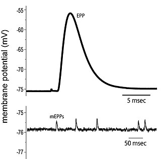
End plate potentials (EPPs) are the voltages which cause depolarization of skeletal muscle fibers caused by neurotransmitters binding to the postsynaptic membrane in the neuromuscular junction. They are called "end plates" because the postsynaptic terminals of muscle fibers have a large, saucer-like appearance. When an action potential reaches the axon terminal of a motor neuron, vesicles carrying neurotransmitters are exocytosed and the contents are released into the neuromuscular junction. These neurotransmitters bind to receptors on the postsynaptic membrane and lead to its depolarization. In the absence of an action potential, acetylcholine vesicles spontaneously leak into the neuromuscular junction and cause very small depolarizations in the postsynaptic membrane. This small response (~0.4mV) is called a miniature end plate potential (MEPP) and is generated by one acetylcholine-containing vesicle. It represents the smallest possible depolarization which can be induced in a muscle.

A nerve conduction study (NCS) is a medical diagnostic test commonly used to evaluate the function, especially the ability of electrical conduction, of the motor and sensory nerves of the human body. These tests may be performed by medical specialists such as clinical neurophysiologists, physical therapists, chiropractors, physiatrists, and neurologists who subspecialize in electrodiagnostic medicine. In the United States, neurologists and physiatrists receive training in electrodiagnostic medicine as part of residency training and in some cases acquire additional expertise during a fellowship in clinical neurophysiology, electrodiagnostic medicine, or neuromuscular medicine. Outside the US, clinical neurophysiologists learn needle EMG and NCS testing.
Motor unit recruitment refers to the activation of additional motor units to accomplish an increase in contractile strength in a muscle. A motor unit consists of one motor neuron and all of the muscle fibers it stimulates. All muscles consist of a number of motor units and the fibers belonging to a motor unit are dispersed and intermingle amongst fibers of other units. The muscle fibers belonging to one motor unit can be spread throughout part, or most of the entire muscle, depending on the number of fibers and size of the muscle. When a motor neuron is activated, all of the muscle fibers innervated by the motor neuron are stimulated and contract. The activation of one motor neuron will result in a weak but distributed muscle contraction. The activation of more motor neurons will result in more muscle fibers being activated, and therefore a stronger muscle contraction. Motor unit recruitment is a measure of how many motor neurons are activated in a particular muscle, and therefore is a measure of how many muscle fibers of that muscle are activated. The higher the recruitment the stronger the muscle contraction will be. Motor units are generally recruited in order of smallest to largest as contraction increases. This is known as Henneman's size principle.

The abductor pollicis brevis is a muscle in the hand that functions as an abductor of the thumb.

Rheobase is a measure of membrane potential excitability. In neuroscience, rheobase is the minimal current amplitude of infinite duration that results in the depolarization threshold of the cell membranes being reached, such as an action potential or the contraction of a muscle. In Greek, the root rhe translates to "current or flow", and basi means "bottom or foundation": thus the rheobase is the minimum current that will produce an action potential or muscle contraction.
Chronaxie is the minimum time required for an electric current double the strength of the rheobase to stimulate a muscle or a neuron. Rheobase is the lowest intensity with indefinite pulse duration which just stimulated muscles or nerves. Chronaxie is dependent on the density of voltage-gated sodium channels in the cell, which affect that cell’s excitability. Chronaxie varies across different types of tissue: fast-twitch muscles have a lower chronaxie, slow-twitch muscles have a higher one. Chronaxie is the tissue-excitability parameter that permits choice of the optimum stimulus pulse duration for stimulation of any excitable tissue. Chronaxie (c) is the Lapicque descriptor of the stimulus pulse duration for a current of twice rheobasic (b) strength, which is the threshold current for an infinitely long-duration stimulus pulse. Lapicque showed that these two quantities (c,b) define the strength-duration curve for current: I = b(1+c/d), where d is the pulse duration. However, there are two other electrical parameters used to describe a stimulus: energy and charge. The minimum energy occurs with a pulse duration equal to chronaxie. Minimum charge (bc) occurs with an infinitely short-duration pulse. Choice of a pulse duration equal to 10c requires a current of only 10% above rheobase (b). Choice of a pulse duration of 0.1c requires a charge of 10% above the minimum charge (bc).
A single nerve fibre will always give a maximum response and producing spikes of the same amplitude when stimulated. If the intensity of the stimulus is increased, the height of the spike always remains the same. In short the propagated impulse in a single fibre cannot be graded by grading the intensity or duration of the stimulus. The nerve fibre gives a maximum response or none at all. This is called the "all or none" principle. It is also Known as all or nothing law.

Group C nerve fibers are one of three classes of nerve fiber in the central nervous system (CNS) and peripheral nervous system (PNS). The C group fibers are unmyelinated and have a small diameter and low conduction velocity, whereas Groups A and B are myelinated. Group C fibers include postganglionic fibers in the autonomic nervous system (ANS), and nerve fibers at the dorsal roots. These fibers carry sensory information.

Denervation is any loss of nerve supply regardless of the cause. If the nerves lost to denervation are part of the neuronal communication to a specific function in the body then altered or a loss of physiological functioning can occur. Denervation can be caused by injury or be a symptom of a disorder like ALS and post-polio syndrome. Additionally, it can be a useful surgical technique to alleviate major negative symptoms, such as in renal denervation. Denervation can have many harmful side effects such as increased risk of infection and tissue dysfunction.
Clinical electrophysiology is the application of electrophysiology principles to medicine. The two main branches of this discipline are electrotherapy and electrophysiologic testing Clinical electrophysiology can be utilized in the study and treatment of various physiological conditions, and most notably in clinical cardiac electrophysiology.
Repetitive nerve stimulation is a variant of the nerve conduction study where electrical stimulation is delivered to a motor nerve repeatedly several times per second. By observing the change in the muscle electrical response (CMAP) after several stimulations, a physician can assess for the presence of a neuromuscular junction disease, and differentiate between presynaptic and postsynaptic conditions. The test was first described by German neurologist Friedrich Jolly in 1895, and is also known as Jolly's test.
A motor pool consists of all individual motor neurons that innervate a single muscle. Each individual muscle fiber is innervated by only one motor neuron, but one motor neuron may innervate several muscle fibers. This distinction is physiologically significant because the size of a given motor pool determines the activity of the muscle it innervates: for example, muscles responsible for finer movements are innervated by motor pools consisting of higher numbers of individual motor neurons. Motor pools are also distinguished by the different classes of motor neurons that they contain. The size, composition, and anatomical location of each motor pool is tightly controlled by complex developmental pathways.
The compound muscle action potential (CMAP) or compound motor action potential is an electromyography investigation . The CMAP idealizes the summation of a group of almost simultaneous action potentials from several muscle fibers in the same area. These are usually evoked by stimulation of the motor nerve. Patients that suffer from critical illness myopathy, which is a frequent cause of weakness seen in patients in hospital intensive care units, have prolonged compound muscle action potential.
Henneman’s size principle describes relationships between properties of motor neurons and the muscle fibers they innervate and thus control, which together are called motor units. Motor neurons with large cell bodies tend to innervate fast-twitch, high-force, less fatigue-resistant muscle fibers, whereas motor neurons with small cell bodies tend to innervate slow-twitch, low-force, fatigue-resistant muscle fibers. In order to contract a particular muscle, motor neurons with small cell bodies are recruited before motor neurons with large cell bodies. It was proposed by Elwood Henneman.

Cutaneous, or skin reflexes, are activated by skin receptors and play a valuable role in locomotion, providing quick responses to unexpected environmental challenges. They have been shown to be important in responses to obstacles or stumbling, in preparing for visually challenging terrain, and for assistance in making adjustments when instability is introduced. In addition to the role in normal locomotion, cutaneous reflexes are being studied for their potential in enhancing rehabilitation therapy (physiotherapy) for people with gait abnormalities.