Nerve glide, also known as nerve flossing or nerve stretching, is an exercise that stretches nerves. It facilitates the smooth and regular movement of peripheral nerves in the body. It allows the nerve to glide freely along with the movement of the joint and relax the nerve from compression. Nerve gliding cannot proceed with injuries or inflammations as the nerve is trapped by the tissue surrounding the nerve near the joint. Thus, nerve gliding exercise is widely used in rehabilitation programs and during the post-surgical period.
Radial, median, sciatic, and ulnar nerves require nerve gliding exercise during the rehabilitation period. The most common conditions that require nerve gliding exercise are carpal tunnel syndrome, cubital tunnel syndrome, radial neuropathy, and so on. Therapists prescribe different nerve gliding exercises in order to maximize the effects by correctly diagnosing the symptoms. Patients feel less pain when there is stretch in nerves, and there should be no aggressive exercise. Without correctly diagnosing symptoms and treatments, it worsens the conditions and nerves. Nerve gliding exercises should be done several times daily, depending on the issue. As patients continuously do nerve gliding exercises, they start to feel less pain after a few weeks.
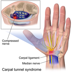
Carpal tunnel syndrome (CTS) is a condition that induces pain when the median nerve passes through the carpal tunnel in the wrist. It occurs when median nerves get irritated, compress, and strengthen. CTS evokes symptoms, including pain, paresthesia, and muscle atrophy. [1] This further leads to chronic pain and economic difficulties for patients as it requires work absence and surgical treatment.[ citation needed ]
Nerve gliding exercise becomes one of the optimal CTS treatments by assisting nerve mobilization. Restoring nerve mobilization would relieve edema and restore adhesion in the carpal tunnel. [2] According to the research, nerve gliding exercise has reduced the pain, decreased sensitive distal latency, and improved the functions that require force to grab. However, inappropriate nerve gliding exercises would worsen the conditions. Neural mobilization via nerve gliding should avoid excessive median nerve stretching when extending fingers in wrist extensions or when otherwise not advised. [3]
Nerve gliding exercise is not an optimal method for every patient. There is limited evidence on the effectiveness of neural gliding. However, the addition of nerve gliding exercise in conservative care accelerates the rehabilitation process and avoids surgical treatment. Further research is required to study the effectiveness of nerve glide physiotherapy and to determine groups that tend to respond better. [4]
Sciatica is low back pain that can extend to the feet. [5] This nerve pain is caused by nerve root irritation or constriction. Sciatica is known as an extremely painful symptom. Nerve glides are a common option for sciatica due to their cost-effectiveness. After performing nerve glides, the Numeric Pain Rating Score (NPRS) rated by patients improved, indicating a reduction in the pain. The nerve glide reduces acute sciatica and improves the range of motion of the hip. When nerve glides were performed along with other therapies, it resulted in a greater reduction in pain. [6] However, the research indicates that there is no statistically significant difference in the results among patients who were treated with nerve glides and other conventional treatments. [7]
Neural gliding is used for the rehabilitation of nerve-related neck and arm pain. The pain initiates from the neck, expanding to the arm. Nerve gliding physical therapy is beneficial in reducing pain intensity, bringing short-term improvements. [8] This treatment was found to manage neural tissue through specific postures and movements of the parts in pain. The stretch reduces nerve mechanosensitive that relieves discomforts, eventually leading to the normal function of the body. However, the long-term effects of nerve gliding exercises still remain unclear. [9]
Cubital tunnel syndrome is a condition that induces pains when ulnar nerves are stretched, pressed, and irritated. This syndrome is also known as "ulnar nerve entrapment". Similar to carpal tunnel syndrome, cubital tunnel syndrome evokes symptoms, including pain, numbness, tingling, and weakness in the hand. [10] Patients with cubital tunnel syndrome start to lose the power of their hands, which becomes hard to grip. The irritation occurs near the elbow, where the cubital tunnel is located. The ulnar nerve on the cubital tunnel is susceptible as the cubital tunnel is made up of soft tissue. Therefore, strong pressure leads to numbness. [11]
Ulnar nerve gliding is recommended to reduce symptoms of cubital tunnel syndrome. Patients with ulnar nerve gliding should stay away from the holding position. Rather, patients must repeat nerve gliding with the range of movement. There are various ulnar nerve gliding methods, which include elbow flexion, wrist extension, head tilt, and arm flexion.[ citation needed ]
Nerve gliding can reduce and strengthen connective tissues resulting in increased hamstring extensibility and passive stiffness. Passive stiffness refers to the resistance elongation that occurs in the joint, tendon, and connective tissue. The acute increase in hamstring extensibility can be seen right after nerve gliding intervention at the maximum range of motion. Nerve glide intervention is found to be slightly more effective than static stretching. The absolute static nerve extensibility was five times greater than the static stretching. While nerve gliding enhances the ability of the hamstring to stretch, the static stretch is more effective in terms of stress relaxation. Unlike static stretching, dynamic stretching shows similar outcomes to nerve gliding exercise. For hamstring flexibility, dynamic stretching targets low extremity muscles, while nerve gliding exercise targets posterior low extremity muscles and neural structures. Although both nerve gliding exercises and dynamic stretching do not lead to huge changes in exercise performance, both stretching methods are essential for pre-exercise stretches to avoid injuries. [12]
An alternative treatment option for carpal tunnel syndrome is low-level laser therapy (LLLT). The research shows that there is a statistically significant improvement in both LLLT and nerve gliding exercises. However, the difference in the effectiveness of LLLT and nerve gliding is not considered to replace nerve gliding physiotherapy. Considering the feasibility of those two treatments in terms of machine availability and cost-effectiveness, nerve gliding exercise remains to be prescribed widely. [13]
The injured or entrapped nerves are sensitive to external stimuli. Thus, nerve gliding, or nerve flossing, must be stopped, or range of motion (ROM) must be reduced once the patient feels pain. Patients' pain must be checked to avoid further irritation and injuries. Continuous nerve gliding enhances the movement of the joints and faster rehabilitation. If there is no further progress in rehabilitation, patients must see doctors or therapists for correct diagnosis. Nerve gliding exercise is not recommended for acute symptoms and severe damage. These cause pulling of nerve roots, which worsens the symptoms and nerve irritations. When the nerve glide is exercised by injured athletes, it has been shown to have no side effects on their sports performance. The measurements of sports performance include bilateral hamstring flexibility, vertical jump height, shuttle run, and dash sprint. This physiotherapy is safe when performed with caution (not going through the pains). Sports athletes can incorporate nerve glides in warm-up sessions. [14]
Carpal tunnel syndrome (CTS) is a nerve compression syndrome associated with the collected signs and symptoms of compression of the median nerve at the carpal tunnel in the wrist. Carpal tunnel syndrome is an idiopathic syndrome but there are environmental, and medical risk factors associated with the condition. CTS can affect both wrists.
The median nerve is a nerve in humans and other animals in the upper limb. It is one of the five main nerves originating from the brachial plexus.
Diabetic neuropathy includes various types of nerve damage associated with diabetes mellitus. The most common form, diabetic peripheral neuropathy, affects 30% of all diabetic patients. Symptoms depend on the site of nerve damage and can include motor changes such as weakness; sensory symptoms such as numbness, tingling, or pain; or autonomic changes such as urinary symptoms. These changes are thought to result from a microvascular injury involving small blood vessels that supply nerves. Relatively common conditions which may be associated with diabetic neuropathy include distal symmetric polyneuropathy; third, fourth, or sixth cranial nerve palsy; mononeuropathy; mononeuropathy multiplex; diabetic amyotrophy; and autonomic neuropathy.
A soft tissue injury is the damage of muscles, ligaments and tendons throughout the body. Common soft tissue injuries usually occur from a sprain, strain, a one-off blow resulting in a contusion or overuse of a particular part of the body. Soft tissue injuries can result in pain, swelling, bruising and loss of function.
Tarsal tunnel syndrome (TTS) is a nerve compression syndrome or nerve entrapment syndrome causing a painful foot condition in which the tibial nerve is entrapped as it travels through the tarsal tunnel. The tarsal tunnel is found along the inner leg behind the medial malleolus. The posterior tibial artery, tibial nerve, and tendons of the tibialis posterior, flexor digitorum longus, and flexor hallucis longus muscles travel in a bundle through the tarsal tunnel. Inside the tunnel, the nerve splits into three segments. One nerve (calcaneal) continues to the heel, the other two continue on to the bottom of the foot. The tarsal tunnel is delineated by bone on the inside and the flexor retinaculum on the outside.
The flexor retinaculum is a fibrous band on the palmar side of the hand near the wrist. It arches over the carpal bones of the hands, covering them and forming the carpal tunnel.
A spinal disc herniation is an injury to the intervertebral disc between two spinal vertebrae, usually caused by excessive strain or trauma to the spine. It may result in back pain, pain or sensation in different parts of the body, and physical disability. The most conclusive diagnostic tool for disc herniation is MRI, and treatment may range from painkillers to surgery. Protection from disc herniation is best provided by core strength and an awareness of body mechanics including good posture.
Ulnar tunnel syndrome, also known as Guyon's canal syndrome or Handlebar palsy, is ulnar neuropathy at the wrist where it passes through the ulnar tunnel. The most common presentation is a palsy of the deep motor branch of the ulnar nerve causing weakness of the interosseous muscles. Ulnar tunnel syndrome is usually caused by a ganglion cyst pressing on the ulnar nerve, other causes include traumas to the wrist and repetitive movements, but often the cause is unknown (idiopathic). Long distance bicycle rides are associated with transient alterations in ulnar nerve function. Sensory loss in the ring and small fingers is usually due to ulnar nerve entrapment at the cubital tunnel near the elbow, which is known as cubital tunnel syndrome, although it can uncommonly be due to compression at the wrist.
Idiopathic ulnar neuropathy at the elbow is a condition where pressure on the ulnar nerve as it passes through the cubital tunnel causes ulnar neuropathy. The symptoms of neuropathy are paresthesia (tingling) and numbness primarily affecting the little finger and ring finger of the hand. Ulnar neuropathy can progress to weakness and atrophy of the muscles in the hand. Symptoms can be alleviated by the use of a splint to prevent the elbow from flexing while sleeping.
Ulnar neuropathy is a disorder involving the ulnar nerve. Ulnar neuropathy may be caused by entrapment of the ulnar nerve with resultant numbness and tingling. It may also cause weakness or paralysis of the muscles supplied by the nerve. Ulnar neuropathy may affect the elbow as cubital tunnel syndrome. At the wrist a similar neuropathy is ulnar tunnel syndrome.
Magnetic resonance neurography (MRN) is the direct imaging of nerves in the body by optimizing selectivity for unique MRI water properties of nerves. It is a modification of magnetic resonance imaging. This technique yields a detailed image of a nerve from the resonance signal that arises from in the nerve itself rather than from surrounding tissues or from fat in the nerve lining. Because of the intraneural source of the image signal, the image provides a medically useful set of information about the internal state of the nerve such as the presence of irritation, nerve swelling (edema), compression, pinch or injury. Standard magnetic resonance images can show the outline of some nerves in portions of their courses but do not show the intrinsic signal from nerve water. Magnetic resonance neurography is used to evaluate major nerve compressions such as those affecting the sciatic nerve (e.g. piriformis syndrome), the brachial plexus nerves (e.g. thoracic outlet syndrome), the pudendal nerve, or virtually any named nerve in the body. A related technique for imaging neural tracts in the brain and spinal cord is called magnetic resonance tractography or diffusion tensor imaging.
Musculoskeletal injury refers to damage of muscular or skeletal systems, which is usually due to a strenuous activity and includes damage to skeletal muscles, bones, tendons, joints, ligaments, and other affected soft tissues. In one study, roughly 25% of approximately 6300 adults received a musculoskeletal injury of some sort within 12 months—of which 83% were activity-related. Musculoskeletal injury spans into a large variety of medical specialties including orthopedic surgery, sports medicine, emergency medicine and rheumatology.
Neural fibrolipoma is an overgrowth of fibro-fatty tissue along a nerve trunk that often leads to nerve compression. These only occur in the extremities, and often affect the median nerve. They are rare, very slow-growing, and their origin is unknown. It is believed that they may begin growth in response to trauma. They are not encapsulated by any sort of covering or sheath around the growth itself, as opposed to other cysts beneath the skin that often are. This means there are loosely defined margins of this lipoma. Despite this, they are known to be benign. Neural fibrolipomas are often more firm and tough to the touch than other lipomas. They are slightly mobile under the skin, and compress with pressure.
Nerve compression syndrome, or compression neuropathy, or nerve entrapment syndrome, is a medical condition caused by chronic, direct pressure on a peripheral nerve. It is known colloquially as a trapped nerve, though this may also refer to nerve root compression. Its symptoms include pain, tingling, numbness and muscle weakness. The symptoms affect just one particular part of the body, depending on which nerve is affected. The diagnosis is largely clinical and can be confirmed with diagnostic nerve blocks. Occasionally imaging and electrophysiology studies aid in the diagnosis. Timely diagnosis is important as untreated chronic nerve compression may cause permanent damage. A surgical nerve decompression can relieve pressure on the nerve but cannot always reverse the physiological changes that occurred before treatment. Nerve injury by a single episode of physical trauma is in one sense an acute compression neuropathy but is not usually included under this heading, as chronic compression takes a unique pathophysiological course.
Injuries to the arm, forearm or wrist area can lead to various nerve disorders. One such disorder is median nerve palsy. The median nerve controls the majority of the muscles in the forearm. It controls abduction of the thumb, flexion of hand at wrist, flexion of digital phalanx of the fingers, is the sensory nerve for the first three fingers, etc. Because of this major role of the median nerve, it is also called the eye of the hand. If the median nerve is damaged, the ability to abduct and oppose the thumb may be lost due to paralysis of the thenar muscles. Various other symptoms can occur which may be repaired through surgery and tendon transfers. Tendon transfers have been very successful in restoring motor function and improving functional outcomes in patients with median nerve palsy.
This article is about physical therapy in carpal tunnel syndrome.
Osborne's ligament, also Osborne's band, Osborne's fascia, Osborne's arcade, arcuate ligament of Osborne, or the cubital tunnel retinaculum, refers to either the connective tissue which spans the humeral and ulnar heads of the flexor carpi ulnaris (FCU) or another distinct tissue located between the olecranon process of the ulna and the medial epicondyle of the humerus. It is named after Geoffrey Vaughan Osborne, a British orthopedic surgeon, who described the eponymous tissue in 1957.
A nerve decompression is a neurosurgical procedure to relieve chronic, direct pressure on a nerve to treat nerve entrapment, a pain syndrome characterized by severe chronic pain and muscle weakness. In this way a nerve decompression targets the underlying pathophysiology of the syndrome and is considered a first-line surgical treatment option for peripheral nerve pain. Despite treating the underlying cause of the disease, the symptoms may not be fully reversible as delays in diagnosis can allow permanent damage to occur to the nerve and surrounding microvasculature. Traditionally only nerves accessible with open surgery have been good candidates, however innovations in laparoscopy and nerve-sparing techniques made nearly all nerves in the body good candidates, as surgical access is no longer a barrier.
Deep gluteal syndrome describes the non-discogenic extrapelvic entrapment of the sciatic nerve in the deep gluteal space. In simpler terms this is sciatica due to nerve irritation in the buttocks rather than the spine or pelvis. It is an extension of non-discogenic sciatic nerve entrapment beyond the traditional model of piriformis syndrome. Where sciatic nerve irritation in the buttocks was once thought of as only piriformis muscle, it is now recognized that there are many other causes. Symptoms are pain or dysthesias in the buttocks, hip, and posterior thigh with or without radiating leg pain. Patients often report pain when sitting. The two most common causes are piriformis syndrome and fibrovascular bands, but many other causes exist. Diagnosis is usually done through physical examination, magnetic resonance imaging, magnetic resonance neurography, and diagnostic nerve blocks. Surgical treatment is an endoscopic sciatic nerve decompression where tissue around the sciatic nerve is removed to relieve pressure.
Nerve entrapment involves a cascade of physiological changes caused by compression and tension. Some of these changes are irreversible. The magnitude and duration of the forces determines the extent of injury. In the acute form, mechanical injury and metabolic blocks impede nerve function. In the chronic form, there is a sequence of changes starting with a breakdown of the blood-nerve-barrier, followed by edema with connective tissue changes, followed by diffuse demyelination, and finally followed by axonmetesis. The injury will often be a mixed lesion where mild/moderate compression is a combination of a metabolic block and neuropraxia, while severe compression combines elements of neuropraxia and axonmetesis.