Neuroimaging intelligence testing

Last updated

Neuroimaging intelligence testing concerns the use of neuroimaging techniques to evaluate human intelligence. Neuroimaging technology has advanced such that scientists hope to use neuroimaging increasingly for investigations of brain function related to IQ.

Contents

IQ testing

Traditional IQ tests observe the test-taker's performance in a standardized battery of samples of behavior. The resulting IQ standard score is the subject of much investigation as psychologists check correlations between IQ and other life outcomes. The Wechsler IQ tests for adults and for children have long been regarded as the "gold standard" in IQ testing. [1]

fMRI data showing regions of activation Functional magnetic resonance imaging.jpg
fMRI data showing regions of activation

Neural bases of intelligence

The varying techniques of imaging-based testing search for different signs of intelligence. The types of intelligence analyzed in this review were fluid intelligence (Gf), general intelligence (g), and crystallized intelligence (Gc). Early studies utilized information from patients with brain damage, noticing changes in intelligence scores that correlated to certain regions of the brain. As imaging technology has improved, so has the ability for deeper neuro-analysis. MRI studies have found that the volume of gray matter correlates to intelligence, providing evidence for generalizations made regarding brain/head-size and intelligence. Additionally, PET and fMRI studies have revealed more information regarding the functionality of certain regions of the brain. By recording and interpreting the brain activity of subjects as they complete a variety of tasks, researchers are able to draw connections between the types of task (and thus, the type of intelligence) that calls on particular areas of the brain. Knowing how parts of the brain are utilized may reveal more information about the structure and hierarchy used in neural development. It also may provide interesting information regarding the pathways of neural signals as they navigate the nervous system. Image-based testing may allow researchers to discover why certain neurons are connected, if they are indeed aligned in a purposeful manner and consequently, how to repair such pathways when they are damaged. [2]

In general, there have been two types of intelligence studies: psychometric and biological. Biological approaches make use of neuroimaging techniques and examine brain function. Psychometrics focuses on mental abilities. Ian Deary and associates suggest that a greater overlap of these techniques will reveal new findings. [3]

Psychometrics

Psychometrics is a field of study specifically dedicated to psychological measurement and involves two main tasks: (i) constructing instruments and procedures for measurement; and (ii) the development and refinement of theoretical approaches to measurement. Brain-based intelligence tests are concerned with both of these aspects. Modern techniques have evolved to focus on a few biological characteristics: Brain ERPs, brain size, and speed of neural conduction. Various instruments have been employed to measure these things.

Brain ERPs allow for the "sequencing" of psychologically interesting processing. These event-related potentials are measured brain responses to specific stimuli, such as sensory, cognitive or motor events. ERPs, when compared to "mental speed," have shown a negative correlation with IQ. Research with ERPs suggests that high IQ individuals have a faster response time in some test conditions, have distinguishable ERP waveforms that are different than those of people with lower IQs, and may have less variability in their ERPs. The lack of variability suggests that individuals with a high IQ will have good scores in a variety of testing situations. [4]

ERPs can be measured using electroencephalography (EEG), which uses electrodes placed on the scalp to measure the electrical activity of the brain. The ERP waveform itself is constructed from the averaged results of many trials (100 or more). The average reduces signal noise from random-brain activity, leaving just the ERP. [5] An advantage of ERPs are that they measure processing between stimulus and response continuously. Having this stream of information makes it possible to see where the brain's electrical activity is being affected by specific stimuli. [6]

Brain size

Using MRI, researchers are able to acquire volumetric measurements of brain size. Some studies have tried to explain the relationship between brain size (meaning volume) and intelligence, specifically in terms of IQ. In general, it has been found that Full Scale IQ and Verbal IQ have a stronger correlation with brain size than Performance IQ. It is thought by some that grey and white matter specifically relate to different IQs (grey matter with Verbal IQ and white matter with Performance IQ), but the results have not been consistent. It has been found that within the cortex, the correlation with IQ was very influenced by the volume of prefrontal grey matter. [4]

A 2009 study examined intracerebral volumetric relationships in twins. Making use of high resolution MRI data, they found strong genetic connections correlations between cerebral structures. Specifically, the study suggests that a strong correlation exists between the tissue type or spatial proximity and genes. By examining the differences or lack thereof between the brain size of twin children, the researchers drew conclusions that individuals that share genes (i.e. twins) will show similar physiological brain properties compared to genetically-unrelated individuals. [7] This study provides evidence of the genetic influence of the brain structure and size, which are believed to both influence intelligence in some way.

Another study in 2006 examined 100 postmortem brains, seeking a relationship between an individual’s Full Scale Wechsler Adult Intelligence Scale score and the volume of their brain regions. Prior to death, the subjects had completed the WAIS test, which measures verbal and visuospatial abilities. The factors considered important to the relationship between brain size and intelligence were age, sex and hemispheric functional lateralization. They found that general verbal ability was correlated with cerebral volume in women and right-handed men. It was not possible to find a relationship between ability and volume in with every group, however. [8]

Neural conduction speed

Nerve conduction velocity (NCV) has been studied, giving varying results. Some have hypothesized that "higher intelligence is associated with better 'neural efficiency.'" A few studies suggested an association between nerve conduction velocity and scores on the Multidimensional Aptitude Battery (MAB). However, other studies have challenged these claims, finding little correlation between nerve conduction velocity and reaction time (RT). [4]

Raven's Progressive Matrices

Raven's Progressive Matrices (RPM) is a test consisting of 60 multiple-choice questions that increase in difficulty. RPM is based on pattern recognition and is a nonverbal group test, requiring the test taker to identify the missing element that completes the pattern. The test is designed to measure reasoning ability. The results of these tests are then paired with the results of imaging studies and relationships are drawn i.e. higher RPM scores and the increased size of a specific brain structure.

Raven's Advanced Progressive Matrices

Raven's Advanced Progressive Matrices (RAPM) is a 36-item test used to measure gF. RAPM tests for differences in novel problem solving and reasoning abilities. Similar to the RPM, subjects complete the pattern, identifying the missing piece of a 3x3 matrix from a list of eight options. [9]

n-Back Working Memory (WM) task

The n-back WM task has commonly utilized to measure cognitive activity during neuroimaging. According to Burgess et al.,

"The n-back task is typically thought to require the updating of information in WM, because, for each sequentially presented item, the participant must judge whether it matches the item presented n trials back (where n is prespecified, and usually equals 1, 2, or 3 items)."

While inside the MRI machine, subjects are asked to complete a variety of tasks. The brain activity is then captured and recorded by using MRI, allowing specific brain responses to be paired with their respective n-back tasks. [9]

Neuroimaging techniques

PET

Positron emission tomography detects gamma rays as they are emitted from a tracer that has been injected into the body. It is useful in neuroimaging because of the assumption that areas of high radioactivity are associated with high brain activity.

CAT Scan

Computed axial tomography (CAT) or computed tomography (CT) creates tomographic images of the body. For neuroimaging studies, computer-processed X-rays are used and the amount of X-ray blockage by different structures is used to generate image 'slices' of the brain. CAT scans are particularly useful for determining the size (volume) of specific structures of the brain. [10]

Global connectivity

A 2012 study from Washington University in St. Louis described the global connectivity of the prefrontal cortex. Global connectivity is the mechanism by which components of the frontoparietal brain network might coordinate control of other tasks. Cole et al. wrote that:

"A lateral prefrontal cortex (LPFC) region's activity was found to predict performance in a high control demand working memory task and also to exhibit high global connectivity. Critically, global connectivity in this LPFC region, involving connections both within and outside the frontoparietal network, showed a highly selective relationship with individual differences in fluid intelligence."

The lateral prefrontal cortex is a region of interest because those who have injuries to that part of the brain often have issues with common, every day tasks such as planning their day. The LPFC is thought to be important for "cognitive control capacity," which can be used to predict future outcomes such as success in school and the workplace. It was found by van den Heuvel et al. that higher intelligence individuals employ more efficient whole-brain network organization. This had led to the thought that cognitive control capacity may be supported by these whole-brain network properties. The 2012 study used a theoretic approach to neuroimage data known as global brain connectivity (GBC) or weighted degree centrality. GBC let the researches look closely at specific regions and their range of connectivity. It was then possible to examine each region's role in human cognitive control and intelligence. The study used fMRI to acquire data and examine each region's connectivity. [11]

Ethical implications

Privacy and confidentiality are major concerns for neuroimaging studies. With high-resolution anatomical images, such as those generated by fMRI, it is possible to identify individual subjects, putting their personal / medical privacy at risk. It is possible to create surface renderings of the brain and face from a volumetric MRI, which can be paired with photographs to identify the individual. [12]

It is becoming more accepted that a neurobiological basis for intelligence exists (at least for reasoning and problem-solving). The success of these intelligence studies present ethical issues. A large concern for the general population is the issue of race and intelligence. While little variation has been found between racial groups, the public perception of intelligence studies has been negatively impacted by concerns of racism. It is important to consider the consequences of studies that investigate intelligence differences in population-groups (racial or ethnic) and if it is ethical to conduct these studies. A study suggesting that one group is biologically more intelligent than another may cause tension. This has made neuroscientists reluctant to investigate individual or group differences in intelligence, as they may be perceived as racist. [13]

See also

Related Research Articles

Working memory is a cognitive system with a limited capacity that can hold information temporarily. It is important for reasoning and the guidance of decision-making and behavior. Working memory is often used synonymously with short-term memory, but some theorists consider the two forms of memory distinct, assuming that working memory allows for the manipulation of stored information, whereas short-term memory only refers to the short-term storage of information. Working memory is a theoretical concept central to cognitive psychology, neuropsychology, and neuroscience.

<span class="mw-page-title-main">Caudate nucleus</span> Structure of the striatum in the basal ganglia of the brain

The caudate nucleus is one of the structures that make up the corpus striatum, which is a component of the basal ganglia in the human brain. While the caudate nucleus has long been associated with motor processes due to its role in Parkinson's disease, it plays important roles in various other nonmotor functions as well, including procedural learning, associative learning and inhibitory control of action, among other functions. The caudate is also one of the brain structures which compose the reward system and functions as part of the cortico–basal ganglia–thalamic loop.

Neuroeconomics is an interdisciplinary field that seeks to explain human decision making, the ability to process multiple alternatives and to follow through on a plan of action. It studies how economic behavior can shape our understanding of the brain, and how neuroscientific discoveries can guide models of economics.

<span class="mw-page-title-main">Brodmann area 9</span> Part of the frontal cortex in the brain of humans and other primates

Brodmann area 9, or BA9, refers to a cytoarchitecturally defined portion of the frontal cortex in the brain of humans and other primates. It contributes to the dorsolateral and medial prefrontal cortex.

Neuroscience and intelligence refers to the various neurological factors that are partly responsible for the variation of intelligence within species or between different species. A large amount of research in this area has been focused on the neural basis of human intelligence. Historic approaches to study the neuroscience of intelligence consisted of correlating external head parameters, for example head circumference, to intelligence. Post-mortem measures of brain weight and brain volume have also been used. More recent methodologies focus on examining correlates of intelligence within the living brain using techniques such as magnetic resonance imaging (MRI), functional MRI (fMRI), electroencephalography (EEG), positron emission tomography and other non-invasive measures of brain structure and activity.

Functional integration is the study of how brain regions work together to process information and effect responses. Though functional integration frequently relies on anatomic knowledge of the connections between brain areas, the emphasis is on how large clusters of neurons – numbering in the thousands or millions – fire together under various stimuli. The large datasets required for such a whole-scale picture of brain function have motivated the development of several novel and general methods for the statistical analysis of interdependence, such as dynamic causal modelling and statistical linear parametric mapping. These datasets are typically gathered in human subjects by non-invasive methods such as EEG/MEG, fMRI, or PET. The results can be of clinical value by helping to identify the regions responsible for psychiatric disorders, as well as to assess how different activities or lifestyles affect the functioning of the brain.

<span class="mw-page-title-main">Prefrontal cortex</span> Part of brain largely responsible for personality, decision making, and social behaviour

In mammalian brain anatomy, the prefrontal cortex (PFC) covers the front part of the frontal lobe of the cerebral cortex. The PFC contains the Brodmann areas BA8, BA9, BA10, BA11, BA12, BA13, BA14, BA24, BA25, BA32, BA44, BA45, BA46, and BA47.

<span class="mw-page-title-main">Orbitofrontal cortex</span> Region of the prefrontal cortex of the brain

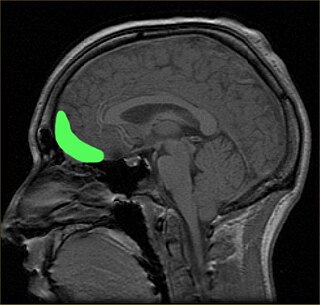
The orbitofrontal cortex (OFC) is a prefrontal cortex region in the frontal lobes of the brain which is involved in the cognitive process of decision-making. In non-human primates it consists of the association cortex areas Brodmann area 11, 12 and 13; in humans it consists of Brodmann area 10, 11 and 47.

Developmental cognitive neuroscience is an interdisciplinary scientific field devoted to understanding psychological processes and their neurological bases in the developing organism. It examines how the mind changes as children grow up, interrelations between that and how the brain is changing, and environmental and biological influences on the developing mind and brain.

<span class="mw-page-title-main">Dorsolateral prefrontal cortex</span> Area of the prefrontal cortex of primates

The dorsolateral prefrontal cortex is an area in the prefrontal cortex of the primate brain. It is one of the most recently derived parts of the human brain. It undergoes a prolonged period of maturation which lasts until adulthood. The DLPFC is not an anatomical structure, but rather a functional one. It lies in the middle frontal gyrus of humans. In macaque monkeys, it is around the principal sulcus. Other sources consider that DLPFC is attributed anatomically to BA 9 and 46 and BA 8, 9 and 10.

<span class="mw-page-title-main">Default mode network</span> Large-scale brain network active when not focusing on an external task

In neuroscience, the default mode network (DMN), also known as the default network, default state network, or anatomically the medial frontoparietal network (M-FPN), is a large-scale brain network primarily composed of the dorsal medial prefrontal cortex, posterior cingulate cortex/precuneus and angular gyrus. It is best known for being active when a person is not focused on the outside world and the brain is at wakeful rest, such as during daydreaming and mind-wandering. It can also be active during detailed thoughts related to external task performance. Other times that the DMN is active include when the individual is thinking about others, thinking about themselves, remembering the past, and planning for the future.

Malleability of intelligence describes the processes by which intelligence can increase or decrease over time and is not static. These changes may come as a result of genetics, pharmacological factors, psychological factors, behavior, or environmental conditions. Malleable intelligence may refer to changes in cognitive skills, memory, reasoning, or muscle memory related motor skills. In general, the majority of changes in human intelligence occur at either the onset of development, during the critical period, or during old age.

<span class="mw-page-title-main">Brain activity and meditation</span>

Meditation and its effect on brain activity and the central nervous system became a focus of collaborative research in neuroscience, psychology and neurobiology during the latter half of the 20th century. Research on meditation sought to define and characterize various practices. Meditation's effect on the brain can be broken up into two categories: state changes and trait changes, respectively alterations in brain activities during the act of meditating and changes that are the outcome of long-term practice.

<span class="mw-page-title-main">Resting state fMRI</span> Type of functional magnetic resonance imaging

Resting state fMRI is a method of functional magnetic resonance imaging (fMRI) that is used in brain mapping to evaluate regional interactions that occur in a resting or task-negative state, when an explicit task is not being performed. A number of resting-state brain networks have been identified, one of which is the default mode network. These brain networks are observed through changes in blood flow in the brain which creates what is referred to as a blood-oxygen-level dependent (BOLD) signal that can be measured using fMRI.

<span class="mw-page-title-main">Biological basis of personality</span>

The biological basis of personality is the collection of brain systems and mechanisms that underlie human personality. Human neurobiology, especially as it relates to complex traits and behaviors, is not well understood, but research into the neuroanatomical and functional underpinnings of personality are an active field of research. Animal models of behavior, molecular biology, and brain imaging techniques have provided some insight into human personality, especially trait theories.

The dorsal nexus is an area within the dorsal medial prefrontal cortex that serves as an intersection point for multiple brain networks. Research suggests it plays a role in the maintenance and manipulation of information, as well as supporting the control of cognitive functions such as behavior, memory, and conflict resolution. Abnormally increased connectivity between these networks through the Dorsal Nexus has been associated with certain types of depression. The activity generated by this abnormally high level of connectivity during a depressive state can be identified through Magnetic resonance imaging (MRI) and Positron emission tomography (PET).

Dynamic functional connectivity (DFC) refers to the observed phenomenon that functional connectivity changes over a short time. Dynamic functional connectivity is a recent expansion on traditional functional connectivity analysis which typically assumes that functional networks are static in time. DFC is related to a variety of different neurological disorders, and has been suggested to be a more accurate representation of functional brain networks. The primary tool for analyzing DFC is fMRI, but DFC has also been observed with several other mediums. DFC is a recent development within the field of functional neuroimaging whose discovery was motivated by the observation of temporal variability in the rising field of steady state connectivity research.

Cognitive humor processing refers to the neural circuitry and pathways that are involved in detecting incongruities of various situations presented in a humorous manner. Over the past decade, many studies have emerged utilizing fMRI studies to describe the neural correlates associated with how a human processes something that is considered "funny". Conceptually, humor is subdivided into two elements: cognitive and affective. The cognitive element, known as humor detection, refers to understanding the joke. Usually, this is characterized by the perceiver attempting to comprehend the disparities between the punch line and prior experience. The affective element, otherwise known as humor appreciation, is involved with enjoying the joke and producing visceral, emotional responses depending on the hilarity of the joke. This ability to comprehend and appreciate humor is a vital aspect of social functioning and is a significant part of the human condition that is relevant from a very early age. Humor comprehension develops in parallel with growing cognitive and language skills during childhood, while its content is mostly influenced by social and cultural factors. A further approach is described which refers to humor as an attitude related to strains. Humorous responses when confronted with troubles are discussed as a skill often associated with high social competence. The concept of humor has also been shown to have therapeutic effects, improving physiological systems such as the immune and central nervous system. It also has been shown to help cope with stress and pain. In sum, humor proves to be a personal resource throughout the life span, and helps support the coping of everyday tasks.

The parieto-frontal integration theory (P-FIT) considers intelligence to relate to how well different brain regions integrate to form intelligent behaviors. The theory proposes that large scale brain networks connect brain regions, including regions within frontal, parietal, temporal, and cingulate cortices, underlie the biological basis of human intelligence. These regions, which overlap significantly with the task-positive network, allow the brain to communicate and exchange information efficiently with one another. Support for this theory is primarily based on neuroimaging evidence, with support from lesion studies. The P-FIT is influential in that it explains the majority of current neuroimaging findings, as well as increasing empirical support for cognition being the result of large-scale brain networks, rather than numerous domain-specific processes or modules. A 2010 review of the neuroscience of intelligence described P-FIT as "the best available answer to the question of where in the brain intelligence resides".

An identity disturbance is a deficiency or inability to maintain one or more major components of identity. These components include a sense of continuity over time; emotional commitment to representations of self, role relationships, core values and self-standards; development of a meaningful world view; and recognition of one's place in the world.

References

  1. Meyer & Weaver 2005, p. 219 Campbell 2006, p. 66 Strauss, Sherman & Spreen 2006, p. 283 Foote 2007, p. 468 Kaufman & Lichtenberger 2006, p. 7 Hunt 2011, p. 12
  2. Gray, J. R.; Thompson, P. M. (2004). "Neurobiology of intelligence: science and ethics". Nature Reviews. Neuroscience. 5 (6): 471–82. doi:10.1038/nrn1405. PMID   15152197. S2CID   2430677.
  3. Deary, Ian J.; Austin, Elizabeth J.; Caryl, Peter G. (1 January 2000). "Testing versus understanding human intelligence". Psychology, Public Policy, and Law. 6 (1): 180–190. doi:10.1037/1076-8971.6.1.180.
  4. 1 2 3 Deary, IJ; Caryl, PG (August 1997). "Neuroscience and human intelligence differences". Trends in Neurosciences. 20 (8): 365–71. doi:10.1016/S0166-2236(97)01070-9. PMID   9246731. S2CID   13295937.
  5. Rugg, edited by Michael D.; Coles, Michael G.H. (1996). Electrophysiology of mind event-related brain potentials and cognition (Reprinted. ed.). Oxford: Oxford University Press. ISBN   9780198524168.{{cite book}}: |first= has generic name (help)
  6. Luck, Steven J. (2005). An introduction to the event-related potential technique . Cambridge, Mass.: MIT Press. pp.  21–23. ISBN   978-0-262-62196-0.
  7. Schmitt, JE; Wallace, GL; Lenroot, RK; Ordaz, SE; Greenstein, D; Clasen, L; Kendler, KS; Neale, MC; Giedd, JN (March 2010). "A twin study of intracerebral volumetric relationships". Behavior Genetics. 40 (2): 114–24. doi:10.1007/s10519-010-9332-6. PMC   3403699 . PMID   20112130.
  8. Witelson, S. F. (26 October 2005). "Intelligence and brain size in 100 postmortem brains: sex, lateralization and age factors". Brain. 129 (2): 386–398. doi: 10.1093/brain/awh696 . PMID   16339797.
  9. 1 2 Burgess, Gregory C.; Gray, Jeremy R.; Conway, Andrew R. A.; Braver, Todd S. (1 January 2011). "Neural mechanisms of interference control underlie the relationship between fluid intelligence and working memory span". Journal of Experimental Psychology: General. 140 (4): 674–692. doi:10.1037/a0024695. PMC   3930174 . PMID   21787103.
  10. Jeeves, Malcolm (1993). Mind fields : reflections on the science of mind and brain. Homebush West, NSW: Anzea Publishers. p. 21. ISBN   9780858925250.
  11. Cole, M. W.; Yarkoni, T.; Repovs, G.; Anticevic, A.; Braver, T. S. (June 2012). "Global connectivity of prefrontal cortex predicts cognitive control and intelligence". J. Neurosci. 32 (26): 8988–99. doi:10.1523/JNEUROSCI.0536-12.2012. PMC   3392686 . PMID   22745498.
  12. Kulynych, J (December 2002). "Legal and ethical issues in neuroimaging research: human subjects protection, medical privacy, and the public communication of research results". Brain and Cognition. 50 (3): 345–57. doi:10.1016/S0278-2626(02)00518-3. PMID   12480482. S2CID   2559078.
  13. Gray, Jeremy R.; Thompson, Paul M. (1 June 2004). "Neurobiology of intelligence: science and ethics". Nature Reviews Neuroscience. 5 (6): 471–482. doi:10.1038/nrn1405. PMID   15152197. S2CID   2430677.