Related Research Articles

The human teeth function to mechanically break down items of food by cutting and crushing them in preparation for swallowing and digesting. Humans have four types of teeth: incisors, canines, premolars, and molars, which each have a specific function. The incisors cut the food, the canines tear the food and the molars and premolars crush the food. The roots of teeth are embedded in the maxilla or the mandible and are covered by gums. Teeth are made of multiple tissues of varying density and hardness.

Cementum is a specialized calcified substance covering the root of a tooth. The cementum is the part of the periodontium that attaches the teeth to the alveolar bone by anchoring the periodontal ligament.

Dentin or dentine is a calcified tissue of the body and, along with enamel, cementum, and pulp, is one of the four major components of teeth. It is usually covered by enamel on the crown and cementum on the root and surrounds the entire pulp. By volume, 45% of dentin consists of the mineral hydroxyapatite, 33% is organic material, and 22% is water. Yellow in appearance, it greatly affects the color of a tooth due to the translucency of enamel. Dentin, which is less mineralized and less brittle than enamel, is necessary for the support of enamel. Dentin rates approximately 3 on the Mohs scale of mineral hardness. There are two main characteristics which distinguish dentin from enamel: firstly, dentin forms throughout life; secondly, dentin is sensitive and can become hypersensitive to changes in temperature due to the sensory function of odontoblasts, especially when enamel recedes and dentin channels become exposed.

The pulp is the part in the centre of a tooth made up of living connective tissues and odontoblasts. The pulp is a part of the dentin–pulp complex (endodontium). The vitality of the dentin-pulp complex, both during health and after injury, depends on pulp cell activity and the signalling processes that regulate the cell's behaviour.

Ameloblasts are cells present only during tooth development that deposit tooth enamel, which is the hard outermost layer of the tooth forming the surface of the crown.
Tomes's processes are a histologic landmark identified on an ameloblast, cells involved in the production of tooth enamel. During the synthesis of enamel, the ameloblast moves away from the enamel, forming a projection surrounded by the developing enamel. Tomes's processes are those projections and give the ameloblast a "picket-fence" appearance under a microscope.
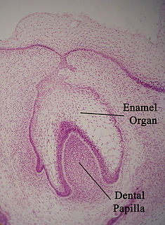
The enamel organ, also known as the dental organ, is a cellular aggregation seen in a developing tooth and it lies above the dental papilla. The enamel organ is responsible for the formation of enamel, initiation of dentine formation, establishment of the shape of a tooth's crown, and establishment of the dentoenamel junction.

Tooth development or odontogenesis is the complex process by which teeth form from embryonic cells, grow, and erupt into the mouth. For human teeth to have a healthy oral environment, all parts of the tooth must develop during appropriate stages of fetal development. Primary (baby) teeth start to form between the sixth and eighth week of prenatal development, and permanent teeth begin to form in the twentieth week. If teeth do not start to develop at or near these times, they will not develop at all, resulting in hypodontia or anodontia.
Amelogenesis is the formation of enamel on teeth and begins when the crown is forming during the advanced bell stage of tooth development after dentinogenesis forms a first layer of dentin. Dentin must be present for enamel to be formed. Ameloblasts must also be present for dentinogenesis to continue.

In embryology and prenatal development, the dental papilla is a condensation of ectomesenchymal cells called odontoblasts, seen in histologic sections of a developing tooth. It lies below a cellular aggregation known as the enamel organ. The dental papilla appears after 8–10 weeks intra uteral life. The dental papilla gives rise to the dentin and pulp of a tooth.
In vertebrates, an odontoblast is a cell of neural crest origin that is part of the outer surface of the dental pulp, and whose biological function is dentinogenesis, which is the formation of dentin, the substance beneath the tooth enamel on the crown and the cementum on the root.
Dentinogenesis is the formation of dentin, a substance that forms the majority of teeth. Dentinogenesis is performed by odontoblasts, which are a special type of biological cell on the outer wall of dental pulps, and it begins at the late bell stage of a tooth development. The different stages of dentin formation after differentiation of the cell result in different types of dentin: mantle dentin, primary dentin, secondary dentin, and tertiary dentin.
Pulpitis is inflammation of dental pulp tissue. The pulp contains the blood vessels, the nerves, and connective tissue inside a tooth and provides the tooth’s blood and nutrients. Pulpitis is mainly caused by bacterial infection which itself is a secondary development of caries. It manifests itself in the form of a toothache.
The dental canaliculi are the blood supply of a tooth. Odontoblast process run in the canaliculi that transverse the dentin layer and are referred as dentinal tubules. The number and size of the canaliculi decrease as the tubules move away from the pulp and toward the enamel or cementum.
The Hydrodynamic or Fluid Movement theory is one of three main theories in dentistry developed to explain dentine hypersensitivity, which is a sharp, transient pain arising from stimuli exposure. It states that different types of stimuli act on exposed dentine, causing increased fluid flow through the dentinal tubules. In response to this movement, mechanoreceptors on the pulp nerves trigger the acute, temporary pain of dentine hypersensitivity.
Korff fibers, also von Korff fibers are thick collageneous fibers in the developing tooth that begin in the dental papilla, spiral between the cells of the odontoblast layer, and form the matrix of the dentin. They are often the first sign of dentin formation. They are 0.1 to 0.2 µm in diameter and take a corkscrew path through the odontoblast layer and become incorporated into the layer of predentin. These fibers are named after German anatomist Karl von Korff. It consist of type 3 collagen, associated, at least initially, with fibronectin.
Dental pertains to the teeth, including dentistry. Topics related to the dentistry, the human mouth and teeth include:
Pulp necrosis is a clinical diagnostic category indicating the death of the pulp and nerves of the pulp chamber and root canal of a tooth which may be due to bacterial sequelae, trauma and chemical or mechanical irritation. It is often the end result of many cases of dental trauma, caries and irreversible pulpitis.

Pulp stones are nodular, calcified masses appearing in either or both the coronal and root portion of the pulp organ in teeth. Pulp stones are not painful unless they impinge on nerves.

Sir John Tomes was an English dental surgeon.
References
- ↑ Lee, Sidney, ed. (1899). . Dictionary of National Biography . Vol. 57. London: Smith, Elder & Co.
- Cate, Arnold Richard Ten (1999). Oral Histology: Development, Structure, and Function. Singapore: Harcourt Asia. ISBN 978-981-4033-17-6. OCLC 52693365.