Related Research Articles

A saccade is a quick, simultaneous movement of both eyes between two or more phases of focal points in the same direction. In contrast, in smooth-pursuit movements, the eyes move smoothly instead of in jumps; it could be associated with a shift in frequency of an emitted signal or a movement of a body part or device. Controlled cortically by the frontal eye fields (FEF), or subcortically by the superior colliculus, saccades serve as a mechanism for focal points, rapid eye movement, and the fast phase of optokinetic nystagmus. The word appears to have been coined in the 1880s by French ophthalmologist Émile Javal, who used a mirror on one side of a page to observe eye movement in silent reading, and found that it involves a succession of discontinuous individual movements.

Esotropia is a form of strabismus in which one or both eyes turn inward. The condition can be constantly present, or occur intermittently, and can give the affected individual a "cross-eyed" appearance. It is the opposite of exotropia and usually involves more severe axis deviation than esophoria. Esotropia is sometimes erroneously called "lazy eye", which describes the condition of amblyopia; a reduction in vision of one or both eyes that is not the result of any pathology of the eye and cannot be resolved by the use of corrective lenses. Amblyopia can, however, arise as a result of esotropia occurring in childhood: In order to relieve symptoms of diplopia or double vision, the child's brain will ignore or "suppress" the image from the esotropic eye, which when allowed to continue untreated will lead to the development of amblyopia. Treatment options for esotropia include glasses to correct refractive errors, the use of prisms, orthoptic exercises, or eye muscle surgery.

The vestibulo-ocular reflex (VOR) is a reflex that acts to stabilize gaze during head movement, with eye movement due to activation of the vestibular system, it is also known as the cervico-ocular reflex. The reflex acts to stabilize images on the retinas of the eye during head movement. Gaze is held steadily on a location by producing eye movements in the direction opposite that of head movement. For example, when the head moves to the right, the eyes move to the left, meaning the image a person sees stays the same even though the head has turned. Since slight head movement is present all the time, VOR is necessary for stabilizing vision: people with an impaired reflex find it difficult to read using print, because the eyes do not stabilise during small head tremors, and also because damage to reflex can cause nystagmus.

The medial longitudinal fasciculus (MLF) is a prominent bundle of nerve fibres which pass within the ventral/anterior portion of periaqueductal gray of the mesencephalon (midbrain). It contains the interstitial nucleus of Cajal, responsible for oculomotor control, head posture, and vertical eye movement.
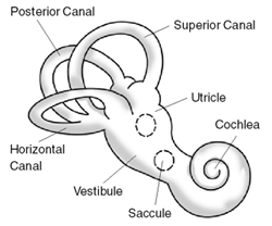
Benign paroxysmal positional vertigo (BPPV) is a disorder arising from a problem in the inner ear. Symptoms are repeated, brief periods of vertigo with movement, characterized by a spinning sensation upon changes in the position of the head. This can occur with turning in bed or changing position. Each episode of vertigo typically lasts less than one minute. Nausea is commonly associated. BPPV is one of the most common causes of vertigo.

Eye movement includes the voluntary or involuntary movement of the eyes. Eye movements are used by a number of organisms to fixate, inspect and track visual objects of interests. A special type of eye movement, rapid eye movement, occurs during REM sleep.

The extraocular muscles, or extrinsic ocular muscles, are the seven extrinsic muscles of the eye in humans and other animals. Six of the extraocular muscles, the four recti muscles, and the superior and inferior oblique muscles, control movement of the eye. The other muscle, the levator palpebrae superioris, controls eyelid elevation. The actions of the six muscles responsible for eye movement depend on the position of the eye at the time of muscle contraction.
Oscillopsia is a visual disturbance in which objects in the visual field appear to oscillate. The severity of the effect may range from a mild blurring to rapid and periodic jumping. Oscillopsia is an incapacitating condition experienced by many patients with neurological disorders. It may be the result of ocular instability occurring after the oculomotor system is affected, no longer holding images steady on the retina. A change in the magnitude of the vestibulo-ocular reflex due to vestibular disease can also lead to oscillopsia during rapid head movements. Oscillopsia may also be caused by involuntary eye movements such as nystagmus, or impaired coordination in the visual cortex and is one of the symptoms of superior canal dehiscence syndrome. Those affected may experience dizziness and nausea. Oscillopsia can also be used as a quantitative test to document aminoglycoside toxicity. Permanent oscillopsia can arise from an impairment of the ocular system that serves to maintain ocular stability. Paroxysmal oscillopsia can be due to an abnormal hyperactivity in the peripheral ocular or vestibular system.

Electronystagmography (ENG) is a diagnostic test to record involuntary movements of the eye caused by a condition known as nystagmus. It can also be used to diagnose the cause of vertigo, dizziness or balance dysfunction by testing the vestibular system. Electronystagmography is used to assess voluntary and involuntary eye movements. It evaluates the cochlear nerve and the oculomotor nerve. The ENG can be used to determine the origin of various eye and ear disorders.
Sensory ataxia is both a symptom and a sign in neurology. It is a form of ataxia caused not by cerebellar dysfunction but by loss of sensory input into the control of movement.

Parinaud's syndrome is a constellation of neurological signs indicating injury to the dorsal midbrain. More specifically, compression of the vertical gaze center at the rostral interstitial nucleus of medial longitudinal fasciculus (riMLF).

The flocculus is a small lobe of the cerebellum at the posterior border of the middle cerebellar peduncle anterior to the biventer lobule. Like other parts of the cerebellum, the flocculus is involved in motor control. It is an essential part of the vestibulo-ocular reflex, and aids in the learning of basic motor skills in the brain.

In medicine, the caloric reflex test is a test of the vestibulo-ocular reflex that involves irrigating cold or warm water or air into the external auditory canal. This method was developed by Robert Bárány, who won a Nobel prize in 1914 for this discovery.

The optokinetic reflex (OKR), also referred to as the optokinetic response, or optokinetic nystagmus (OKN), is a compensatory reflex that supports visual image stabilization. The purpose of OKR is to prevent motion blur on the retina that would otherwise occur when an animal moves its head or navigates through its environment. This is achieved by the reflexive movement of the eyes in the same direction as image motion, so as to minimize the relative motion of the visual scene on the eye. OKR is best evoked by slow, rotational motion, and operates in coordination with several complementary reflexes that also support image stabilization, including the vestibulo-ocular reflex (VOR).
Conjugate gaze palsies are neurological disorders affecting the ability to move both eyes in the same direction. These palsies can affect gaze in a horizontal, upward, or downward direction. These entities overlap with ophthalmoparesis and ophthalmoplegia.
The term gaze is frequently used in physiology to describe coordinated motion of the eyes and neck. The lateral gaze is controlled by the paramedian pontine reticular formation (PPRF). The vertical gaze is controlled by the rostral interstitial nucleus of medial longitudinal fasciculus and the interstitial nucleus of Cajal.
Nystagmus is a condition of involuntary eye movement. People can be born with it but more commonly acquire it in infancy or later in life. In many cases it may result in reduced or limited vision.

A vision disorder is an impairment of the sense of vision.

Vestibulocerebellar syndrome, also known as vestibulocerebellar ataxia, is a progressive neurological disorder that causes a variety of medical problems. Initially symptoms present as periodic attacks of abnormal eye movements but may intensify to longer-lasting motor incapacity. The disorder has been localized to the vestibulocerebellum, specifically the flocculonodular lobe. Symptoms of vestibulocerebellar syndrome may appear in early childhood but the full onset of neurological symptoms including nystagmus, ataxia, and tinnitus does not occur until early adulthood. To date, vestibulocerebellar syndrome has only been identified in three families but has affected multiple generations within them. Based on the familial pedigrees it has been characterized as an autosomal dominant disorder, although the exact genetic locus has not been identified. It has been found to be genetically distinct from other seemingly similar forms of neurological syndromes such as episodic ataxia types 1 and 2. Due to its rarity, however, little is known about specific details of the pathology or long-term treatment options. There is currently no cure for vestibulocerebellar syndrome, although some drug therapies have been effective in alleviating particular symptoms of the disorder.
Childhood cataract is cataract that occurs at birth or in childhood. It may be congenital or acquired.
References
- ↑ Hain, Timothy C. (1 August 2022). "Pendular Nystagmus". dizziness-and-balance.com. Archived from the original on 18 January 2022. Retrieved 8 December 2023.
- ↑ Nguyen, Huy D.; Azzez, Leen Saad; Bailey, Lucy (22 June 2021). "Acquired Nystagmus: Background, Pathophysiology, Epidemiology" . Medscape . Archived from the original on 1 June 2023. Retrieved 8 December 2023.
- ↑ Barton, Jason JS (13 January 2022). "Pendular nystagmus" . UpToDate . Archived from the original on 23 September 2022. Retrieved 8 December 2023.