Cognitive neuroscience is the scientific field that is concerned with the study of the biological processes and aspects that underlie cognition, with a specific focus on the neural connections in the brain which are involved in mental processes. It addresses the questions of how cognitive activities are affected or controlled by neural circuits in the brain. Cognitive neuroscience is a branch of both neuroscience and psychology, overlapping with disciplines such as behavioral neuroscience, cognitive psychology, physiological psychology and affective neuroscience. Cognitive neuroscience relies upon theories in cognitive science coupled with evidence from neurobiology, and computational modeling.

Functional neuroimaging is the use of neuroimaging technology to measure an aspect of brain function, often with a view to understanding the relationship between activity in certain brain areas and specific mental functions. It is primarily used as a research tool in cognitive neuroscience, cognitive psychology, neuropsychology, and social neuroscience.
Statistical parametric mapping (SPM) is a statistical technique for examining differences in brain activity recorded during functional neuroimaging experiments. It was created by Karl Friston. It may alternatively refer to software created by the Wellcome Department of Imaging Neuroscience at University College London to carry out such analyses.
The first neuroimaging technique ever is the so-called ‘human circulation balance’ invented by Angelo Mosso in the 1880s and able to non-invasively measure the redistribution of blood during emotional and intellectual activity. Then, in the early 1900s, a technique called pneumoencephalography was set. This process involved draining the cerebrospinal fluid from around the brain and replacing it with air, altering the relative density of the brain and its surroundings, to cause it to show up better on an x-ray, and it was considered to be incredibly unsafe for patients. A form of magnetic resonance imaging (MRI) and computed tomography (CT) were developed in the 1970s and 1980s. The new MRI and CT technologies were considerably less harmful and are explained in greater detail below. Next came SPECT and PET scans, which allowed scientists to map brain function because, unlike MRI and CT, these scans could create more than just static images of the brain's structure. Learning from MRI, PET and SPECT scanning, scientists were able to develop functional MRI (fMRI) with abilities that opened the door to direct observation of cognitive activities.

Neuroimaging or brain imaging is the use of various techniques to either directly or indirectly image the structure, function, or pharmacology of the nervous system. It is a relatively new discipline within medicine, neuroscience, and psychology. Physicians who specialize in the performance and interpretation of neuroimaging in the clinical setting are neuroradiologists. Neuroimaging falls into two broad categories:
Brain mapping is a set of neuroscience techniques predicated on the mapping of (biological) quantities or properties onto spatial representations of the brain resulting in maps.

Talairach coordinates, also known as Talairach space, is a 3-dimensional coordinate system of the human brain, which is used to map the location of brain structures independent from individual differences in the size and overall shape of the brain. It is still common to use Talairach coordinates in functional brain imaging studies and to target transcranial stimulation of brain regions. However, alternative methods such as the MNI Coordinate System have largely replaced Talairach for stereotaxy and other procedures.
The Organization for Human Brain Mapping (OHBM) is an organization of scientists with the main aim of organizing an annual meeting.

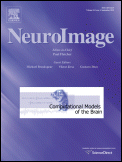
NeuroImage is a peer-reviewed scientific journal covering research on neuroimaging, including functional neuroimaging and functional human brain mapping. The current Editor in Chief is Michael Breakspear. Abstracts from the annual meeting of the Organization for Human Brain Mapping have been published as supplements to the journal. Members of the Organization for Human Brain Mapping are eligible for reduced subscription rates. In 2012, Elsevier launched an online-only, open access sister journal to NeuroImage, entitled NeuroImage: Clinical.

The University of Texas Health Science Center at San Antonio Department of Radiology is the second largest academic department in Radiological Sciences in the United States. Its Graduate Program in Radiological Sciences offers graduate training in various tracks, including Medical Physics, Radiation biology, Medical Health Physics, and Neuroimaging. In addition the educational enterprise includes an accredited radiology residency program and a number of fellowships.
Anders Martin Dale is a prominent neuroscientist and Professor of Radiology, Neurosciences, Psychiatry, and Cognitive Science at the University of California, San Diego (UCSD), and is one of the world's leading developers of sophisticated computational neuroimaging techniques. He is the founding Director of the Center for Multimodal Imaging Genetics (CMIG) at UCSD.

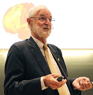
Marcus E. Raichle is an American neurologist at the Washington University School of Medicine in Saint Louis, Missouri. He is a professor in the Department of Radiology with joint appointments in Neurology, Neurobiology and Biomedical Engineering. His research over the past 40 years has focused on the nature of functional brain imaging signals arising from PET and fMRI and the application of these techniques to the study of the human brain in health and disease. He received the Kavli Prize in Neuroscience “for the discovery of specialized brain networks for memory and cognition", together with Brenda Milner and John O’Keefe in 2014.

Resting state fMRI is a method of functional magnetic resonance imaging (fMRI) that is used in brain mapping to evaluate regional interactions that occur in a resting or task-negative state, when an explicit task is not being performed. A number of resting-state conditions are identified in the brain, one of which is the default mode network. These resting brain state conditions are observed through changes in blood flow in the brain which creates what is referred to as a blood-oxygen-level dependent (BOLD) signal that can be measured using fMRI.
The following outline is provided as an overview of and topical guide to brain mapping:
Paul Thompson is a professor of neurology at the Imaging Genetics Center (IGC) at the University of Southern California. Thompson obtained a bachelor's degree in Greek and Latin languages and mathematics from Oxford University. He also earned a master's degree in mathematics from Oxford and a PhD degree in neuroscience from University of California, Los Angeles.

Professor Rajendra D Badgaiyan is an Indian-American psychiatrist and cognitive neuroscientist. He is best known for developing a new neuroimaging technique for detection of acute changes in concentration of dopamine released in the live human brain during performance of a cognitive. behavioral or emotional task.

Russell "Russ" Alan Poldrack is an American psychologist and neuroscientist. He is a professor of Psychology at Stanford University, member of the Stanford Neuroscience Institute and director of the Stanford Center for Reproducible Neuroscience.

Wolfgang Grodd is a German radiologist and professor emeritus of the University of Tübingen. He is known for his scientific works on the development and application of structural and functional magnetic resonance imaging in metabolic diseases, sensorimotor representation, language production and cognitive processing, cerebellum and thalamus. Currently, Grodd is research scientist at the Max Planck Institute for Biological Cybernetics.

Arthur W. Toga is an American neuroscientist and the director of the Laboratory of Neuro Imaging (LONI) and the Mark and Mary Stevens Neuroimaging and Informatics Institute within the Keck School of Medicine of the University of Southern California. He is also the Ghada Irani Chair in Neuroscience and provost professor of ophthalmology, neurology, psychiatry and the behavioral sciences, radiology and engineering.
Julie A. Fiez is a cognitive neuroscientist known for her research on the neural basis of speech, language, reading, working memory, and learning in healthy and patient populations. She is Professor of Psychology and Neuroscience at the Learning Research and Development Center and the Center for the Neural Basis of Cognition at the University of Pittsburgh. She is also Adjunct Faculty in the Department of Psychology at Carnegie Mellon University.


