Polychromasia

Last updated
Polychromasia
Other namesPolychromatophilia
Polychromasie-6.JPG
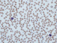
Polychromatic red blood cells appear bluish-gray on the blood smear.

Polychromasia is a disorder where there is an abnormally high number of immature red blood cells found in the bloodstream as a result of being prematurely released from the bone marrow during blood formation (poly- refers to many, and -chromasia means color.) These cells are often shades of grayish-blue. Polychromasia is usually a sign of bone marrow stress as well as immature red blood cells. 3 types are recognized, with types 1 and 2 being referred to as 'young red blood cells' and type 3 as 'old red blood cells'. Giemsa stain is used to distinguish all three types of blood smears. [1] The young cells will generally stain gray or blue in the cytoplasm. These young red blood cells are commonly called reticulocytes. All polychromatophilic cells are reticulocytes, however, not all reticulocytes are polychromatophilic. In the old blood cells, the cytoplasm either stains a light orange or does not stain at all.

Contents

Causes

Red blood cells can be released prematurely by a number of mechanisms. Premature release of red blood cells is usually caused due to damage of the bone marrow due to underlying causes as well as in response to the stimulation of hormones in strong association with anemia. Erythropoetin, a hormone made by the kidneys, controls the production of red blood cells as well as the rate at which they are released from the bone marrow. When these levels of erythropoetin rise, they signal the release of immature red blood cells into the bloodstream and is linked to anemia. Damaged bone marrow can also lead to polychromasia. The most common cause of bone marrow damage is penetration by cancer cells, either from the bone marrow itself or as a consequence of metastasis from another part of the body. [2]

Iron-deficiency anemia blood film Iron deficiency anemia blood film.jpg
Iron-deficiency anemia blood film

Association with anemia

Normocytic anemia is the most commonly seen type of anemia. This type of anemia is usually caused by the underproduction of blood cells as well as hemolysis. Anemia can be caused by either overproduction or underproduction of red blood cells, as well as the production of defective blood cells. Because there are more red blood cells needed in the body at that moment, they are released prematurely, leading to polychromasia. [3] [ citation needed ]

Association with reticulocytosis

There is a slight correlation between polychromasia and reticulocytosis. It is much easier to test for polychromasia in blood cells than to perform special staining for reticulocytosis. If polychromasia is found in the blood cells, the reticulocyte count is taken to detect further disease or stress. If a low count of reticulocytes is found, it usually indicates bone marrow stress. If a high reticulocyte count is found, it is usually linked to hemolysis, but a Coombs test may be performed in this case to rule out immune-mediated hemolysis. [4] Polychromasia can also be seen in blood smears when there is a normal reticulocyte count. This can be caused by infiltration of the bone marrow due to tumors as well as fibrosis, or scarring, of the marrow.[ citation needed ]

Embryology

The formation of red blood cells is commonly known as hematopoiesis. Up to the first 60 days of life, the yolk sac is the main source of hematopoiesis. The liver is then used as the main hematopoietic organ of the embryo until near birth, where it is then taken over by the bone marrow. [5] Most red blood cells are released into the blood as reticulocytes. Polychromasia occurs when the immature reticulocytes of the bone marrow are released, resulting in a grayish blue color of the cells. This color is seen because of the ribosomes still left on the immature blood cells, which are not found on mature red blood cells. These cells still contain a nucleus as well due to the early release, which is not needed in mature blood cells because their only function is to carry oxygen in the blood. The life span of a typical red blood cell is acknowledged to be approximately 120 days, and the time period of a reticulocyte found in the blood to be one day. The percentage of reticulocytes calculated to be in the blood at any given time indicates the rapidity of the red blood cell turnover in a healthy patient. The number of reticulocytes, however, reflects the amount of erythropoiesis that has occurred on any certain day. [6] The absolute number of reticulocytes is referred to as the reticulocyte index and is calculated by adjusting the reticulocyte percentage by the ratio of observed hematocrit to expected hematocrit to get the 'corrected' reticulocyte count.[ citation needed ]

Diagnosis

Polychromasia can be detected through the use of stains that will change the color of the red blood cells that are affected. Under certain conditions, these red blood cells are shown to have an affinity for basic stains, contrary to the usual acid stains used. Polychromatic cells usually stain dark blue or gray and are distinguishable from normal blood cells mostly by a slight change in color.[ citation needed ]

History

In 1890, research done by William Henry Howell indicated that certain red blood cells found both in fetal circulation and bone marrow (of a cat) had unusual granulation. These granules are also called Grawitz granules. In most instances, he found that these granules were connected by a network of sorts. The cells that had this granulation were found in blood and tissues that had been freshly stained without undergoing fixation. Howell was the first to describe these blood cells as being of the prototype stippling, which meant granular degeneration of the red blood cells. [7] In 1893, Max Askanazy, who was studying the blood of an anemic patient, discovered granulation in the blood cells that were polychromatic. Later studies were done by other scientists also showed the same results in other forms of anemia. This pattern of granulation was also seen in several types of toxic poisoning, especially lead poisoning. However, other research has shown that there has been stippling found in normal blood cells as well. Stippling is supposed to be one of the earliest symptoms of lead poisoning, although most scientists now regard it as a degenerative condition, along with polychromasia.[ citation needed ]

Related Research Articles

<span class="mw-page-title-main">Hemolysis</span> Rupturing of red blood cells and release of their contents

Hemolysis or haemolysis, also known by several other names, is the rupturing (lysis) of red blood cells (erythrocytes) and the release of their contents (cytoplasm) into surrounding fluid. Hemolysis may occur in vivo or in vitro.

<span class="mw-page-title-main">Anemia</span> Medical condition

Anemia or anaemia is a blood disorder in which the blood has a reduced ability to carry oxygen due to a lower than normal number of red blood cells, or a reduction in the amount of haemoglobin. When anemia comes on slowly, the symptoms are often vague, such as tiredness, weakness, shortness of breath, headaches, and a reduced ability to exercise. When anemia is acute, symptoms may include confusion, feeling like one is going to pass out, loss of consciousness, and increased thirst. Anemia must be significant before a person becomes noticeably pale. Additional symptoms may occur depending on the underlying cause. Preoperative anemia can increase the risk of needing a blood transfusion following surgery. Anemia can be temporary or long term and can range from mild to severe.

<span class="mw-page-title-main">Complete blood count</span> Routine laboratory test of blood cells

A complete blood count (CBC), also known as a full blood count (FBC), is a set of medical laboratory tests that provide information about the cells in a person's blood. The CBC indicates the counts of white blood cells, red blood cells and platelets, the concentration of hemoglobin, and the hematocrit. The red blood cell indices, which indicate the average size and hemoglobin content of red blood cells, are also reported, and a white blood cell differential, which counts the different types of white blood cells, may be included.

<span class="mw-page-title-main">Reticulocyte</span> Blood cells

Reticulocytes are immature red blood cells (RBCs). In the process of erythropoiesis, reticulocytes develop and mature in the bone marrow and then circulate for about a day in the blood stream before developing into mature red blood cells. Like mature red blood cells, in mammals, reticulocytes do not have a cell nucleus. They are called reticulocytes because of a reticular (mesh-like) network of ribosomal RNA that becomes visible under a microscope with certain stains such as new methylene blue and Romanowsky stain.

<span class="mw-page-title-main">Blood smear</span> Stained blood on microscope slide

A blood smear, peripheral blood smear or blood film is a thin layer of blood smeared on a glass microscope slide and then stained in such a way as to allow the various blood cells to be examined microscopically. Blood smears are examined in the investigation of hematological (blood) disorders and are routinely employed to look for blood parasites, such as those of malaria and filariasis.

<span class="mw-page-title-main">Hereditary spherocytosis</span> Medical condition

Hereditary spherocytosis (HS) is a congenital hemolytic disorder, wherein a genetic mutation coding for a structural membrane protein phenotype leads to a spherical shaping of erythrocytic cellular morphology. As erythrocytes are sphere-shaped (spherocytosis), rather than the normal biconcave disk-shaped, their morphology interferes with these cells' abilities to be flexible during circulation throughout the entirety of the body - arteries, arterioles, capillaries, venules, veins, and organs. This difference in shape also makes the red blood cells more prone to rupture under osmotic and/or mechanical stress. Cells with these dysfunctional proteins are degraded in the spleen, which leads to a shortage of erythrocytes resulting in hemolytic anemia.

<span class="mw-page-title-main">Paroxysmal nocturnal hemoglobinuria</span> Medical condition

Paroxysmal nocturnal hemoglobinuria (PNH) is a rare, acquired, life-threatening disease of the blood characterized by destruction of red blood cells by the complement system, a part of the body's innate immune system. This destructive process occurs due to deficiency of the red blood cell surface protein DAF, which normally inhibits such immune reactions. Since the complement cascade attacks the red blood cells within the blood vessels of the circulatory system, the red blood cell destruction (hemolysis) is considered an intravascular hemolytic anemia. Other key features of the disease, such as the high incidence of venous blood clot formation, are incompletely understood.

<span class="mw-page-title-main">Hemolytic anemia</span> Medical condition

Hemolytic anemia or haemolytic anaemia is a form of anemia due to hemolysis, the abnormal breakdown of red blood cells (RBCs), either in the blood vessels or elsewhere in the human body (extravascular). This most commonly occurs within the spleen, but also can occur in the reticuloendothelial system or mechanically. Hemolytic anemia accounts for 5% of all existing anemias. It has numerous possible consequences, ranging from general symptoms to life-threatening systemic effects. The general classification of hemolytic anemia is either intrinsic or extrinsic. Treatment depends on the type and cause of the hemolytic anemia.

<span class="mw-page-title-main">Erythropoiesis</span> Process which produces red blood cells

Erythropoiesis is the process which produces red blood cells (erythrocytes), which is the development from erythropoietic stem cell for mature red blood cell.

<span class="mw-page-title-main">Megaloblastic anemia</span> Medical condition

Megaloblastic anemia is a type of macrocytic anemia. An anemia is a red blood cell defect that can lead to an undersupply of oxygen. Megaloblastic anemia results from inhibition of DNA synthesis during red blood cell production. When DNA synthesis is impaired, the cell cycle cannot progress from the G2 growth stage to the mitosis (M) stage. This leads to continuing cell growth without division, which presents as macrocytosis. Megaloblastic anemia has a rather slow onset, especially when compared to that of other anemias. The defect in red cell DNA synthesis is most often due to hypovitaminosis, specifically vitamin B12 deficiency or folate deficiency. Loss of micronutrients may also be a cause.

<span class="mw-page-title-main">Sideroblastic anemia</span> Medical condition

Sideroblastic anemia, or sideroachrestic anemia, is a form of anemia in which the bone marrow produces ringed sideroblasts rather than healthy red blood cells (erythrocytes). In sideroblastic anemia, the body has iron available but cannot incorporate it into hemoglobin, which red blood cells need in order to transport oxygen efficiently. The disorder may be caused either by a genetic disorder or indirectly as part of myelodysplastic syndrome, which can develop into hematological malignancies.

The reticulocyte production index (RPI), also called a corrected reticulocyte count (CRC), is a calculated value used in the diagnosis of anemia. This calculation is necessary because the raw reticulocyte count is misleading in anemic patients. The problem arises because the reticulocyte count is not really a count but rather a percentage: it reports the number of reticulocytes as a percentage of the number of red blood cells. In anemia, the patient's red blood cells are depleted, creating an erroneously elevated reticulocyte count.

Autoimmune hemolytic anemia (AIHA) occurs when antibodies directed against the person's own red blood cells (RBCs) cause them to burst (lyse), leading to an insufficient number of oxygen-carrying red blood cells in the circulation. The lifetime of the RBCs is reduced from the normal 100–120 days to just a few days in serious cases. The intracellular components of the RBCs are released into the circulating blood and into tissues, leading to some of the characteristic symptoms of this condition. The antibodies are usually directed against high-incidence antigens, therefore they also commonly act on allogenic RBCs. AIHA is a relatively rare condition, with an incidence of 5-10 cases per 1 million persons per year in the warm-antibody type and 0.45 to 1.9 cases per 1 million persons per year in the cold antibody type. Autoimmune hemolysis might be a precursor of later onset systemic lupus erythematosus.

Myelophthisic anemia is a severe type of anemia found in some people with diseases that affect the bone marrow. Myelophthisis refers to the displacement of hemopoietic bone-marrow tissue by fibrosis, tumors, or granulomas. The word comes from the roots myelo-, which refers to bone marrow, and phthysis, shrinkage or atrophy.

The term macrocytic is from Greek words meaning "large cell". A macrocytic class of anemia is an anemia in which the red blood cells (erythrocytes) are larger than their normal volume. The normal erythrocyte volume in humans is about 80 to 100 femtoliters. In metric terms the size is given in equivalent cubic micrometers. The condition of having erythrocytes which are too large, is called macrocytosis. In contrast, in microcytic anemia, the erythrocytes are smaller than normal.

Red blood cell indices are blood tests that provide information about the hemoglobin content and size of red blood cells. Abnormal values indicate the presence of anemia and which type of anemia it is.

<span class="mw-page-title-main">Pappenheimer bodies</span>

Pappenheimer bodies are abnormal basophilic granules of iron found inside red blood cells on routine blood stain. They are a type of inclusion body composed of ferritin aggregates, or mitochondria or phagosomes containing aggregated ferritin. They appear as dense, blue-purple granules within the red blood cell and there are usually only one or two, located in the cell periphery. They stain on a Romanowsky stain because clumps of ribosomes are co‐precipitated with the iron‐containing organelles.

Normocytic anemia is a type of anemia and is a common issue that occurs for men and women typically over 85 years old. Its prevalence increases with age, reaching 44 percent in men older than 85 years. The most common type of normocytic anemia is anemia of chronic disease.

<span class="mw-page-title-main">White blood cell</span> Type of cells of the immunological system

White blood cells, also called leukocytes or leucocytes, are the cells of the immune system that are involved in protecting the body against both infectious disease and foreign invaders. All white blood cells are produced and derived from multipotent cells in the bone marrow known as hematopoietic stem cells. Leukocytes are found throughout the body, including the blood and lymphatic system.

<span class="mw-page-title-main">Nucleated red blood cell</span>

A nucleated red blood cell (NRBC), also known by several other names, is a red blood cell that contains a cell nucleus. Almost all vertebrate organisms have hemoglobin-containing cells in their blood, and with the exception of mammals, all of these red blood cells are nucleated. In mammals, NRBCs occur in normal development as precursors to mature red blood cells in erythropoiesis, the process by which the body produces red blood cells. NRBCs are normally found in the bone marrow of humans of all ages and in the blood of fetuses and newborn infants. After infancy, RBCs normally contain a nucleus only during the very early stages of the cell's life, and the nucleus is ejected as a normal part of cellular differentiation before the cell is released into the bloodstream. Thus, if NRBCs are identified on an adult's complete blood count or peripheral blood smear, it suggests that there is a very high demand for the bone marrow to produce RBCs, and immature RBCs are being released into circulation. Possible pathologic causes include anemia, myelofibrosis, thalassemia, miliary tuberculosis, cancers involving bone marrow, and in chronic hypoxemia.

References

  1. Thomas, Lyell J. (1937). "On the Life Cycle of Contracaecum Spiculigerum". The Journal of Parasitology. 23 (4): 429–31. JSTOR   3272243.
  2. "What Is Polychromasia?". WebMD. Retrieved 28 June 2021.
  3. "Anemia: Overview". The Lecturio Medical Concept Library. Retrieved 28 June 2021.
  4. Camitta, Bruce M.; Slye, Rebecca Jean (2012). "Optimizing Use of the Complete Blood Count". Pediatria Polska. 87 (1): 72–77. doi: 10.1016/S0031-3939(12)70597-8 .
  5. Bleyl, Steven B., Philip R. Brauer, and Philippa H. Francis-West. "Development of Yolk Sac and Chorionic Cavity." Larsen's Human Embryology. By Gary C. Schoenwolf. 4th ed. Philadelphia: Churchill Livingstone, 2009. 58. Print.
  6. Bessman JD (1990). "156 Reticulocytes". In Walker HK, Hall WD, Hurst JW (eds.). Clinical Methods: The History, Physical, and Laboratory Examinations (3rd ed.). Butterworths. ISBN   0-409-90077-X. PMID   21250107. NBK264.
  7. Hawes, John B. (1909). "A Study of the Reticulated Red Blood Corpuscle by Means of Vital Staining Methods; Its Relation to Polychromatophilia and Stippling". Boston Medical and Surgical Journal. 15: 493–9. doi:10.1056/NEJM190910071611501.