Related Research Articles

The human teeth function to mechanically break down items of food by cutting and crushing them in preparation for swallowing and digesting. Humans have four types of teeth: incisors, canines, premolars, and molars, which each have a specific function. The incisors cut the food, the canines tear the food and the molars and premolars crush the food. The roots of teeth are embedded in the maxilla or the mandible and are covered by gums. Teeth are made of multiple tissues of varying density and hardness.

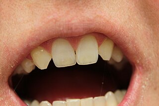
In mammalian oral anatomy, the canine teeth, also called cuspids, dog teeth, or fangs, eye teeth, vampire teeth, or vampire fangs, are the relatively long, pointed teeth. They can appear more flattened however, causing them to resemble incisors and leading them to be called incisiform. They developed and are used primarily for firmly holding food in order to tear it apart, and occasionally as weapons. They are often the largest teeth in a mammal's mouth. Individuals of most species that develop them normally have four, two in the upper jaw and two in the lower, separated within each jaw by incisors; humans and dogs are examples. In most species, canines are the anterior-most teeth in the maxillary bone.

Incisors are the front teeth present in most mammals. They are located in the premaxilla above and on the mandible below. Humans have a total of eight. Opossums have 18, whereas armadillos have none.
The premolars, also called premolar teeth, or bicuspids, are transitional teeth located between the canine and molar teeth. In humans, there are two premolars per quadrant in the permanent set of teeth, making eight premolars total in the mouth. They have at least two cusps. Premolars can be considered transitional teeth during chewing, or mastication. They have properties of both the canines, that lie anterior and molars that lie posterior, and so food can be transferred from the canines to the premolars and finally to the molars for grinding, instead of directly from the canines to the molars.
Hypodontia is defined as the developmental absence of one or more teeth excluding the third molars. It is one of the most common dental anomalies, and can have a negative impact on function, and also appearance. It rarely occurs in primary teeth and the most commonly affected are the adult second premolars and the upper lateral incisors. It usually occurs as part of a syndrome that involves other abnormalities and requires multidisciplinary treatment.

In orthodontics, a malocclusion is a misalignment or incorrect relation between the teeth of the upper and lower dental arches when they approach each other as the jaws close. The English-language term dates from 1864; Edward Angle (1855-1930), the "father of modern orthodontics", popularised it. The word "malocclusion" derives from occlusion, and refers to the manner in which opposing teeth meet.

Tooth eruption is a process in tooth development in which the teeth enter the mouth and become visible. It is currently believed that the periodontal ligament plays an important role in tooth eruption. The first human teeth to appear, the deciduous (primary) teeth, erupt into the mouth from around 6 months until 2 years of age, in a process known as "teething". These teeth are the only ones in the mouth until a person is about 6 years old creating the primary dentition stage. At that time, the first permanent tooth erupts and begins a time in which there is a combination of primary and permanent teeth, known as the mixed dentition stage, which lasts until the last primary tooth is lost. Then, the remaining permanent teeth erupt into the mouth during the permanent dentition stage.
Dental anatomy is a field of anatomy dedicated to the study of human tooth structures. The development, appearance, and classification of teeth fall within its purview. Tooth formation begins before birth, and the teeth's eventual morphology is dictated during this time. Dental anatomy is also a taxonomical science: it is concerned with the naming of teeth and the structures of which they are made, this information serving a practical purpose in dental treatment.
Occlusion, in a dental context, means simply the contact between teeth. More technically, it is the relationship between the maxillary (upper) and mandibular (lower) teeth when they approach each other, as occurs during chewing or at rest.

Overjet is the extent of horizontal (anterior-posterior) overlap of the maxillary central incisors over the mandibular central incisors. In class II malocclusion the overjet is increased as the maxillary central incisors are protruded.

Crossbite is a form of malocclusion where a tooth has a more buccal or lingual position than its corresponding antagonist tooth in the upper or lower dental arch. In other words, crossbite is a lateral misalignment of the dental arches.

A lingual arch is an orthodontic device which connects two molars in the upper or lower dental arch. The lower lingual arch (LLA) has an archwire adapted to the lingual side of the lower teeth. In the upper arch the archwire is usually connecting the two molars passing through the palatal vault, and is commonly referred as "Transpalatal Arch" (TPA). The TPA was originally described by Robert Goshgarian in 1972. TPAs could possibly be used for maintaining transverse arch widths, anchorage in extraction case, prevent buccal tipping of molars during Burstonian segmented arch mechanics, transverse anchorage and space maintenance.
Serial extraction is the planned extraction of certain deciduous teeth and specific permanent teeth in an orderly sequence and predetermined pattern to guide the erupting permanent teeth into a more favorable position.
Dentition analyses are systems of tooth and jaw measurement used in orthodontics to understand arch space and predict any malocclusion. Example systems of dentition analysis are listed below.
Bolton Analysis is a tooth analysis developed by Wayne A. Bolton to determine the discrepancy between size of maxillary and mandibular teeth. This analysis helps to determine the optimum interarch relationship. This analysis measures the Mesio-distal width of each tooth and is divided into two analyses.
Moyer's mixed dentition analysis was created in 1971 by Robert Moyers. This an analysis that is used in dentistry to predict the size of the permanent teeth by measuring the size of the permanent teeth. The analysis usually requires a dental cast, Boley's gauge and a Probability Chart.
Tanaka and Johnston analysis is a mixed dentition analysis which allows one to estimate the space available in an arch for the permanent teeth to erupt. This analysis was developed by Marvin M. Tanaka and Lysle E. Johnston in 1974 after they conducted a study on 506 orthodontic patients done in Cleveland at the Case Western Reserve University School of Dental Medicine.
Molar distalization is a process in the field of Orthodontics which is used to move molar teeth, especially permanent first molars, distally (backwards) in an arch. This procedure is often used in treatment of patients who have Class 2 malocclusion. The cause is often the result of loss of E space in an arch due to early loss of primary molar teeth and mesial (forward) migration of the molar teeth. Sometimes molars are distalized to make space for other impacted teeth, such as premolars or canines, in the mouth.
Open bite is a type of orthodontic malocclusion which has been estimated to occur in 0.6% of the people in the United States. This type of malocclusion has no vertical overlap or contact between the anterior incisors. The term "open bite" was coined by Carevelli in 1842.
Orthodontic indices are one of the tools that are available for orthodontists to grade and assess malocclusion. Orthodontic indices can be useful for an epidemiologist to analyse prevalence and severity of malocclusion in any population.
References
- ↑ Mühlberg, G.; Nedelko, U.; Weiskopf, J. (1969-10-01). "[Evaluation of Pont's index with special reference to the mesiodistal distance of the lateral teeth. Statistical analysis of 417 eugnathic dentitions]". Deutsche Stomatologie. 19 (10): 775–783. ISSN 0012-0790. PMID 5262916.
- ↑ Joondeph, Donald (1970). "Pont's Index: A clinical Evaluation". Angle Orthodontist. 40 (2): 112–8. PMID 5266011 . Retrieved 17 January 2016.