Spleen pain

Last updated
The human spleen is located in the upper left abdomen, behind the stomach Illu spleen.jpg
The human spleen is located in the upper left abdomen, behind the stomach

Spleen pain is a pain felt from the left upper quadrant of the abdomen or epigastrium where the human spleen is located or neighboring. [1]

Contents

Etiology

Splenomegaly

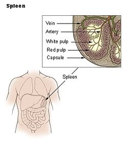
Splenomegaly can result in hematologic disturbances and abdominal pain and can increase the risk for splenic rupture, which also causes spleen pain, from blunt trauma, resulting in life-threatening internal bleeding. [2] Many conditions can cause splenomegaly, such as various infections, liver disease, and cancer. [2]

Splenic infarction

Abdominal pain remains the leading chief complaint in patients diagnosed with a splenic infarct. [1] Evaluation of patients who present with abdominal pain requires a broad differential approach. [3]

Lab evaluation may help rule in other causes of abdominal pain. Elevated liver function tests, bilirubin or lipase, may suggest a hepatobiliary or pancreatic source for pain. Leukocytosis and elevated lactate dehydrogenase (LDH) may be found in splenic infarction. However, these results lack specificity to splenic infarct. [3]

Radiographic testing is required to detect this rare illness. In the hyperacute phase of infarction, abdominal CT scan performed with intravenous contrast is the imaging modality of choice in suspected splenic infarction. Splenic infarct appears as a wedge-shaped area of splenic tissue with the apex pointed toward the hilum and the base of the splenic capsule. As the infarction matures, the affected tissue may normalize, liquefy or become contracted or scarred. Abdominal ultrasound has also been used to detect splenic infarction. Ultrasound findings of the hypoechoic wedge-shaped region of splenic tissue indicate infarction. Evolution of infarction may appear as hyperechoic with retraction of the splenic capsule. [3] [4]

Splenic abscess

Fever is the most common symptom of splenic abscess, followed by abdominal pain and a tender mass on palpation of the left upper quadrant of the abdomen. The common signs and symptoms described of a splenic abscess include the triad of fever, left upper quadrant tenderness, and leukocytosis is present only in one-third of the cases. [5]

Splenic rupture

Trauma is the most common mechanism of splenic rupture, and blunt trauma involving the left-upper quadrant, left rib cage, or left flank should raise suspicion for splenic involvement. The absence of substantial trauma cannot exclude the possibility of splenic injury, as individuals with a history of splenomegaly require less force for traumatic rupture. [6] [7]

Severally ill patients may present with hypovolemic shock manifesting as tachycardia, hypotension, and pallor. Other findings include tenderness to palpation in the left upper quadrant, generalized peritonitis, or referred pain to the left shoulder (Kehr sign). [8] Kehr sign is a rare finding and should increase the suspicion of the peritoneal process and possible splenic rupture. Some patients also report pleuritic left-sided chest pain in the setting of a ruptured spleen. Caution should be used early in the evaluation of patients with a concerning mechanism of injury, since few symptoms may be present early in the course of splenic rupture. [7] [9]

It is important to ask focused questions regarding previous surgical history, hepatic disease process, recent infections, anticoagulant, aspirin or nonsteroidal anti-inflammatory drugs usage and bleeding disorders. Visual inspection for signs of external trauma such as abrasions, lacerations, contusions, and classic seatbelt sign on the abdomen is helpful. The absence of external visual findings does not exclude intra-abdominal pathology, as up to 20% of patients with intra-abdominal injury may not display these findings upon initial examination. Also, examination on arrival may not reveal severe tenderness, rigidity, or abdominal distention in spite of rupture. [10] Physical examination may also be limited in a patient with altered mental status or distracting injuries. [11] Therefore, the physical exam alone is not always sensitive when evaluating a patient with a splenic rupture. [7]

Related Research Articles

<span class="mw-page-title-main">Spleen</span> Organ recycling old red blood cells and also housing lymphocytes

The spleen is an organ found in almost all vertebrates. Similar in structure to a large lymph node, it acts primarily as a blood filter. The word spleen comes from Ancient Greek σπλήν (splḗn).

<span class="mw-page-title-main">Peritonitis</span> Medical condition

Peritonitis is inflammation of the localized or generalized peritoneum, the lining of the inner wall of the abdomen and cover of the abdominal organs. Symptoms may include severe pain, swelling of the abdomen, fever, or weight loss. One part or the entire abdomen may be tender. Complications may include shock and acute respiratory distress syndrome.

<span class="mw-page-title-main">Splenectomy</span> Surgical removal of the spleen

A splenectomy is the surgical procedure that partially or completely removes the spleen. The spleen is an important organ in regard to immunological function due to its ability to efficiently destroy encapsulated bacteria. Therefore, removal of the spleen runs the risk of overwhelming post-splenectomy infection, a medical emergency and rapidly fatal disease caused by the inability of the body's immune system to properly fight infection following splenectomy or asplenia.

<span class="mw-page-title-main">Abdominal pain</span> Stomach aches

Abdominal pain, also known as a stomach ache, is a symptom associated with both non-serious and serious medical issues. Since the abdomen contains most of the body's vital organs, it can be an indicator of a wide variety of diseases. Given that, approaching the examination of a person and planning of a differential diagnosis is extremely important.

<span class="mw-page-title-main">Infarction</span> Tissue death due to inadequate blood supply

Infarction is tissue death (necrosis) due to inadequate blood supply to the affected area. It may be caused by artery blockages, rupture, mechanical compression, or vasoconstriction. The resulting lesion is referred to as an infarct (from the Latin infarctus, "stuffed into").

<span class="mw-page-title-main">Splenomegaly</span> Enlargement of the spleen

Splenomegaly is an enlargement of the spleen. The spleen usually lies in the left upper quadrant (LUQ) of the human abdomen. Splenomegaly is one of the four cardinal signs of hypersplenism which include: some reduction in number of circulating blood cells affecting granulocytes, erythrocytes or platelets in any combination; a compensatory proliferative response in the bone marrow; and the potential for correction of these abnormalities by splenectomy. Splenomegaly is usually associated with increased workload, which suggests that it is a response to hyperfunction. It is therefore not surprising that splenomegaly is associated with any disease process that involves abnormal red blood cells being destroyed in the spleen. Other common causes include congestion due to portal hypertension and infiltration by leukemias and lymphomas. Thus, the finding of an enlarged spleen, along with caput medusae, is an important sign of portal hypertension.

<span class="mw-page-title-main">Abdominal examination</span> Physical examination of abdomen

An abdominal examination is a portion of the physical examination which a physician or nurse uses to clinically observe the abdomen of a patient for signs of disease. The abdominal examination is conventionally split into four different stages: first, inspection of the patient and the visible characteristics of their abdomen. Auscultation (listening) of the abdomen with a stethoscope. Palpation of the patient's abdomen. Finally, percussion (tapping) of the patient's abdomen and abdominal organs. Depending on the need to test for specific diseases such as ascites, special tests may be performed as a part of the physical examination. An abdominal examination may be performed because the physician suspects a disease of the organs inside the abdominal cavity (including the liver, spleen, large or small intestines), or simply as a part of a complete physical examination for other conditions. In a complete physical examination, the abdominal exam classically follows the respiratory examination and cardiovascular examination.

<span class="mw-page-title-main">Splenic injury</span> Injury to the spleen

A splenic injury, which includes a ruptured spleen, is any injury to the spleen. The rupture of a normal spleen can be caused by trauma, such as a traffic collision.

<span class="mw-page-title-main">Blunt trauma</span> Trauma to the body without penetration of the skin

Blunt trauma, also known as blunt force trauma or non-penetrating trauma, describes a physical trauma due to a forceful impact without penetration of the body's surface. Blunt trauma stands in contrast with penetrating trauma, which occurs when an object pierces the skin, enters body tissue, and creates an open wound. Blunt trauma occurs due to direct physical trauma or impactful force to a body part. Such incidents often occur with road traffic collisions, assaults, sports-related injuries, and are notably common among the elderly who experience falls.

Valentino's syndrome is pain presenting in the right lower quadrant of the abdomen caused by a duodenal ulcer with perforation through the retroperitoneum.

<span class="mw-page-title-main">Castell's sign</span> Medical sign assessed to evaluate splenomegaly

Castell's sign is a medical sign assessed to evaluate splenomegaly and typically part of an abdominal examination. It is an alternative physical examination maneuver to percussion over Traube's space.

<span class="mw-page-title-main">Hypochondrium</span> Upper left and right sections of the abdomen

In anatomy, the division of the abdomen into regions can employ a nine-region scheme. The hypochondrium refers to the two hypochondriac regions in the upper third of the abdomen; the left hypochondrium and right hypochondrium. They are located on the lateral sides of the abdominal wall respectively, inferior to (below) the thoracic cage, being separated by the epigastrium.

An acute abdomen refers to a sudden, severe abdominal pain. It is in many cases a medical emergency, requiring urgent and specific diagnosis. Several causes need immediate surgical treatment.

<span class="mw-page-title-main">Splenic infarction</span> Medical condition

Splenic infarction is a condition in which blood flow supply to the spleen is compromised, leading to partial or complete infarction in the organ. Splenic infarction occurs when the splenic artery or one of its branches are occluded, for example by a blood clot.

<span class="mw-page-title-main">Hemorrhagic infarct</span> Medical condition

A hemorrhagic infarct is determined when hemorrhage is present around an area of infarction. Simply stated, an infarction is an area of dead tissue or necrosis. When blood escapes outside of the vessel (extravasation) and re-perfuses back into the tissue surrounding the infarction, the infarction is then termed a hemorrhagic infarct (infarction). Hemorrhagic infarcts can occur in any region of the body, such as the head, trunk and abdomen-pelvic regions, typically arising from their arterial blood supply being interrupted by a blockage or compression of an artery.

<span class="mw-page-title-main">Epiploic appendagitis</span> Medical condition

Epiploic appendagitis (EA) is an uncommon, benign, self-limiting inflammatory process of the epiploic appendices. Other, older terms for the process include appendicitis epiploica and appendagitis, but these terms are used less now in order to avoid confusion with acute appendicitis.

<span class="mw-page-title-main">Blunt splenic trauma</span>

Blunt splenic trauma occurs when a significant impact to the spleen from some outside source damages or ruptures the spleen. Treatment varies depending on severity, but often consists of embolism or splenectomy.

<span class="mw-page-title-main">Abdominal trauma</span> Medical condition

Abdominal trauma is an injury to the abdomen. Signs and symptoms include abdominal pain, tenderness, rigidity, and bruising of the external abdomen. Complications may include blood loss and infection.

<span class="mw-page-title-main">Quadrants and regions of abdomen</span> Anatomical subdivision scheme

The human abdomen is divided into quadrants and regions by anatomists and physicians for the purposes of study, diagnosis, and treatment. The division into four quadrants allows the localisation of pain and tenderness, scars, lumps, and other items of interest, narrowing in on which organs and tissues may be involved. The quadrants are referred to as the left lower quadrant, left upper quadrant, right upper quadrant and right lower quadrant. These terms are not used in comparative anatomy, since most other animals do not stand erect.

Splenosis is the result of spleen tissue breaking off the main organ and implanting at another site inside the body. This is called heterotopic autotransplantation of the spleen. It most commonly occurs as a result of traumatic splenic rupture or abdominal surgery. Depending on the location of the spleen, the new piece usually implants in another part of the abdominal cavity. Single case reports also describe splenosis in the thoracic cavity, in subcutaneous tissue, in the liver or in the cranial cavity. Splenosis must be distinguished from the presence of additional spleens, which are innate and are the result of differences in embryological development. Additionally, splenosis must be differentiated from malignant tumors which may look similar when imaged.

References

  1. 1 2 Ami, S; Meital, A; Ella, K; Abraham, K (2015-09-11). "Acute Splenic Infarction at an Academic General Hospital Over 10 Years: Presentation, Etiology, and Outcome". Medicine. 94 (36): e1363. doi:10.1097/MD.0000000000001363. PMC   4616622 . PMID   26356690. Most patients presented with abdominal pain (84%), often felt in the left upper quadrant or epigastrium.
  2. 1 2 Zambrano, Laura D.; Samson, Olivia; Phares, Christina; Jentes, Emily; Weinberg, Michelle; Goers, Matthew; Kachur, S. Patrick; McDonald, Robert; Morawski, Bozena; Njuguna, Henry; Bakhsh, Yasser; Laws, Rebecca; Peak, Corey; Iverson, Sally Ann; Bezold, Carla; Allkhenfr, Hayder; Horth, Roberta; Yang, Jun; Miller, Susan; Kacka, Michael; Davids, Abby; Mortimer, Margaret; Khan, Nomana; Stauffer, William; Marano, Nina (2018-12-14). "Unresolved Splenomegaly in Recently Resettled Congolese Refugees ― Multiple States, 2015–2018". Morbidity and Mortality Weekly Report. 67 (49). Centers for Disease Control MMWR Office: 1358–1362. doi:10.15585/mmwr.mm6749a2. ISSN   0149-2195. PMC   6300079 . PMID   30543602.
  3. 1 2 3 Chapman, J; Bhimji, SS (2018), "article-29380", Splenic Infarcts, Treasure Island (FL): StatPearls Publishing, PMID   28613652 , retrieved 2019-02-27
  4. Hwang, JH; Lee, CS (2014-12-03). "Malaria-Induced Splenic Infarction". The American Journal of Tropical Medicine and Hygiene. 91 (6): 1094–1100. doi:10.4269/ajtmh.14-0190. PMC   4257629 . PMID   25294615.
  5. Mathew, G; Bhimji, SS (2018), "article-29377", Splenic Abscess, Treasure Island (FL): StatPearls Publishing, PMID   30137831 , retrieved 2019-02-28
  6. Reinhold, Gregory; Melonakos, Tina; Lyman, Daniel (2017-07-28). "A Near Fatal Sneeze Spontaneous Splenic Rupture: A Case Report and Review of the Literature". Clinical Practice and Cases in Emergency Medicine. 1 (3). Western Journal of Emergency Medicine: 190–193. doi:10.5811/cpcem.2017.2.32847. ISSN   2474-252X. PMC   5965167 . PMID   29849293.
  7. 1 2 3 Akoury, T; Whetstone, DR (2018), "article-29383", Splenic Rupture, Treasure Island (FL): StatPearls Publishing, PMID   30247826 , retrieved 2019-02-28
  8. Söyüncü, S; Bektaş, F; Cete, Y (2012). "Traditional Kehr's sign: Left shoulder pain related to splenic abscess". Turkish Journal of Trauma & Emergency Surgery. 18 (1): 87–8. doi: 10.5505/tjtes.2012.04874 . ISSN   1306-696X. PMID   22290058.
  9. Oh, JS; Mentzer, CJ; Abuzeid, AM; Holsten, SB (2016). "Delayed Splenic Rupture with Normal Admission CT Scan after Blunt Trauma". The American Surgeon. 82 (8): e223–4. ISSN   0003-1348. PMID   27657577.
  10. Blaivas, M; Quinn, J (1998). "Diagnosis of spontaneous splenic rupture with emergency ultrasonography". Annals of Emergency Medicine. 32 (5): 627–30. doi:10.1016/S0196-0644(98)70046-0. ISSN   0196-0644. PMID   9795331.
  11. Monti, Jonathan D. (2016). "A rare cause of abdominal pain and hypotension in pregnancy". JAAPA. 29 (11). Ovid Technologies (Wolters Kluwer Health): 31–34. doi:10.1097/01.jaa.0000499629.82493.d1. ISSN   1547-1896. PMID   27787274.