Standard Event System

Last updated
Standard Event System character depiction Standard Event System.png
Standard Event System character depiction

The "Standard Event System" (SES) to Study Vertebrate Embryos was developed in 2009 to establish a common language in comparative embryology. [1] Homologous developmental characters are defined therein and should be recognisable in all vertebrate embryos. The SES includes a protocol on how to describe and depict vertebrate embryonic characters. The SES was initially developed for external developmental characters of organogenesis, particularly for turtle embryos. However, it is expandable both taxonomically and in regard to anatomical or molecular characters. This article should act as an overview on the species staged with SES and document the expansions of this system. New entries need to be validated based on the citation of scientific publications. The guideline on how to establish new SES-characters and to describe species can be found in the original paper of Werneburg (2009). [1]

Contents

SES-characters are used to reconstruct ancestral developmental sequences in evolution such as that of the last common ancestor of placental mammals. [2] Also the plasticity of developmental characters can be documented and analysed.

SES-staged species

Overview on the vertebrate species staged with SES.

Major taxon 1Major taxon 2SpeciesReference
Lissamphibia Caudata Ambystoma mexicanum [3]
Testudines Pleurodira Emydura subglobosa [4]
Testudines Cryptodira Graptemys nigrinoda [3]
Testudines Cryptodira Kinosternum scorpionoides [5]
Testudines Cryptodira Apalone spinifera , Caretta caretta , Carettochelys insculpta , Chelonia mydas , Chelydra serpentina , Chrysemys picta , Dermochelys coriacea , Eretmochelys imbricata , Lepidochelys olivacea , Natator depressus , Pelodiscus sinensis , Testudo hermanni , Trachemys scripta [3]
Crocodilia Alligatoridae Alligator mississippiensis [3]
Squamata Lacertidae Zootoca vivipara [3]
Squamata Serpentes Bothropoides jararaca [6]
Squamata Gymnophthalmidae Calyptommatus sinebranchiatus , Nothobachia ablephara [7]
Squamata Varanidae Varanus panoptes [8]
Squamata Iguanidae Pogona vitticeps [9]
Aves Galliformes Gallus gallus [3]
Mammalia Monotremata Tachyglossus aculeatus [10]
Mammalia Monotremata Ornithorhynchus anatinus [11]
Mammalia Marsupialia Didelphis virginiana [3]
Mammalia Marsupialia Monodelphis domestica , Sminthopsis macroura , Dasyurus hallucatus , Dasyurus viverrinus , Phascolarctos cinereus , Petauroides volans , Trichosurus vulpecula , Bettongia gaimardi , Macropus eugenii [11]
Mammalia Marsupialia Caenolestes fuliginosus [12]
Mammalia Xenarthra Dasypus hybridus [3]
Mammalia Xenarthra Bradypus tridactylus [11]
Mammalia Afrotheria Echinops telfairi , Tenrec ecaudatus [13]
Mammalia Afrotheria Elephas maximus , Loxodonta africana [11]
Mammalia Laurasiatheria Talpa occidentalis , Talpa europaea , Erinaceus europaeus , Erinaceus roumanicus , Suncus murinus , Crocidura russula , Neomys fodiens , Sorex araneus , Pecari tajacu , Sus scrofa domesticus , Rangifer tarandus , Capreolus capreolus , Bos primigenius taurus , Capra aegagrus hircus , Ovis aries , Phocoena phocoena , Delphinus delphis , Stenella longirostris , Stenella attenuata , Hipposideros armiger , Hipposideros pratti , Molossus rufus , Myotis lucifugus , Myotis myotis , Pipistrellus abramus , Scotophilus kuhlii , Miniopterus natalensis , Miniopterus schreibersii , Carollia perspicillata , Equus ferus caballus , Manis javanica , Felis catus , Mustela putorius , Canis lupus familiaris [11]
Mammalia Laurasiatheria Artibeus fimbriatus , Artibeus lituratus , Artibeus obscurus , Carollia perspicillata , Eumops patagonicus , Hipposideros armiger , Hipposideros pratti , Miniopterus natalensis , Miniopterus schreibersii , Molossus rufus , Myotis albescens , Myotis lucifugus , Myotis myotis , Pipistrellus abramus , Rhinolophus ferrumequinum , Rousettus aegyptiacus , Rousettus amplexicaudatus , Scotophilus kuhlii , Vespertilio sinensis , Mus musculus [14]
Mammalia Boreoeutheria Oryctolagus cuniculus , Spermophilus citellus , Cavia porcellus , Cricetulus barabensis , Mesocricetus auratus , Myodes glareolus , Meriones unguiculatus , Acomys dimidiatus , Apodemus sylvaticus , Apodemus flavicollis , Apodemus agrarius , Rattus norvegicus , Tupaia javanica , Tupaia belangeri , Galago senegalensis , Nycticebus coucang , Tarsius tarsier , Callithrix jacchus , Erythrocebus patas , Papio cynocephalus , Macaca mulatta , Homo sapiens [11]
Mammalia Boreoeutheria Mus musculus [13]

SES-characters

New SES-characters are continuously described in new publications. Currently, characters of organogenesis are described for Vertebrata (V), Gnathostomata (G), Tetrapoda (T), Amniota (A), Sauropsida (S), Squamata (SQ), Mammalia (M), and Monotremata (MO). In total, 166 SES-characters are currently defined.

Character complexCharacterCharacter descriptionReferenceIllustration
A. Egg (V01).A1. Egg lay (V01a).Most authors begin to create their staging tables shortly after or around egg lay time. [1]
Standard Event System character depiction Character A1. Egg lay (V01a).png
Standard Event System character depiction
B. Blastula (V02).B1. Blastoporus (V02a).The blastoporus begins to form as a lip and is later on visible as a clear depression in the blastula. [1]
Standard Event System character depiction Character B1. Blastoporus (V02a).png
Standard Event System character depiction
C. Neural tube (V03).C1. Primitive streak present (V03a).The neural plate starts folding. The lateral primitive folds border the primitive groove. [1]
Standard Event System character depiction C1. Primitive streak present (V03a).png
Standard Event System character depiction
C. Neural tube (V03).C2. Neural folds begin to close (V03b).The dorsal borders of the neural folds come in touch and begin to form the neural tube that encloses the neural tube. The anterior and posterior regions of the primitive streak remain open. [1]
Standard Event System character depiction C2. Neural folds begin to close (V03b).png
Standard Event System character depiction
C. Neural tube (V03).C3. Anterior neuropore closed (V03c).The neural tube closes anteriorly by final caudal fusion of the primitive folds. [1]
Standard Event System character depiction C3. Anterior neuropore closed (V03c).png
Standard Event System character depiction
C. Neural tube (V03).C4. Posterior neuropore closed (V03d).The neural tube closes posteriorly by final caudal fusion of the primitive folds. [1]
Standard Event System character depiction C4. Posterior neuropore closed (V03d).png
Standard Event System character depiction
D. Somites.D1. Somites hard to count (V04a).Correlated to the internal differentiation and partly due to the carapace forming in turtles somite borders become blurred in specific regions until somites are completely inconspicuous. [1]
Standard Event System character depiction Character D1. Somites hard to count (V04a).png
Standard Event System character depiction
D. Somites.D2. 1-5 somite pairs (V04b).The total number of somite pairs is count, filled in a formula for each specimen and afterwards grouped within the somite cluster of five somites each. Seldom the left and the right side show a different number of mesodermal segments. By definition the row with the maximum is to be count. Often a somite pair underlies a forming process. This one should also be counted. [1]
Standard Event System character depiction D2ff. somite pairs (V04b ff).png
Standard Event System character depiction
D. Somites.D3. 6-10 somite pairs (V04c).compare to character D2 [1] see D2
D. Somites.D4. 11-15 somite pairs (V04d).compare to character D2 [1] see D2
D. Somites.D5. 16-20 somite pairs (V04e).compare to character D2 [1] see D2
D. Somites.D6. 21-25 somite pairs (V04f).compare to character D2 [1] see D2
D. Somites.D7. 26-30 somite pairs (V04g).compare to character D2 [1] see D2
D. Somites.D8. 31-35 somite pairs (V04h).compare to character D2 [1] see D2
D. Somites.D9. 36-40 somite pairs (V04i).compare to character D2 [1] see D2
D. Somites.D10. 41-45 somite pairs (V04j).compare to character D2 [1] see D2
D. Somites.D12. 51-55 somite pairs (V04l).compare to character D2 [1] see D2
D. Somites.D13. 56-60 somite pairs (V04m).compare to character D2 [1] see D2
D. Somites.D14. 61- somite pairs (V04n).Sixty one or more somite pairs, character list to be continued if needed. [6] see D2
E. Head (V05)E1. Head bulbus (V05a)The head is formed as a distinct "bulbus" and a slight strangling (neck) restricts the head region from the thoracal region. [1]
Standard Event System character depiction E1. Head bulbus (V05a).png
Standard Event System character depiction
E. Head (V05)E2. Anterior cephalic projection (V05b).The neck (pharyngeal area) elongates and is at least as long as broad. [1]
Standard Event System character depiction E2. Anterior cephalic projection (V05b).png
Standard Event System character depiction
E. Head (V05)E3. Head projection disappeared (V05c).The forming of a mesencephalic (or/and a diencephalic) projection in the posterodorsal region of the head is characterized by a continuing growth and can not be defined as a distinguishable event. But the disappearance of the structure resulting in a flat occipital head region can be well defined and is possibly associated to skull ossification. [1]
Standard Event System character depiction E3. Head projection disappeared (V05c).png
Standard Event System character depiction
F. Nose (V06)F1. Olfactory placode (V06a).The nasal placode occurs. [1]
Standard Event System character depiction F1. Olfactory placode (V06a).png
Standard Event System character depiction
F. Nose (V06)F2. External nares (V06b).Deep furrows between the frontonasal and maxillary processes develop and are visible as invaginations in the nasal region. [1]
Standard Event System character depiction F2. External nares (V06b).png
Standard Event System character depiction
F. Nose (V06)F3. Nostrils formed (V06c).The external nares are closed and nostrils are formed. [11]
Standard Event System character depiction F3. Nostrils formed (V06c).png
Standard Event System character depiction
G. Ear (V07).G1. Otic placode (V07a).The otic placode occurs. [1]
Standard Event System character depiction G1. Otic placode (V07a).png
Standard Event System character depiction
G. Ear (V07).G2. Otic vesicle (V07b).The ear is formed by an invagination and a capsule is forming that becomes large in turtles. [1]
Standard Event System character depiction G2. Otic vesicle (V07b).png
Standard Event System character depiction
G. Ear (V07).G3. Otic capsule inconspicuous (V07c).The skin becomes intransparent and the otic capsule is no longer visible. [1]
Standard Event System character depiction G3. Otic capsule inconspicuous (V07c).png
Standard Event System character depiction
G. Ear (V07).G4. Pinna fold (V07d).A pinna fold is formed in mammals. [10]
Standard Event System character depiction G4. Pinna fold (V07d).png
Standard Event System character depiction
G. Ear (V07).G5. Tragus (V07e).N.N. [14] N.N.
H. Eye (V08).H1. Optic vesicle (V08a).An optic vesicle forms lateral of the prosencephalic region. It can be mistaken for the trigeminal ganglion which is proportional enlarged in Tachyglossus or Monodelphis at this early period of development. But the ganglion lies more caudally. [1]
Standard Event System character depiction H1. Optic vesicle (V08a).png
Standard Event System character depiction
H. Eye (V08).H2. Lens placode (V08b).The lens placode forms in the middle of the optic cup and primarily has only an indistinct contour. [1]
Standard Event System character depiction H2. Lens placode (V08b).png
Standard Event System character depiction
H. Eye (V08).H3. Optic fissure (V08c).The optic or choroid fissure represents the blood vessel agglomeration, which supplies the developing lens. It forms a clear streak between the lens and the ventral most curvature of the optic cup. When the lens is completely formed the optic fissure disappears, which is a fluent process that can not be defined as a distinct event. [1]
Standard Event System character depiction H4. Contour lens-iris (V08d).png
Standard Event System character depiction
H. Eye (V08).H4. Contour lens/iris (V08d).The lens is completely formed and shows a distinct contour. Parallel the iris gets its contour but is often hard to distinguish from the lens. [1] see H3
H. Eye (V08).H5. Pupil forms (V08e).In the middle of the optic cup/lens region the pupil forms. That reflects the complete formation of the iris musculature. [1]
Standard Event System character depiction H5. Pupil forms (V08e).png
Standard Event System character depiction
H. Eye (V08).H6. Scleral papillae (V08f).Between the lens and the border of the optic cup circularly scleral bones occur. [1]
Standard Event System character depiction H7. Scleral papillae inconspicuous (V08g).png
Standard Event System character depiction
H. Eye (V08).H7. Scleral papillae inconspicuous (V08g).The scleral papillae become inconspicuous. [1] see H6
I. Ribs (V09).I1. Rib primordial visible (V09a).Rib primordia are visible through the carapace or skin for a short time of development. [1]
Standard Event System character depiction I1. Rib primordial visible (V09a).png
Standard Event System character depiction
J. Heart (V10).J1. Ventricle bulbus (V10a).Description The developing heart (ventricle) primordium forms a small bulging in the ventral thoracal region. [1]
Standard Event System character depiction J1. Ventricle bulbus (V10a).png
Standard Event System character depiction
J. Heart (V10).J2. Thoracal bulbus disappeared (V10b).During development the ventricle and the liver form a common thoracal bulbus which disappears when the plastron forms in turtles or ribs ossify. [1]
Standard Event System character depiction J2. Thoracal bulbus disappeared (V10b).png
Standard Event System character depiction
J. Heart (V10).J3. Ventricle S-shaped (V10c).The ventricle forms a curvature that gives the ventricle an S-shaped look. [1]
Standard Event System character depiction J3. Ventricle S-shaped (V10c).png
Standard Event System character depiction
K. Tail (V11).K1. Tail bud (V11a).Although in many references a tail is arbitrarily described very early or in association to the development to the hind limb bud here the occurrence of the tail bud is defined as a distinct constriction of the caudal body region. [1]
Standard Event System character depiction K1. Tail bud (V11a).png
Standard Event System character depiction
L. Limbs (V12).L1. Forelimb ridge (V12a).It is a lateral eruption in the anterior part of body wall that is wider (anterior-posterior) than long (proximal-distal). [1]
Standard Event System character depiction Forelimb ridge (V12a).png
Standard Event System character depiction
L. Limbs (V12).L2. Hind limb ridge (V12o).It is a lateral eruption in the posterior part of body wall that is wider (anterior-posterior) than long (proximal-distal). [10] see L1.
L. Limbs (V12).L3. Forelimb bud (V12b).The forelimb bud is about as broad as wide. [1]
Standard Event System character depiction L3. Forelimb bud (V12b).png
Standard Event System character depiction
L. Limbs (V12).L4. Hind limb bud (V12s).The hind limb bud is about as broad as wide. [1] see L3
L. Limbs (V12).L5. Forelimb elongated (V12c).The forelimb bud is longer (proximal-distal) than wide (anterior-posterior). [1]
Standard Event System character depiction L5. Forelimb elongated (V12c).tif
Standard Event System character depiction
L. Limbs (V12).L6. Hind limb elongated (V12p).The hind limb bud is longer (proximal-distal) than wide (anterior-posterior). [10] see L5
L. Limbs (V12).L7. Forelimb AER (V12d).On the distal part of the forelimb bud an apical ectodermal ridge (AER) is formed in a horizontal longitude. Often it is only visible as a slightly eruption and in a particular angle of view. [1]
Standard Event System character depiction L7. Forelimb AER (V12d).png
Standard Event System character depiction
L. Limbs (V12).L8. Hindlimb AER (V12e).On the distal part of the hindlimb bud an apical ectodermal ridge (AER) is formed in a horizontal longitude. [1] see L7
L. Limbs (V12).L9. Forelimb elbow (V12f).The forelimb develops an elbow. [1]
Standard Event System character depiction L9. Forelimb elbow (V12f).png
Standard Event System character depiction
L. Limbs (V12).L10. Hind limb knee (V12t).The hind limb develops an knee. [1] see L9
L. Limbs (V12).L11. Forelimb paddle (V12g).The forelimb is thicker distally than proximally. Paddle shaped. [1]
Standard Event System character depiction L11. Forelimb paddle (V12g).png
Standard Event System character depiction
L. Limbs (V12).L12. Hind limb paddle (V12h).The hind limb is thicker distally than proximally. Paddle shaped. [1] see L11
L. Limbs (V12).L13. Forelimb digital plate (V12i).In the distal region of the forelimb a round digital plate is formed by flattening of its paddle like end in a horizontal plane. The digital plate is clearly separated from the tube shaped leg by a surrounding step. [1]
Standard Event System character depiction L13. Forelimb digital plate (V12i).png
Standard Event System character depiction
L. Limbs (V12).L14. Hindlimb digital plate (V12j).In the distal region of the hindlimb a digital plate forms. [1] see L13
L. Limbs (V12).L15. Digital grooves forelimb (V12k).On the forelimb digital grooves/ridges are visible. Sometimes grooves develop simultaneously, sometimes only one or two ridges are visible first. [1]
Standard Event System character depiction L15. Digital grooves forelimb (V12k).png
Standard Event System character depiction
L. Limbs (V12).L16. Digital grooves hind limb (V12u).On the hind limb digital grooves/ridges are visible. Sometimes grooves develop simultaneously, sometimes only one or two ridges are visible first. [1] see L15
L. Limbs (V12).L17. Digital serration forelimb (V12l).The periphery of the digital plate is slightly serrated on the forelimb [1]
Standard Event System character depiction L19. Finger (V12m).png
Standard Event System character depiction
L. Limbs (V12).L18. Digital serration hind limb (V12v).The periphery of the digital plate is slightly serrated on the hind limb. [1] see L17
L. Limbs (V12).L19. Finger (V12m).At least one forelimb phalanx projects beyond the digital web and is longer than wide. [1] N.N.
L. Limbs (V12).L20. Toe (V12w).At least one hind limb phalanx projects beyond the digital web and is longer than wide. [1]
Standard Event System character depiction L20. Toe (V12w).png
Standard Event System character depiction
L. Limbs (V12).L21. First claw forelimb (V12n).At least one claw develops on the forelimb. [1]
Standard Event System character depiction L21. First claw forelimb (V12n).png
Standard Event System character depiction
L. Limbs (V12).L22. First claw hind limb (V12x).At least one claw develops on the hind limb. [1] see L21
L. Limbs (V12).L23. Hind limb sporn (V12q).On the medial face of the hindlimb the poison sporn of Monotremata is visible. [10]
Standard Event System character depiction L23. Hind limb sporn (V12q).png
Standard Event System character depiction
L. Limbs (V12).L24. Hind limb sporn pouch (V12r).On the medial face of the hindlimb a skin fold is formed where the poison sporn of Monotremata can be retracted. [10] N.N.
L. Limbs (V12).L25. Membranes between the fingers are completely gone (V12s).see character name [11]
Standard Event System character depiction L25. Membranes between the fingers are completely gone (V12s).png
Standard Event System character depiction
L. Limbs (V12).L26. Membranes between the toes are completely gone (V12t).see character name [11] see L25
L. Limbs (V12).L27. Separate phalanges are visible in the fingers (V12u).see character name [11]
Standard Event System character depiction L27. Separate phalanges are visible in the fingers (V12u).png
Standard Event System character depiction
L. Limbs (V12).L28. Separate phalanges are visible in the toes (V12v).see character name [11] see L27
M. Scales, etc. (V13).M1. Head scales (V13a).Scales on the dorsum of the head occur. The forming of scutes on throat and lower eyelid are encoded separately. Due feathers and scales are assumed to be homologous the scale characters are also applicable to bird development. Mammalian hears are formed differently and are not regarded here. [1]
Standard Event System character depiction M2. Throat scales (V13b).png
Standard Event System character depiction
M. Scales, etc. (V13).M2. Throat scales (V13b).Scales between the lower jaw and the ventral neck region occur. [1]
Standard Event System character depiction M3. Lower eyelid scales (V13c).png
Standard Event System character depiction
M. Scales, etc. (V13).M3. Lower eyelid scales (V13c).Scales on the lower eyelid occur. [1]
Standard Event System character depiction M3. Lower eyelid scales (V13c).png
Standard Event System character depiction
M. Scales, etc. (V13).M4. Neck scales (V13d).Scales on the neck occur. This character is sometimes difficult to define in cryptodire turtles due to the head/neck retraction. [1]
Standard Event System character depiction M4. Neck scales (V13d).png
Standard Event System character depiction
M. Scales, etc. (V13).M5. Back scales (V13e).Scales on the back (the dorsal) of the trunk. Scales on the carapace in turtles are encoded separately. [1]
Standard Event System character depiction M5. Back scales (V13e).png
Standard Event System character depiction
M. Scales, etc. (V13).M6. Forelimb scales proximal (V13f).The first occurrence of scales on a limb. Generally on the upper part of the forelimb. [1]
Standard Event System character depiction M6. Forelimb scales proximal (V13f).png
Standard Event System character depiction
M. Scales, etc. (V13).M7. Forelimb scales distal (V13g).The forelimb is completely covered by scales, which includes the cover of the whole leg and the digital region. [1] see M6
M. Scales, etc. (V13).M8. Hind limb scales proximal (V13h).The first occurrence of scales on a limb. Generally on the upper part of the forelimb. [1]
Standard Event System character depiction M8. Hind limb scales proximal (V13f).png
Standard Event System character depiction
M. Scales, etc. (V13).M9. Hind limb scales distal (V13i).The forelimb is completely covered by scales, which includes the cover of the whole leg and the digital region. [1] see M8
M. Scales, etc. (V13).M10. Tail scales (V13j).Scales on the tail occur. [1]
Standard Event System character depiction M10. Tail scales (V13h).png
Standard Event System character depiction
M. Scales, etc. (V13).M11. Carapace scales (V13k).Horny scutes on the turtle carapace develop already in the egg. They are difficult to demonstrate in photographs. [1]
Standard Event System character depiction M11. Carapace scales (V13i).png
Standard Event System character depiction
N. Hatch / birth / leaving pouch / weaning (V14).N1. Hatch (V14a).Mostly, but not necessarily the hatch represents the end of the embryonic, organ differentiating period in egg laying vertebrates. [1]
Standard Event System character depiction N1. Hatch (V14a).png
Standard Event System character depiction
N. Hatch / birth / leaving pouch / weaning (V14).N2. Birth (V14b).The fetus is born. [1]
Standard Event System character depiction N2. Birth (V14b).png
Standard Event System character depiction
N. Hatch / birth / leaving pouch / weaning (V14).N3. Leaving pouch (V14c).In marsupials, the young are leaving the pouch for the first time. [11]
Standard Event System character depiction N3. Leaving pouch (V14c).jpg
Standard Event System character depiction
N. Hatch / birth / leaving pouch / weaning (V14).N4. Weaning (V14d).End of lactation. [11]
Standard Event System character depiction N4. Weaning (V14d).gif
Standard Event System character depiction
O. Maxillary process of the mandibular arch (G01).O1. Maxillary bud (G01a).The maxillary process of the mandibular arch occurs as a bud posterior to the eye. Often it is clearly seen as the anterodorsal process of the first pharyngeal arch. [1]
Standard Event System character depiction O1. Maxillary bud (G01a).png
Standard Event System character depiction
|-
O. Maxillary process of the mandibular arch (G01).O2. Maxillary process posterior to eye (G01b).The maxillary process lies posterior to the eye for a long period. First, when a clear rostrad development of the maxillary process is recognizable, its position at the level of the posterior margin of the optic cup should be noted. For identifying the level of the maxillary process in respect to the eye the ventral border of the telencephalic/diencephalic head region must be orientated horizontally. [1]
Standard Event System character depiction O2. Maxillary process posterior to eye (G01b).png
Standard Event System character depiction
O. Maxillary process of the mandibular arch (G01).O3. Maxillary process midline of eye (G01c).The tip of the maxillary process is located at the level of the optic (choroid) fissure, around the midline of the eye. [1]
Standard Event System character depiction O3. Maxillary process midline of eye (G01c).png
Standard Event System character depiction
O. Maxillary process of the mandibular arch (G01).O4. Maxillary process anterior to lens (G01d).The tip of the maxillary process is located beyond the optic fissure and is situated around the level of the anterior borders of pupil, iris, lens and scleral papillae. [1]
Standard Event System character depiction O4. Maxillary process anterior to lens (G01d).png
Standard Event System character depiction
O. Maxillary process of the mandibular arch (G01).O5. Maxillary process anterior to eye (G01e).The tip of the maxillary process is located beyond the level of the anterior border of the eye up to the posterior margin of frontonasal process. [1]
Standard Event System character depiction O5. Maxillary process anterior to eye (G01e).png
Standard Event System character depiction
O. Maxillary process of the mandibular arch (G01).O6. Maxillary process fuses with frontonasal process (G01f).The maxillary process fuses with the frontonasal process and both form a more or less consistent upper jaw. [1]
Standard Event System character depiction O6. Maxillary process posterior to eye (G01f).png
Standard Event System character depiction
P. Mandibular process (G02).P1. Mandibular arch bud (G02a).The first pharyngeal (mandibular) arch is generally the first pharyngeal arch to occur as a bud. It forms later on a dorsal maxillary and a ventral mandibular process. [1]
Standard Event System character depiction P1. Mandibular arch bud (G02a).png
Standard Event System character depiction
P. Mandibular process (G02).P2. Mandibular process of the mandibular arch posterior eye (G02b).The mandibular process lays posterior to the eye for a long period. First, when a clear rostrad development of the mandibular process is recognizable, its position around the level of the posterior margin of the optic cup should be noted. For defining the level of the mandibular process in respect to the eye the ventral border of the telencephalic/diencephalic head region must be orientated horizontally. [1]
Standard Event System character depiction P2. Mandibular process of the mandibular arch posterior eye (G02b).png
Standard Event System character depiction
P. Mandibular process (G02).P3. Mandibular arch bud posterior lens (G02c).The tip of the mandibular process is situated around the level of the posterior margins of scleral papillae, lens, iris and pupil. [1]
Standard Event System character depiction P3. Mandibular arch bud posterior lens (G02c).png
Standard Event System character depiction
P. Mandibular process (G02).P4. Mandibular arch bud midline eye (G02d).The tip of the mandibular process is located around the level of the optic (choroid) fissure, around the midline of the eye. [1]
Standard Event System character depiction P4. Mandibular arch bud midline eye (G02d).png
Standard Event System character depiction
P. Mandibular process (G02).P5. Mandibular arch bud anterior lens (G02e).The tip of the mandibular process is located beyond the optic fissure and is situated around the level of the anterior borders of pupil, iris, lens and scleral papillae. [1]
Standard Event System character depiction P5. Mandibular arch bud anterior lens (G02e).png
Standard Event System character depiction
P. Mandibular process (G02).P6. Mandibular arch bud anterior eye (G02f).The tip of the mandibular process is situated at the level around the anterior border of the eye. [1]
Standard Event System character depiction P6. Mandibular arch bud anterior eye (G02f).png
Standard Event System character depiction
P. Mandibular process (G02).P7. Mandibular arch bud level frontonasal groove (G02g).The tip of the mandibular process is situated at the level of the frontonasal process. [1]
Standard Event System character depiction P7. Mandibular arch bud level frontonasal groove (G02g).png
Standard Event System character depiction
P. Mandibular process (G02).P8. Mandibular arch bud occlusion point = jaw closure (G02h).The tip of the mandibular process comes in contact with the anterior part of the upper jaw. [1]
Standard Event System character depiction P8. Mandibular arch bud occlusion point = jaw closure (G02h).png
Standard Event System character depiction
Q. Postmandibular pharyngeal arches (G03).Q1. Second arch bud (G03a).The second pharyngeal arch, the hyoid arch occurs as a bud. [1]
Standard Event System character depiction Q1. Second arch bud (G03a).png
Standard Event System character depiction
Q. Postmandibular pharyngeal arches (G03).Q2. Hyoid flap (G03e).The second (hyoid) arch develops an opercular flap that covers at least one pharyngeal slit. [1]
Standard Event System character depiction Q2. Hyoid flap (G03e).png
Standard Event System character depiction
Q. Postmandibular pharyngeal arches (G03).Q3. Third arch bud (G03b).The third pharyngeal arch occurs as a bud. [1] see Q1
Q. Postmandibular pharyngeal arches (G03).Q4. Fourth arch bud (G03c).The fourth pharyngeal arch occurs as a bud. [1] see Q1
Q. Postmandibular pharyngeal arches (G03).Q5. Fifth arch bud (G03d).The fifth pharyngeal arch occurs as a bud. [1] see Q1
R. Pharyngeal slits (G04).R1. First pharyngeal slit (G04a).The first pharyngeal slit, which is the lateral opening of the 1st pharyngeal pouch, occurs between the 1st and the 2nd pharyngeal arch. [1]
Standard Event System character depiction R1. First pharyngeal slit (G04a).png
Standard Event System character depiction
R. Pharyngeal slits (G04).R2. Second pharyngeal slit (G04b).The second pharyngeal slit occurs between the 2nd and the 3rd pharyngeal arch. [1] see R1
R. Pharyngeal slits (G04).R3. Third pharyngeal slit (G04c).The third pharyngeal slit occurs between the 3rd and the 4th pharyngeal arch. [1] see R1
R. Pharyngeal slits (G04).R4. Fourth pharyngeal slit (G04d).The 4th pharyngeal slit occurs between the 4th and the 5th pharyngeal arch. [1] see R1
R. Pharyngeal slits (G04).R5. Pharyngeal slits closed (G04e).All pharyngeal slits are closed. [1]
Standard Event System character depiction R5. Pharyngeal slits closed (G04e).png
Standard Event System character depiction
S. Urogenital papillae (G05).S1. Urogenital papilla bud (G05a).Between the hind limbs an urogenital papillae occurs as a distinct bud. [1]
Standard Event System character depiction S1. Urogenital papilla bud (G05a).png
Standard Event System character depiction
S. Urogenital papillae (G05).S2. Urogenital papilla bud inconspicuous (G05b).The urogenital organs are drawn into the body and the papillae becomes inconspicuous. [1]
Standard Event System character depiction S2. Urogenital papilla bud inconspicuous (G05b).png
Standard Event System character depiction
T. Neck (T01).T1. Cervical flexure 90° (T01a).During development of different species a 90° cervical flexure can occur and reverse several times. Only the first occurrence of a 90° cervical flexure is to be noted. [1]
Standard Event System character depiction T1. Cervical flexure 90deg (T01a).png
Standard Event System character depiction
T. Neck (T01).T2. Cervical flexure disappeared (T01b).Only the final disappearance of the cervical flexure is to be noted. [1]
Standard Event System character depiction T2. Cervical flexure disappeared (T01b).png
Standard Event System character depiction
T. Neck (T01).T3. Wrinkles on neck (T01c).The first occurrence of wrinkles on the dorsum of the neck. [1] see T2
T. Neck (T01).T4. Nuchal fold (T01d).A nuchal fold is forming in the rhombencephalic region. [11]
Standard Event System character depiction T4. Nuchal fold (T01d).png
Standard Event System character depiction
U. Eyelids (A01).U1. Lower lid appears (A01a).First occurrence of the lower eyelid. This should not be confused with the remainder of the maxillary process that fused with the frontonasal process. [1]
Standard Event System character depiction U1. Lower lid appears (A01a).png
Standard Event System character depiction
U. Eyelids (A01).U2. Eyelid begun overgrow (A01b).The lower eyelid has begun overgrowing the eye but does not reach the scleral papillae yet. [1]
Standard Event System character depiction U2. Eyelid begun overgrow (A01b).png
Standard Event System character depiction
U. Eyelids (A01).U3. Eyelid at level of scleral papillae (A01c).The lower eyelid has grown dorsad up to the lower margin of the iris or is at least around the lower curvature of the scleral papillae. [1]
Standard Event System character depiction U3. Eyelid at level of scleral papillae (A01c).png
Standard Event System character depiction
U. Eyelids (A01).U4. Eyelid ventral to lens (A01d).The lower eyelid reaches the ventral margin of the lens. [1]
Standard Event System character depiction U4. Eyelid ventral to lens (A01d).png
Standard Event System character depiction
U. Eyelids (A01).U5. Eyelid covers eye (A01e).The lower eyelid covers at least half of the eye/pupil. In reptiles the eye sometimes never closes completely. [1]
Standard Event System character depiction U5. Eyelid covers eye (A01e).png
Standard Event System character depiction
U. Eyelids (A01).U6. Membrana nictitans (A01f).The membrana nictitans, the "third eyelid", occurs in the anterior angle of the eye. [1]
Standard Event System character depiction U6. Membrana nictitans (A01f).png
Standard Event System character depiction
U. Eyelids (A01).U7. Eyelid open again (A01g).The eyelids open after closure. [11]
Standard Event System character depiction U7. Eyelid open again (A01g).png
Standard Event System character depiction
U. Eyelids (A01).U8. Epitrichium covering eye (A01h).In marsupials the eye is covered by the epitrichium. [11]
Standard Event System character depiction U8. Epitrichium covering eye (A01h).png
Standard Event System character depiction
V. Caruncle (A02).V1. Caruncle appears (A02a).The caruncle (egg tooth) is first visible as a medial calcification of the skin that covers the symphysis of the maxillaries and lies between the nasal openings. Later on it enlarges and fuses with the maxillaries to get a mechanical support for slashing the egg while hatching. [1]
Standard Event System character depiction W1. Ramphothecae appear (S01a).png
Standard Event System character depiction
W. Ramphothecae (S01).Ramphothecae appear (S01a).Horny ramphothecae are sometimes described as "lips" or "labia" in literature and form distinct "beaks" in turtles or birds. [1]
Standard Event System character depiction W1. Ramphothecae appear (S01a).png
Standard Event System character depiction
X. Carapace (S02a).X1. Carapacial ridge (S02a).On the lateral side of the trunk and between fore- and hind limb a distinct horizontal carapacial ridge occurs in turtle development. [1]
Standard Event System character depiction X1. Carapacial ridge (S02a).png
Standard Event System character depiction
X. Carapace (S02a).X2. Longitudinal carapacial ridge (S02b).The capacial ridge of turtles elongates beyond the roots of the limbs and forms a lateral longitudinal border of the already recognizable carapace. [1]
Standard Event System character depiction X2. Longitudinal carapacial ridge (S02b).png
Standard Event System character depiction
X. Carapace (S02a).X3. Carapace not reaching anterior (S02c).The anterior border of the turtle carapace is not yet defined. [1]
Standard Event System character depiction X3. Carapace not reaching anterior (S02c).png
Standard Event System character depiction
X. Carapace (S02a).X4. Carapace clearly limited (S02d).The turtle carapace is now clearly limited around its periphery. [1]
Standard Event System character depiction X4. Carapace clearly limited (S02d).png
Standard Event System character depiction
X. Carapace (S02a).X5. Carapace posterior projection (S02e).The posterior carapacial edge of turtles projects beyond the root of the tail. [1]
Standard Event System character depiction X5. Carapace posterior projection (S02e).png
Standard Event System character depiction
X. Carapace (S02a).X6. Carapace irregular (S02f).The turtle carapace becomes irregular around its periphery. [1]
Standard Event System character depiction X6. Carapace irregular (S02f).png
Standard Event System character depiction
Y. Hair follicles (M04).Y1. Back follicles (M04a).Follicles occur on the back. [11]
Standard Event System character depiction Y1. Hair follicles (M04).png
Standard Event System character depiction
Y. Hair follicles (M04).Y2. Belly follicles (M04b).First follicles occur on the belly. [11] see Y1
Y. Hair follicles (M04).Y3. Whole belly follicles (M04c).Follicles occur on the whole belly. [11] see Y1
Y. Hair follicles (M04).Y4. Forelimb follicles proximal (M04d).Follicles occur on the proximal part of the forelimb. [11] see Y1
Y. Hair follicles (M04).Y5. Forelimb follicles distal (M04e).Follicles occur on the distal part of the forelimb. [11] see Y1
Y. Hair follicles (M04).Y6. Hind limb follicles proximal (M04f).Follicles occur on the proximal part of the hind limb. [11] see Y1
Y. Hair follicles (M04).Y7. Hind limb follicles distal (M04g).Follicles occur on the distal part of the hind limb. [11] see Y1
Y. Hair follicles (M04).Y8. Tail follicles (M04h).Follicles occur on the tail. [11] see Y1
Y. Hair follicles (M04).Y9. Neck follicles (M04i).Follicles occur on the neck (not throat). [11] see Y1
Y. Hair follicles (M04).Y10. Head follicles (M04j).Follicles occur on the head (except for snout region). [11] see Y1
Y. Hair follicles (M04).Y11. Throat follicles (M04k).Follicles occur on the throat. [11] see Y1
Y. Hair follicles (M04).Y12. Snout follicles (M04l)Vibrissal follicles on the snout occur. [11] see Y1
Z. Hairs (M01).Z1. Back hairs (M01a).Hairs occur on the back but not on the tail, the neck or the top of the head. [10]
Standard Event System character depiction Z1. Back hairs (M01a).png
Standard Event System character depiction
Z. Hairs (M01).Z2. Belly hairs (M01b).First hairs occur on the belly. [10] see Z1
Z. Hairs (M01).Z3. Whole belly hairs (M01c).The whole belly is covered by hairs. [10] see Z1
Z. Hairs (M01).Z4. Forelimb hairs proximal (M01d).The proximal part of the forelimb is covered by hairs. [10]
Standard Event System character depiction Z4. Forelimb hairs proximal (M01d).png
Standard Event System character depiction
Z. Hairs (M01).Z5. Forelimb hairs distal (M01e).The distal part of the forelimb is covered by hairs. [10] see Z4
Z. Hairs (M01).Z6. Hind limb hairs proximal (M01g).The proximal part of the hind limb is covered by hairs. [10]
Standard Event System character depiction Z6. Hind limb hairs proximal (M01g).png
Standard Event System character depiction
Z. Hairs (M01).Z7. Hind limb hairs distal (M01h).The distal part of the hind limb is covered by hairs. [10] see Z6
Z. Hairs (M01).Z8. Tail hairs (M01f).Hairs occur on the tail. [10] see Z6
Z. Hairs (M01).Z9. Neck hairs (M01i).Hairs occur on the neck (not throat). [10]
Standard Event System character depiction Z9. Neck hairs (M01i).png
Standard Event System character depiction
Z. Hairs (M01).Z10. Head hairs (M01j).Hairs occur on the head (except for snout region). [10] see Z6
Z. Hairs (M01).Z11. Throat hairs (M01k).Hairs occur on the throat. [10] see Z6
Z. Hairs (M01).Z12. Snout hairs (M01k).Vibrissal hairs occur on the snout. [10] see Z6
AA. Trunk coiling (M02).AA1. Trunk coiling (M02a).The trunk is coiled by about 180°. [10]
Standard Event System character depiction AA1. Trunk coiling (M02a).png
Standard Event System character depiction
AA. Trunk coiling (M02).AA2. Trunk coiling disappeared (M02b).The 180° coiling of the trunk finally disappears. [10] see AA1
AB. Marsupium (M03).AB1. Marsupium anlage (M03a).The pouch (marsupium) develops as a distinct field on the belly. [10]
Standard Event System character depiction AB1. Marsupium anlage (M03a).png
Standard Event System character depiction
AB. Marsupium (M03).AB2. Marsupium dent (M03b).The marsupial anlage, previously visible as a distinct field, now dents to a concave buckling into the belly. [10]
Standard Event System character depiction AB2. Marsupium dent (M03b).png
Standard Event System character depiction
AC. Monotreme beak (MO01).AC1. Monotreme beak (MO01a).The jaws narrow to a distinct beaklike snout and are clearly separated from the head, i.e. by a skin fold. [10]
Standard Event System character depiction AC1. Monotreme beak (MO01a).png
Standard Event System character depiction
AD. Hemipenes (SQ01).AD1. Hemipenes appears (SQ01a).The everted hemipenes are seen as paired structures sticking out on each side of the cloaca. Depending on the taxa, different forms are possible. The hemipenes are only found in males. [6]
Standard Event System character depiction AD1. Hemipenes appears (Sq01a).png
Standard Event System character depiction
AD. Hemipenes (SQ01).AD2. Hemipenes disappeared (SQ01b).The hemipenes are inverted and thus no longer visible from the outside. [6]
Standard Event System character depiction AD2. Hemipenes disappeared (Sq01b).png
Standard Event System character depiction
AE. Mammary anlage (M04).AE1. Mammary anlage (M04a).The mammary organ is visible externally. [11]
Standard Event System character depiction AE1. Mammary anlage (M04a).png
Standard Event System character depiction
AF. Tongue protruding (T02).AF1. Tongue protruding (T02a).The tongue protrudes over the borders of the snout. [11]
Standard Event System character depiction AF1. Tongue protruding (T02a).png
Standard Event System character depiction
AG. Patagium (flying membrane) (M05).AG1. Plagiopatagium (M05a).At the flank of chiropterans, the anlage of a plagiopatagium is present between forelimb and hind limb. [11]
Standard Event System character depiction AG1. Plagiopatagium (M05a).png
Standard Event System character depiction
AG. Patagium (flying membrane) (M05).AG2. Uropatagium (M05b).Between the hind limbs of chiropterans, the anlage of an uropatagium is present. [11] see AG1
AG. Patagium (flying membrane) (M05).AG3. Propatagium (M05c).N.N. [14] see AG1
AG. Patagium (flying membrane) (M05).AG4. Chiropatagium (M05d).N.N. [14] N.N.
AG. Patagium (flying membrane) (M05).AG5. Free thumb (M05e).N.N. [14] N.N.
AG. Patagium (flying membrane) (M05).AG6. Calcar (M05f).N.N. [14] N.N.
AH. Pigmentation.AH1. Claw pigmentation.N.N. [14] N.N.
AH. Pigmentation.AH2. Body pigmentation.N.N. [14] N.N.

Related Research Articles

<span class="mw-page-title-main">Embryo drawing</span> Illustration of embryos in their developmental sequence

Embryo drawing is the illustration of embryos in their developmental sequence. In plants and animals, an embryo develops from a zygote, the single cell that results when an egg and sperm fuse during fertilization. In animals, the zygote divides repeatedly to form a ball of cells, which then forms a set of tissue layers that migrate and fold to form an early embryo. Images of embryos provide a means of comparing embryos of different ages, and species. To this day, embryo drawings are made in undergraduate developmental biology lessons.

<span class="mw-page-title-main">Embryo</span> Multicellular diploid eukaryote in its earliest stage of development

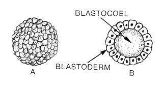
An embryo is an initial stage of development of a multicellular organism. In organisms that reproduce sexually, embryonic development is the part of the life cycle that begins just after fertilization of the female egg cell by the male sperm cell. The resulting fusion of these two cells produces a single-celled zygote that undergoes many cell divisions that produce cells known as blastomeres. The blastomeres are arranged as a solid ball that when reaching a certain size, called a morula, takes in fluid to create a cavity called a blastocoel. The structure is then termed a blastula, or a blastocyst in mammals.

<span class="mw-page-title-main">Mesoderm</span> Middle germ layer of embryonic development

The mesoderm is the middle layer of the three germ layers that develops during gastrulation in the very early development of the embryo of most animals. The outer layer is the ectoderm, and the inner layer is the endoderm.

<span class="mw-page-title-main">Embryology</span> Branch of biology studying prenatal biology

Embryology is the branch of animal biology that studies the prenatal development of gametes, fertilization, and development of embryos and fetuses. Additionally, embryology encompasses the study of congenital disorders that occur before birth, known as teratology.

<span class="mw-page-title-main">Blastulation</span> Sphere of cells formed during early embryonic development in animals

Blastulation is the stage in early animal embryonic development that produces the blastula. In mammalian development the blastula develops into the blastocyst with a differentiated inner cell mass and an outer trophectoderm. The blastula is a hollow sphere of cells known as blastomeres surrounding an inner fluid-filled cavity called the blastocoel. Embryonic development begins with a sperm fertilizing an egg cell to become a zygote, which undergoes many cleavages to develop into a ball of cells called a morula. Only when the blastocoel is formed does the early embryo become a blastula. The blastula precedes the formation of the gastrula in which the germ layers of the embryo form.

<span class="mw-page-title-main">Gastrulation</span> Stage in embryonic development in which germ layers form

Gastrulation is the stage in the early embryonic development of most animals, during which the blastula, or in mammals the blastocyst is reorganized into a multilayered structure known as the gastrula. Before gastrulation, the embryo is a continuous epithelial sheet of cells; by the end of gastrulation, the embryo has begun differentiation to establish distinct cell lineages, set up the basic axes of the body, and internalized one or more cell types including the prospective gut.

<span class="mw-page-title-main">Amniote</span> Clade of tetrapods including reptiles, birds and mammals

Amniotes belong to the clade Amniota, a clade of tetrapod vertebrates that comprises sauropsids and synapsids. They are distinguished from the other living tetrapod clade—the lissamphibians—by the development of three extraembryonic membranes, thicker and more keratinized skin, and costal respiration.

<span class="mw-page-title-main">Notochord</span> Flexible rod-shaped body found in embryos of all chordates

In anatomy, the notochord is a flexible rod which is similar in structure to the stiffer cartilage. If a species has a notochord at any stage of its life cycle, it is, by definition, a chordate. The notochord consists of inner, vacuolated cells covered by fibrous and elastic sheaths, lies along the anteroposterior axis, is usually closer to the dorsal than the ventral surface of the embryo, and is composed of cells derived from the mesoderm.

<span class="mw-page-title-main">Long-beaked echidna</span> Genus of monotremes

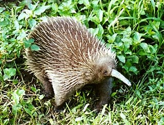
The long-beaked echidnas make up one of the two extant genera of echidnas, spiny monotremes that live in New Guinea; the other being the short-beaked echidna. There are three living species and one extinct species in this genus. The extinct species were present in Australia. Echidnas are one of the two types of mammals that lay eggs, the other being the platypus. The echidnas retain reptilian features such as egg-laying but display mammalian features such as fur and lactation.

<span class="mw-page-title-main">Neurula</span> Embryo at the early stage of development in which neurulation occurs

A neurula is a vertebrate embryo at the early stage of development in which neurulation occurs. The neurula stage is preceded by the gastrula stage; consequentially, neurulation is preceded by gastrulation. Neurulation marks the beginning of the process of organogenesis.

<span class="mw-page-title-main">Pharyngeal arch</span> Embryonic precursor structures in vertebrates

The pharyngeal arches, also known as visceral arches, are structures seen in the embryonic development of vertebrates that are recognisable precursors for many structures. In fish, the arches are known as the branchial arches, or gill arches.

<span class="mw-page-title-main">Placentation</span> Formation and structure of the placenta

Placentation refers to the formation, type and structure, or arrangement of the placenta. The function of placentation is to transfer nutrients, respiratory gases, and water from maternal tissue to a growing embryo, and in some instances to remove waste from the embryo. Placentation is best known in live-bearing mammals (theria), but also occurs in some fish, reptiles, amphibians, a diversity of invertebrates, and flowering plants. In vertebrates, placentas have evolved more than 100 times independently, with the majority of these instances occurring in squamate reptiles.

In embryology, Carnegie stages are a standardized system of 23 stages used to provide a unified developmental chronology of the vertebrate embryo.

<span class="mw-page-title-main">Epiblast</span> Embryonic inner cell mass tissue that forms the embryo itself, through the three germ layers

In amniote embryonic development, the epiblast is one of two distinct cell layers arising from the inner cell mass in the mammalian blastocyst, or from the blastula in reptiles and birds, the other layer is the hypoblast. It drives the embryo proper through its differentiation into the three primary germ layers, ectoderm, mesoderm and endoderm, during gastrulation. The amniotic ectoderm and extraembryonic mesoderm also originate from the epiblast.

<span class="mw-page-title-main">Red-bellied short-necked turtle</span> Species of turtle

The red-bellied short-necked turtle, also known commonly as the pink-bellied side-necked turtle and the Jardine River turtle, is a species of turtle in the family Chelidae. The species is native to Australia and New Guinea. There are two recognized subspecies.

<span class="mw-page-title-main">Premaxilla</span> Cranial bones at the very tip of the upper jaw of many animals

The premaxilla is one of a pair of small cranial bones at the very tip of the upper jaw of many animals, usually, but not always, bearing teeth. In humans, they are fused with the maxilla. The "premaxilla" of therian mammals has been usually termed as the incisive bone. Other terms used for this structure include premaxillary bone or os premaxillare, intermaxillary bone or os intermaxillare, and Goethe's bone.

<span class="mw-page-title-main">Rosa Beddington</span> English developmental biologist

Rosa Susan Penelope Beddington FRS was a British biologist whose career had a major impact on developmental biology.

von Baers laws (embryology) Foundational theory of embryonic development (published 1828)

In developmental biology, von Baer's laws of embryology are four rules proposed by Karl Ernst von Baer to explain the observed pattern of embryonic development in different species.

<span class="mw-page-title-main">Postparietal</span> Fish skull bones

Postparietals are cranial bones present in fish and many tetrapods. Although initially a pair of bones, many lineages possess postparietals which were fused into a single bone. The postparietals were dermal bones situated along the midline of the skull, behind the parietal bones. They formed part of the rear edge of the skull roof, and the lateral edge of each postparietal often contacts the tabular and supratemporal bones. In fish, the postparietals are elongated, typically the largest components of the skull roof. Tetrapods possessed shorter postparietals, which were reduced further and shifted towards the braincase in amniotes. At several points in synapsid evolution, the postparietals fused to each other and the tabulars during embryological development. This fusion produces the interparietal bone, which is inherited by mammals. Postparietals are common in extinct amphibians and early reptiles. However, most living amphibians and living reptiles lack postparietal bones, with a few exceptions.

This glossary of developmental biology is a list of definitions of terms and concepts commonly used in the study of developmental biology and related disciplines in biology, including embryology and reproductive biology, primarily as they pertain to vertebrate animals and particularly to humans and other mammals. The developmental biology of invertebrates, plants, fungi, and other organisms is treated in other articles; e.g. terms relating to the reproduction and development of insects are listed in Glossary of entomology, and those relating to plants are listed in Glossary of botany.

References

  1. 1 2 3 4 5 6 7 8 9 10 11 12 13 14 15 16 17 18 19 20 21 22 23 24 25 26 27 28 29 30 31 32 33 34 35 36 37 38 39 40 41 42 43 44 45 46 47 48 49 50 51 52 53 54 55 56 57 58 59 60 61 62 63 64 65 66 67 68 69 70 71 72 73 74 75 76 77 78 79 80 81 82 83 84 85 86 87 88 89 90 91 92 93 94 95 96 97 98 99 100 101 102 103 104 105 106 107 108 109 110 111 112 113 114 115 116 Werneburg (2009). A Standard System to Study Vertebrate Embryos. PLoS ONE 4(6): e5887. http://www.plosone.org/article/info%3Adoi%2F10.1371%2Fjournal.pone.0005887
  2. Werneburg and Spiekman (2016). Mammalian embryology and organogenesis. From gametes to weaning. In: Zachos F., Asher R. (eds.). Mammalia. Series: The Handbook of Zoology / Handbuch der Zoologie. De Gruyter, Berlin
  3. 1 2 3 4 5 6 7 8 Werneburg I and Sánchez-Villagra MS (2009). Timing of organogenesis support basal position of turtles in the amniote tree of life. BMC Evolutionary Biology, 9:82 http://www.biomedcentral.com/1471-2148/9/82
  4. Werneburg I, Hugi J, Müller J, Sánchez-Villagra MS (2009). Embryogenesis and ossification of Emydura subglobosa (Testudines, Pleurodira, Chelidae) and patterns of turtle development. Developmental Dynamics, Volume 238, Issue 11 Pages 2770-2786 http://onlinelibrary.wiley.com/doi/10.1002/dvdy.22104/abstract
  5. Nunes Silva R and Sobral Sampaio F (2014). Immunoreactivity of Mel1a-like melatonin receptor and NRH: Quinone reductase enzyme (QR2) in testudine whole embryo and in developing whole retinas Trends in Developmental Biology 8:39-46.
  6. 1 2 3 4 Polachowski KM and Werneburg I (2013). Late embryos and bony skull development in Bothropoides jararaca (Serpentes, Viperidae). Zoology
  7. Roscito and Rodriges (2012). Embryonic development of the fossorial gymnophthalmid lizards Nothobachia ablephara and Calyptommatus sinebranchiatus. Zoology 115:302-318
  8. Werneburg et al. (2015). Bony skull development in the Argus monitor (Squamata, Varanidae, Varanus panoptes) with comments on developmental timing and adult anatomy. Zoology 118(4):255-280
  9. Ollonen, J., Da Silva, F.O., Mahlow, K. and Di-Poï, N., 2018. Skull development, ossification pattern, and adult shape in the emerging lizard model organism Pogona vitticeps: a comparative analysis with other squamates. Frontiers in physiology, 9, p.278.
  10. 1 2 3 4 5 6 7 8 9 10 11 12 13 14 15 16 17 18 19 20 21 22 23 Werneburg I and Sánchez-Villagra MR (2011). The early development of the echidna, Tachyglossus aculeatus (Mammalia: Monotremata) and patterns of mammalian development. Acta Zoologica. 82(1) http://onlinelibrary.wiley.com/doi/10.1111/j.1463-6395.2009.00447.x/abstract
  11. 1 2 3 4 5 6 7 8 9 10 11 12 13 14 15 16 17 18 19 20 21 22 23 24 25 26 27 28 29 30 31 32 Werneburg I, Laurin M, Koyabu D, Sánchez-Villagra (2016). Evolution of organogenesis and the origin of altriciality in mammals. Evolution and Development
  12. González B, Soria-Escobar AM, Rojas-Díaz V, Pustovrh MC, Monsalve LS, Rougier GW (2020). The embryo of the silky shrew opossum, Caenolestes fuliginosus (Tomes, 1863): First description of the embryo of Paucituberculata. Journal of Morphology 2020: 1-12
  13. 1 2 Werneburg I, Tzika AC, Hautier L, Asher RJ, Milinkovitch MC, Sánchez-Villagra MR (2013). Development and embryonic staging in non-model organisms: the case of an afrotherian mammal. The Journal of Anatomy 222:2-18
  14. 1 2 3 4 5 6 7 8 Taro Nojiri, Dai Fukui, Ingmar Werneburg, Takashi Saitoh, Hideki Endo, Daisuke Koyabu (2021). Embryonic staging of bats with special reference to Vespertilio sinensis and its cochlear development. Development Dynamics, DOI10.1002/dvdy.325