Structured light plethysmography

Last updated
Structured light plethysmography
Purposenoninvasive method for collecting accurate representations of chest and abdominal wall movement

Structured Light Plethysmography (SLP) technology is a noninvasive method for collecting accurate representations of chest and abdominal wall movement. A checkerboard pattern of light is projected from a light projector onto the chest of an individual. Movements of the grid are viewed by two digital cameras, digitalised, and processed to form a 3D model and can be interrogated to assess lung function. The system has been tested on over 70 adults (data presented at clinical meetings). SLP is simple to use, accurate and cost effective, is self-calibrating and does not require the use of plastic consumables, reducing cost, risk of cross infection and the device's carbon footprint. In conjunction with the Cambridge Veterinary School, proof of concept studies have indicated that the device is sensitive enough to noninvasively pick up respiratory movements in domestic animals (cats and dogs).

Contents

Patient Scan Horizontal.jpg

A totally “non-invasive” technique using structured light to measure pulmonary function was developed as long ago as the mid-1980s by a London group at the Royal Brompton Hospital working in close association with IBM (1-4). The technique used the distortion with movement of a structured pattern of light to calculate a volume or change in volume of a textured surface. At that time structured light pattern was shone on to the anterior chest and abdominal wall surfaces, and movie camera film footage was taken and sent to IBM for processing. The digital information was used to compute automatically the volume of the trunk and the position of any point on its surface and its cross-sectional shape at any level. The group found that linear dimensions could be calculated to within 0.5mm and a cross-sectional area within 5% and volume within 3-7%. The group suggested that non-invasive technique measurements were of sufficient accuracy to be tried in clinical practice. However, probably due to technology limitations, data processing and storage, the methodology was never commercialised.

Seated patient preparing for chest SLP scan Male Patient SLP.jpg
Seated patient preparing for chest SLP scan

Independently the Royal Brompton group of the 1980s, a Cambridge (UK) consortium of clinicians and engineers developed a system in 2009 that has revisited structure light pattern as a noninvasive method for collecting accurate representations of chest and abdominal wall movement. The methodology has several advantages: there are no fluorescent markers required to define chest or abdominal surface and the hardware can be minimalized to 2 digital cameras and a digital projector when imaging the anterior surface of the body. The projector shines a grid of black and white squares from superior iliac crest to clavicle; the subject can wear a plain t-shirt of any colour. The 2 digital cameras image the grid on chest and abdomen and the software extracts 2 sets of 2D image positions of the grid points and stereo vision is used to reconstruct these grid points to form a 3D representation of the chest and abdominal wall surface. The group has tried anything from one grid point to 2000 and is current working on roughly 200 to 300. The thoracic volume is calculated from the volume beneath the reconstructed virtual surface and can be plotted in real time. Calibration is internal: the relative position of cameras and shape of the subject is auto-calibrated at reconstruction time; and externally by placing an object of reference size in front of the cameras. This can be a sheet of paper of known size and potentially has now been transferred to projected points within the grid itself. The group have now collected data on up to 70 adults and 5 children and have also collected data from small mammals. They have compared simultaneous measurements of tidal breathing followed by full inspiration and force expiration by pneumotach and SLP. The pneumotach data was obtained using a laptop based spirometer and exported for analysis using J-scope software. Extremely good correlations have been obtained from tidal breathing and forced expiration manoeuvres. Tidal breathing correlations have shown to be 0.99 for individual data sets and for n=70 the mean correlation was 0.92 at an SD of 0.04. For forced expiration the correlation was shown to be 0.98, n=70 mean correlation was 0.98 with an SD of 0.12. For forced expirations inspiratory manoeuvres correlation coefficients for peak flow were r² 0.84, FEV1 0.95, FEF75 0.76 and FEF50 0.69.

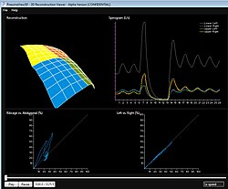
Patient Example Outputs

Healthy Patient.jpg Healthy Patient SLP Scan

Thoracic Patient.jpg Thoracic Patient SLP Scan

Diseased Patient.jpg Diseased Patient SLP Scan

Being totally non-invasive SLP allows respiratory monitoring without the need for a face mask or mouth piece, allowing for normal respiratory patterns to be recorded. Data output can be configured to allow for the comparisons between selected regions of interest such as chest and abdomen. Further analysis uses spectral analysis techniques to monitor trends and changes in respiratory function and alterations in regional chest dynamics. The group has a number of studies on-going .

The first application of this technology has been developed to measure spirometry by PneumaCare Ltd (www.pneumacare.com), The device - PneumaScan has been developed by PneumaCare in partnership with Cambridge University, Addenbrookes NHS Hospital Trust and Plextek Limited (a leading UK device specialist consultancy). It is capable of recording tidal breathing and spirometry parameters and is self calibrating.

Further reading

1. Optical mapping of the thoraco-abdominal wall: AJ Peacock, MDL Morgan, S Gourlay, C Turton, DM Dension. Lung Function Unit, Brompton Hospital, London, and UK Science Centre, IBM, Winchester Thorax 1984 39: 93-100

2. Optical measurement of the change in trunk volume with breathing. Peacock A, Gourlay A, Denison D.Bull Eur Physiopathol Respir. 1985 Mar-Apr;21(2):125-9.

3. Measuring the human chest with structured lighting J.R.T. Lewis and T. Sopwith Pattern Recognition Letters Volume 4, Issue 5, October 1986, Pages 359-36

4. J. R. T. Lewis, T. Sopwith: Three-dimensional surface measurement by microcomputer. Image Vision Comput. 4(3): 159-166 (1986) 5. Structured light Plethysmography in infants and Children – a pilot study. Juliet Usher-Smith, Richard Wareham, J Lasenby, J Cameron, P Bridge, R Iles RCPCH York 2009 Arch Dis Child 2009;94(Suppl I):A38–A40

2. Structured light plethysmography (SLP) compared to spirometry: R. Wareham, J. Lasenby, J. Cameron, P. D. Bridge, R. Iles (Cambridge, United Kingdom) E-communication : 350 ERS 2009

3. Spectral analysis of respiratory flow signals from spirometry and a novel structured light imaging system. Peter Bridge1, Richard Iles1, Rich Wareham2, Jonathan Cameron2, Juliet Usher-Smith1, Ward Hills3, Gareth Roberts3, Willem de Boer2, Joan Lasenby2 E-communication : 1858 ERS 2009

4. Comparison of Forced Expiratory Volumes Measured with Structured Light Plethysmography (SLP) and Spirometry Eugene Lau, Douglas Brand, Peter Bridge, Richard Iles, Rich Wareham, Jonathan Cameron, Juliet Usher-Smith, Ward Hills, Gareth Roberts, Willem de Boer, Joan Lasenby. BTS 2009 Thorax 2009;64:A51-A54

5. Comparison of Tidal Breathing with Structured Light Plethysmography (SLP) and Spirometry Eugene Lau, Douglas Brand, Peter Bridge, Richard Iles, Rich Wareham, Jonathan Cameron, Juliet Usher-Smith, Ward Hills, Gareth Roberts, Willem de Boer, Joan Lasenby. BTS 2009 Thorax 2009;64:A51-A54

6. Spectral analysis of regional respiratory flow signals using structured light plethysmography: S. Ahmed1, P. Bridge2, J. Usher-Smith2, R. Wareham3, J. Cameron3, J. Lasenby3, R. Iles2; 1Cambridge University Medical School - Cambridge/UK, 2Addenbrookes NHS Trust, Cambridge University Hospitals - Cambridge/UK, 3Cambridge University - Cambridge/UK. ATS 2010 Am J Respir Crit Care Med 181;2010:A2170

7. Tidal breathing parameters measured by structured light plethysmography (slp) and spirometry: D. Brand1, E. Lau1, J. Cameron2, R. Wareham2, J. Usher-Smith3, P. Bridge3, J. Lasenby2, R. Iles3; 1University of Cambridge School of Clinical Medicine - Cambridge/UK, 2Cambridge University, Department of Engineering - Cambridge/UK, 3Addenbrookes NHS Trust, Cambridge University Hospitals - Cambridge/UK Am J Respir Crit Care Med 181;2010:A2171 ATS 2010

8. SLP: A Zero-Contact Non-Invasive Method for Pulmonary Function Testing Richard Iles, Rich Wareham, Jonathan Cameron, Juliet Usher-Smith, Ward Hills, Gareth Roberts, Willem de Boer, Joan Lasenby BMVC 2010

9. Structured light plethysmography with pneumotachography. Symposium : Physiological plasters and visiology: the visionary future for lung function? ERS 2010

10. Measurement of lung volumes in real time, using optical plethysmography: Postgraduate Course: WS1 Educational Workshop: Noninvasive assessment of respiratory function in children. ERS 2010

Related Research Articles

<span class="mw-page-title-main">Lung</span> Primary organ of the respiratory system

The lungs are the primary organs of the respiratory system in humans and most other animals, including some snails and a small number of fish. In mammals and most other vertebrates, two lungs are located near the backbone on either side of the heart. Their function in the respiratory system is to extract oxygen from the air and transfer it into the bloodstream, and to release carbon dioxide from the bloodstream into the atmosphere, in a process of gas exchange. The pleurae, which are thin, smooth, and moist, serve to reduce friction between the lungs and chest wall during breathing, allowing for easy and effortless movements of the lungs.

<span class="mw-page-title-main">Respiratory system</span> Biological system in animals and plants for gas exchange

The respiratory system is a biological system consisting of specific organs and structures used for gas exchange in animals and plants. The anatomy and physiology that make this happen varies greatly, depending on the size of the organism, the environment in which it lives and its evolutionary history. In land animals the respiratory surface is internalized as linings of the lungs. Gas exchange in the lungs occurs in millions of small air sacs; in mammals and reptiles these are called alveoli, and in birds they are known as atria. These microscopic air sacs have a very rich blood supply, thus bringing the air into close contact with the blood. These air sacs communicate with the external environment via a system of airways, or hollow tubes, of which the largest is the trachea, which branches in the middle of the chest into the two main bronchi. These enter the lungs where they branch into progressively narrower secondary and tertiary bronchi that branch into numerous smaller tubes, the bronchioles. In birds the bronchioles are termed parabronchi. It is the bronchioles, or parabronchi that generally open into the microscopic alveoli in mammals and atria in birds. Air has to be pumped from the environment into the alveoli or atria by the process of breathing which involves the muscles of respiration.

<span class="mw-page-title-main">Lung volumes</span> Volume of air in the lungs

Lung volumes and lung capacities refer to the volume of air in the lungs at different phases of the respiratory cycle.

<span class="mw-page-title-main">Obesity hypoventilation syndrome</span> Condition in which severely overweight people fail to breathe rapidly or deeply enough

Obesity hypoventilation syndrome (OHS) is a condition in which severely overweight people fail to breathe rapidly or deeply enough, resulting in low oxygen levels and high blood carbon dioxide (CO2) levels. The syndrome is often associated with obstructive sleep apnea (OSA), which causes periods of absent or reduced breathing in sleep, resulting in many partial awakenings during the night and sleepiness during the day. The disease puts strain on the heart, which may lead to heart failure and leg swelling.

Exhalation is the flow of the breath out of an organism. In animals, it is the movement of air from the lungs out of the airways, to the external environment during breathing. This happens due to elastic properties of the lungs, as well as the internal intercostal muscles which lower the rib cage and decrease thoracic volume. As the thoracic diaphragm relaxes during exhalation it causes the tissue it has depressed to rise superiorly and put pressure on the lungs to expel the air. During forced exhalation, as when blowing out a candle, expiratory muscles including the abdominal muscles and internal intercostal muscles generate abdominal and thoracic pressure, which forces air out of the lungs.

<span class="mw-page-title-main">Spirometry</span> Pulmonary function test

Spirometry is the most common of the pulmonary function tests (PFTs). It measures lung function, specifically the amount (volume) and/or speed (flow) of air that can be inhaled and exhaled. Spirometry is helpful in assessing breathing patterns that identify conditions such as asthma, pulmonary fibrosis, cystic fibrosis, and COPD. It is also helpful as part of a system of health surveillance, in which breathing patterns are measured over time.

<span class="mw-page-title-main">Plethysmograph</span>

A plethysmograph is an instrument for measuring changes in volume within an organ or whole body. The word is derived from the Greek "plethysmos", and "graphein".

Optoelectronic plethysmography is a method to evaluate ventilation through an external measurement of the chest wall surface motion.

<span class="mw-page-title-main">Diaphragmatic breathing</span> Form of breathing done by contracting the diaphragm

Diaphragmatic breathing, abdominal breathing, belly breathing, or deep breathing, is breathing that is done by contracting the diaphragm, a muscle located horizontally between the thoracic cavity and abdominal cavity. Air enters the lungs as the diaphragm strongly contracts, but unlike during traditional relaxed breathing (eupnea) the intercostal muscles of the chest do minimal work in this process. The belly also expands during this type of breathing to make room for the contraction of the diaphragm.

<span class="mw-page-title-main">Pneumomediastinum</span> Abnormal presence of gas in the thorax

Pneumomediastinum is pneumatosis in the mediastinum, the central part of the chest cavity. First described in 1819 by René Laennec, the condition can result from physical trauma or other situations that lead to air escaping from the lungs, airways, or bowel into the chest cavity. In underwater divers it is usually the result of pulmonary barotrauma.

<span class="mw-page-title-main">Functional residual capacity</span> Volume of air in the lungs at the end of passive expiration

Functional residual capacity (FRC) is the volume of air present in the lungs at the end of passive expiration. At FRC, the opposing elastic recoil forces of the lungs and chest wall are in equilibrium and there is no exertion by the diaphragm or other respiratory muscles.

<span class="mw-page-title-main">Incentive spirometer</span> Handheld device to improve lung function

An incentive spirometer is a handheld medical device used to help patients improve the functioning of their lungs. By training patients to take slow and deep breaths, this simplified spirometer facilitates lung expansion and strengthening. Patients inhale through a mouthpiece, which causes a piston inside the device to rise. This visual feedback helps them monitor their inspiratory effort. Incentive spirometers are commonly used after surgery or other illnesses to prevent pulmonary complications.

<span class="mw-page-title-main">Obstructive lung disease</span> Category of respiratory disease characterized by airway obstruction

Obstructive lung disease is a category of respiratory disease characterized by airway obstruction. Many obstructive diseases of the lung result from narrowing (obstruction) of the smaller bronchi and larger bronchioles, often because of excessive contraction of the smooth muscle itself. It is generally characterized by inflamed and easily collapsible airways, obstruction to airflow, problems exhaling, and frequent medical clinic visits and hospitalizations. Types of obstructive lung disease include; asthma, bronchiectasis, bronchitis and chronic obstructive pulmonary disease (COPD). Although COPD shares similar characteristics with all other obstructive lung diseases, such as the signs of coughing and wheezing, they are distinct conditions in terms of disease onset, frequency of symptoms, and reversibility of airway obstruction. Cystic fibrosis is also sometimes included in obstructive pulmonary disease.

<span class="mw-page-title-main">Pulmonary function testing</span> Test to evaluate respiratory system

Pulmonary function testing (PFT) is a complete evaluation of the respiratory system including patient history, physical examinations, and tests of pulmonary function. The primary purpose of pulmonary function testing is to identify the severity of pulmonary impairment. Pulmonary function testing has diagnostic and therapeutic roles and helps clinicians answer some general questions about patients with lung disease. PFTs are normally performed by a pulmonary function technician, respiratory therapist, respiratory physiologist, physiotherapist, pulmonologist, or general practitioner.

Pulmonary hygiene, formerly referred to as pulmonary toilet, is a set of methods used to clear mucus and secretions from the airways. The word pulmonary refers to the lungs. The word toilet, related to the French toilette, refers to body care and hygiene; this root is used in words such as toiletry that also relate to cleansing.

<span class="mw-page-title-main">Respiratory inductance plethysmography</span>

Respiratory inductance plethysmography (RIP) is a method of evaluating pulmonary ventilation by measuring the movement of the chest and abdominal wall.

Modes of mechanical ventilation are one of the most important aspects of the usage of mechanical ventilation. The mode refers to the method of inspiratory support. In general, mode selection is based on clinician familiarity and institutional preferences, since there is a paucity of evidence indicating that the mode affects clinical outcome. The most frequently used forms of volume-limited mechanical ventilation are intermittent mandatory ventilation (IMV) and continuous mandatory ventilation (CMV). There have been substantial changes in the nomenclature of mechanical ventilation over the years, but more recently it has become standardized by many respirology and pulmonology groups. Writing a mode is most proper in all capital letters with a dash between the control variable and the strategy.

Intermittent Mandatory Ventilation (IMV) refers to any mode of mechanical ventilation where a regular series of breaths are scheduled but the ventilator senses patient effort and reschedules mandatory breaths based on the calculated need of the patient. Similar to continuous mandatory ventilation in parameters set for the patients pressures and volumes but distinct in its ability to support a patient by either supporting their own effort or providing support when patient effort is not sensed. IMV is frequently paired with additional strategies to improve weaning from ventilator support or to improve cardiovascular stability in patients who may need full life support.

Intermittent positive pressure breathing (IPPB) is a respiratory therapy treatment for people who are hypoventilating. While not a preferred method due to cost, IPPB is used to expand the lungs, deliver aerosol medications, and in some circumstances ventilate the patient.

<span class="mw-page-title-main">Deep inspiration breath-hold</span> Method of delivering radiotherapy

Deep inspiration breath-hold (DIBH) is a method of delivering radiotherapy while limiting radiation exposure to the heart and lungs. It is used primarily for treating left-sided breast cancer.