
A ganglion is a group of neuron cell bodies in the peripheral nervous system. In the somatic nervous system this includes dorsal root ganglia and trigeminal ganglia among a few others. In the autonomic nervous system there are both sympathetic and parasympathetic ganglia which contain the cell bodies of postganglionic sympathetic and parasympathetic neurons respectively.

In biology, the nervous system is the highly complex part of an animal that coordinates its actions and sensory information by transmitting signals to and from different parts of its body. The nervous system detects environmental changes that impact the body, then works in tandem with the endocrine system to respond to such events. Nervous tissue first arose in wormlike organisms about 550 to 600 million years ago. In vertebrates it consists of two main parts, the central nervous system (CNS) and the peripheral nervous system (PNS). The CNS consists of the brain and spinal cord. The PNS consists mainly of nerves, which are enclosed bundles of the long fibers or axons, that connect the CNS to every other part of the body. Nerves that transmit signals from the brain are called motor nerves or efferent nerves, while those nerves that transmit information from the body to the CNS are called sensory nerves or afferent. Spinal nerves are mixed nerves that serve both functions. The PNS is divided into three separate subsystems, the somatic, autonomic, and enteric nervous systems. Somatic nerves mediate voluntary movement. The autonomic nervous system is further subdivided into the sympathetic and the parasympathetic nervous systems. The sympathetic nervous system is activated in cases of emergencies to mobilize energy, while the parasympathetic nervous system is activated when organisms are in a relaxed state. The enteric nervous system functions to control the gastrointestinal system. Both autonomic and enteric nervous systems function involuntarily. Nerves that exit from the cranium are called cranial nerves while those exiting from the spinal cord are called spinal nerves.

The sympathetic nervous system (SNS) is one of the three divisions of the autonomic nervous system, the others being the parasympathetic nervous system and the enteric nervous system. The enteric nervous system is sometimes considered part of the autonomic nervous system, and sometimes considered an independent system.

Cephalization is an evolutionary trend in which, over many generations, the mouth, sense organs, and nerve ganglia become concentrated at the front end of an animal, producing a head region. This is associated with movement and bilateral symmetry, such that the animal has a definite head end. This led to the formation of a highly sophisticated brain in three groups of animals, namely the arthropods, cephalopod molluscs, and vertebrates.

A pseudounipolar neuron is a type of neuron which has one extension from its cell body. This type of neuron contains an axon that has split into two branches. A single process arises from the cell body and then divides into an axon and a dendrite. They develop embryologically as bipolar in shape, and are thus termed pseudounipolar instead of unipolar.

The supraesophageal ganglion is the first part of the arthropod, especially insect, central nervous system. It receives and processes information from the first, second, and third metameres. The supraesophageal ganglion lies dorsal to the esophagus and consists of three parts, each a pair of ganglia that may be more or less pronounced, reduced, or fused depending on the genus:

The ventral nerve cord is a major structure of the invertebrate central nervous system. It is the functional equivalent of the vertebrate spinal cord. The ventral nerve cord coordinates neural signaling from the brain to the body and vice versa, integrating sensory input and locomotor output. Because arthropods have an open circulatory system,decapitated insects can still walk, groom, and mate - illustrating that the circuitry of the ventral nerve cord is sufficient to perform complex motor programs without brain input.

The superior cervical ganglion (SCG) is part of the autonomic nervous system (ANS); more specifically, it is part of the sympathetic nervous system, a division of the ANS most commonly associated with the fight or flight response. The ANS is composed of pathways that lead to and from ganglia, groups of nerve cells. A ganglion allows a large amount of divergence in a neuronal pathway and also enables a more localized circuitry for control of the innervated targets. The SCG is the only ganglion in the sympathetic nervous system that innervates the head and neck. It is the largest and most rostral (superior) of the three cervical ganglia. The SCG innervates many organs, glands and parts of the carotid system in the head.

The mouthparts of arthropods have evolved into a number of forms, each adapted to a different style or mode of feeding. Most mouthparts represent modified, paired appendages, which in ancestral forms would have appeared more like legs than mouthparts. In general, arthropods have mouthparts for cutting, chewing, piercing, sucking, shredding, siphoning, and filtering. This article outlines the basic elements of four arthropod groups: insects, myriapods, crustaceans and chelicerates. Insects are used as the model, with the novel mouthparts of the other groups introduced in turn. Insects are not, however, the ancestral form of the other arthropods discussed here.

The (pan)arthropod head problem is a long-standing zoological dispute concerning the segmental composition of the heads of the various arthropod groups, and how they are evolutionarily related to each other. While the dispute has historically centered on the exact make-up of the insect head, it has been widened to include other living arthropods such as chelicerates, myriapods, crustaceans; and fossil forms, such as the many arthropods known from exceptionally preserved Cambrian faunas. While the topic has classically been based on insect embryology, in recent years a great deal of developmental molecular data has become available. Dozens of more or less distinct solutions to the problem, dating back to at least 1897, have been published, including several in the 2000s.
Insect physiology includes the physiology and biochemistry of insect organ systems.

In arthropods, the maxillae are paired structures present on the head as mouthparts in members of the clade Mandibulata, used for tasting and manipulating food. Embryologically, the maxillae are derived from the 4th and 5th segment of the head and the maxillary palps; segmented appendages extending from the base of the maxilla represent the former leg of those respective segments. In most cases, two pairs of maxillae are present and in different arthropod groups the two pairs of maxillae have been variously modified. In crustaceans, the first pair are called maxillulae.

The lumbar ganglia are paravertebral ganglia located in the inferior portion of the sympathetic trunk. The lumbar portion of the sympathetic trunk typically has 4 lumbar ganglia. The lumbar splanchnic nerves arise from the ganglia here, and contribute sympathetic efferent fibers to the nearby plexuses. The first two lumbar ganglia have both white and gray rami communicates.

The nervous system of gastropods consists of a series of paired ganglia connected by major nerve cords, and a number of smaller branching nerves. It is sometimes called ganglionic.
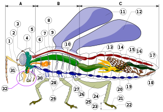
Insect morphology is the study and description of the physical form of insects. The terminology used to describe insects is similar to that used for other arthropods due to their shared evolutionary history. Three physical features separate insects from other arthropods: they have a body divided into three regions, have three pairs of legs, and mouthparts located outside of the head capsule. It is this position of the mouthparts which divides them from their closest relatives, the non-insect hexapods, which includes Protura, Diplura, and Collembola.

Pseudunela cornuta is a species of minute sea slug, an acochlidian, a shell-less marine and temporarily brackish gastropod mollusk in the family Pseudunelidae. Adults are about 3 mm long and live in the spaces between sand grains.
The cephalon is the head section of an arthropod. It is a tagma, i.e., a specialized grouping of arthropod segments. The word cephalon derives from the Greek κεφαλή (kephalē), meaning "head".

The labrum is a flap-like structure that lies immediately in front of the mouth in almost all extant Euarthropoda. The most conspicuous exceptions are the Pycnogonida, which probably are chelicerate-relatives. In entomology, the labrum amounts to the "upper lip" of an insect mouth, the corresponding "lower lip" being the labium.
The evolution of nervous systems dates back to the first development of nervous systems in animals. Neurons developed as specialized electrical signaling cells in multicellular animals, adapting the mechanism of action potentials present in motile single-celled and colonial eukaryotes. Primitive systems, like those found in protists, use chemical signalling for movement and sensitivity; data suggests these were precursors to modern neural cell types and their synapses. When some animals started living a mobile lifestyle and eating larger food particles externally, they developed ciliated epithelia, contractile muscles and coordinating & sensitive neurons for it in their outer layer.
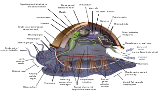
A circumesophageal or circumpharyngeal nerve ring is an arrangement of nerve ganglia around the esophagus/ pharynx of an animal. It is a common feature of nematodes, molluscs, and many other invertebrate animals, though it is absent in all vertebrate animals and is not structurally possible in simpler ones such as water bears.
