Synthesis of nanoparticles by fungi

Last updated

Throughout human history, fungi have been utilized as a source of food and harnessed to ferment and preserve foods and beverages. In the 20th century, humans have learned to harness fungi to protect human health (antibiotics, anti-cholesterol statins, and immunosuppressive agents), while industry has utilized fungi for large scale production of enzymes, acids, and biosurfactants. [1] With the advent of modern nanotechnology in the 1980s, fungi have remained important by providing a greener alternative to chemically synthesized nanoparticle. [2]

Contents

Background

SEM image of fungal hyphae and fungal derived silver nanoparticles showing a large conglomeration made up of individual nanoparticles with fungal hyphae (dark areas) in background. Fungal nanoparticles -conglomerate with smaller nanoparticles.tif
SEM image of fungal hyphae and fungal derived silver nanoparticles showing a large conglomeration made up of individual nanoparticles with fungal hyphae (dark areas) in background.

A nanoparticle is defined as having one dimension 100 nm or less in size. Environmentally toxic or biologically hazardous reducing agents are typically involved in the chemical synthesis of nanoparticles [2] so there has been a search for greener production alternatives. [3] [4] Current research has shown that microorganisms, plant extracts, and fungi can produce nanoparticles through biological pathways. [2] [3] [5] The most common nanoparticles synthesized by fungi are silver and gold, however fungi have been utilized in the synthesis other types of nanoparticles including zinc oxide, platinum, magnetite, zirconia, silica, titanium, and cadmium sulfide and cadmium selenide quantum dots.

Silver nanoparticle production

Synthesis of silver nanoparticles has been investigated utilizing many ubiquitous fungal species including Trichoderma , [6] [7] Fusarium , [8] Penicillium , [9] Rhizoctonia ,[ citation needed ] Pleurotus and Aspergillus . [10] Extracellular synthesis has been demonstrated by Trichoderma virde, T. reesei, Fusarium oxysporm, F. semitectum, F. solani, Aspergillus niger, A. flavus, [11] A. fumigatus, A. clavatus, Pleurotus ostreatus , Cladosporium cladosporioides, [6] Penicillium brevicompactum , P. fellutanum, an endophytic Rhizoctonia sp., Epicoccum nigrum , Chrysosporium tropicum, and Phoma glomerata, while intracellular synthesis was shown to occur in a Verticillium [12] species, and in Neurospora crassa .

Gold nanoparticle production

Synthesis of gold nanoparticles has been investigated utilizing Fusarium, [13] Neurospora, [14] Verticillium , yeasts, [15] [16] and Aspergillus. Extracellular gold nanoparticle synthesis was demonstrated by Fusarium oxysporum, Aspergillus niger, and cytosolic extracts from Candida albican. Intracellular gold nanoparticle synthesis has been demonstrated by a Verticillum species, V. luteoalbum, [17]

Miscellaneous nanoparticle production

In addition to gold and silver, Fusarium oxysporum has been used to synthesize zirconia, titanium, cadmium sulfide and cadmium selenide nanosize particles. Cadmium sulfide nanoparticles have also been synthesized by Trametes versicolor , Schizosaccharomyces pombe , and Candida glabrata. [18] The white-rot fungus Phanerochaete chrysosporium has also been demonstrated to be able to synthesize elemental selenium nanoparticles. [19]

Culture techniques and conditions

Culture techniques and media vary depending upon the requirements of the fungal isolate involved, however the general procedure consist of the following: fungal hyphae are typically placed in liquid growth media and placed in shake culture until the fungal culture has increased in biomass. The fungal hyphae are removed from the growth media, washed with distilled water to remove the growth media, placed in distilled water and incubated on shake culture for 24 to 48 hours. The fungal hyphae are separated from the supernatant, and an aliquot of the supernatant is added to 1.0 mM ion solution. The ion solution is then monitored for 2 to 3 days for the formation of nanoparticles. Another common culture technique is to add washed fungal hyphae directly into 1.0 mM ion solution instead of utilizing the fungal filtrate. Silver nitrate is the most widely used source of silver ions, but silver sulfate has also been utilized.[ citation needed ] Choloroauric acid is generally used as the source of gold ions at various concentrations (1.0 mM [13] and 250 mg to 500 mg [17] of Au per liter). Cadmium sulfide nanoparticle synthesis for F. oxysporum was conducted using a 1:1 ratio of Cd2+ and SO42− at a 1 mM concentration. [20] Gold nanoparticles can vary in shape and size depending on the pH of the ion solution. [17] Gericke and Pinches (2006) reported that for V. luteoalbum small (cc.10 nm) spherical gold nanoparticles are formed at pH 3, larger (spherical, triangular, hexagon and rods) gold nanoparticles are formed at pH 5, and at pH 7 to pH 9 the large nanoparticles tend to lack a defined shape. Temperature interactions for both silver and gold nanoparticles were similar; a lower temperature resulted in larger nanoparticles while higher temperatures produced smaller nanoparticles. [17]

Analytical techniques

Visual observations

For externally synthesized silver nanoparticles the silver ion solution generally becomes brownish in color, [7] [8] [9] but this browning reaction may be absent.[ citation needed ] For fungi that synthesize intracellular silver nanoparticles, the hyphae darken to a brownish color while the solution remains clear. In both cases the browning reaction is attributed to the surface plasmon resonance of the metallic nanoparticles. [6] [21] For external gold nanoparticle production, the solution color can vary depending on the size of the gold nanoparticles; smaller particles appear pink while large particles appear purple. Intracellular gold nanoparticle synthesis typically turns the hyphae purple while the solution remains clear. Externally synthesized cadmium sulfide nanoparticles were reported to make the solution color appear bright yellow. [20]

Analytical tools

Scanning electron microscopy (SEM), transmission electron microscopy (TEM), energy dispersive analysis of X-ray (EDX), UV-vis spectroscopy, and X-ray diffraction are used to characterize different aspects of nanoparticles. Both SEM and TEM can be used to visualize the location, size, and morphology of the nanoparticles, while UV-vis spectroscopy can be used to confirm the metallic nature, size and aggregation level. Energy dispersive analysis of X-ray is used to determine elemental composition, and X-ray diffraction is used to determine chemical composition and crystallographic structure. UV-Vis absorption peaks for silver, gold, and cadmium sulfide nanoparticles can vary depending on particle size: 25-50 nm silver particles peak ca. 415 nm, gold nanoparticles 30-40 nm peak ca. 450 nm, while a cadmium sulfide absorption edge ca. 450 is indicative of quantum size particles. [20] Larger nanoparticle of each type will have UV-Vis absorption peaks or edges that shift to longer wavelengths while smaller nanoparticles will have UV-Vis absorption peaks or edges that shift to shorter wavelengths.

Formation mechanisms

Gold and silver

SEM image of fungal derived silver nanoparticles stabilized by a capping agent. SEM image of fungal derived silvernanoparticles.tif
SEM image of fungal derived silver nanoparticles stabilized by a capping agent.

Nitrate reductase was suggested to initiate nanoparticle formation by many fungi including Penicillium species, while several enzymes, α-NADPH-dependent reductases, nitrate-dependent reductases and an extracellular shuttle quinone, were implicated in silver nanoparticle synthesis for Fusarium oxysporum. Jain et al. (2011) indicated that silver nanoparticle synthesis for A. flavus occurs initially by a "33kDa" protein followed by a protein (cystein and free amine groups) electrostatic attraction which stabilizes the nanoparticle by forming a capping agent. [11] Intracellular silver and gold nanoparticle synthesis is not fully understood but similar fungal cell wall surface electrostatic attraction, reduction, and accumulation has been proposed. [20] External gold nanoparticle synthesis by P. chrysosporium was attributed to laccase, while intracellular gold nanoparticle synthesis was attributed to ligninase. [20]

Cadmium sulfide

Cadmium sulfide nanoparticle synthesis by yeast involves sequestration of Cd2+ by glutathione-related peptides followed by reduction within the cell. Ahmad et al. (2002) reported that cadmium sulfide nanoparticle synthesis by Fusarium oxysporum was based on a sulfate reductase (enzyme) process.

Related Research Articles

<span class="mw-page-title-main">Mold</span> Wooly, dust-like fungal structure or substance

A mold or mould is one of the structures that certain fungi can form. The dust-like, colored appearance of molds is due to the formation of spores containing fungal secondary metabolites. The spores are the dispersal units of the fungi. Not all fungi form molds. Some fungi form mushrooms; others grow as single cells and are called microfungi.

<i>Fusarium oxysporum</i> Species of fungus

Fusarium oxysporum, an ascomycete fungus, comprises all the species, varieties and forms recognized by Wollenweber and Reinking within an infrageneric grouping called section Elegans. It is part of the family Nectriaceae.

<i>Aspergillus fumigatus</i> Species of fungus

Aspergillus fumigatus is a species of fungus in the genus Aspergillus, and is one of the most common Aspergillus species to cause disease in individuals with an immunodeficiency.
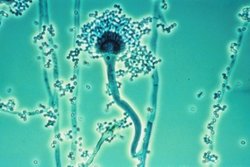
<span class="mw-page-title-main">Mycoremediation</span> Process of using fungi to degrade or sequester contaminants in the environment

Mycoremediation is a form of bioremediation in which fungi-based remediation methods are used to decontaminate the environment. Fungi have been proven to be a cheap, effective and environmentally sound way for removing a wide array of contaminants from damaged environments or wastewater. These contaminants include heavy metals, organic pollutants, textile dyes, leather tanning chemicals and wastewater, petroleum fuels, polycyclic aromatic hydrocarbons, pharmaceuticals and personal care products, pesticides and herbicides in land, fresh water, and marine environments.

<span class="mw-page-title-main">Gliotoxin</span> Chemical compound

Gliotoxin is a sulfur-containing mycotoxin that belongs to a class of naturally occurring 2,5-diketopiperazines produced by several species of fungi, especially those of marine origin. It is the most prominent member of the epipolythiopiperazines, a large class of natural products featuring a diketopiperazine with di- or polysulfide linkage. These highly bioactive compounds have been the subject of numerous studies aimed at new therapeutics. Gliotoxin was originally isolated from Gliocladium fimbriatum, and was named accordingly. It is an epipolythiodioxopiperazine metabolite that is one of the most abundantly produced metabolites in human invasive Aspergillosis (IA).

<i>Fusarium solani</i> Species of fungus

Fusarium solani is a species complex of at least 26 closely related filamentous fungi in the division Ascomycota, family Nectriaceae. It is the anamorph of Nectria haematococca. It is a common soil fungus and colonist of plant materials. Fusarium solani is implicated in plant disease as well as human disease notably infection of the cornea of the eye.

Pathogenic fungi are fungi that cause disease in humans or other organisms. Although fungi are eukaryotic, many pathogenic fungi are microorganisms. Approximately 300 fungi are known to be pathogenic to humans; their study is called "medical mycology". Fungal infections kill more people than either tuberculosis or malaria—about 2 million people per year.

<span class="mw-page-title-main">Fungivore</span> Organism that consumes fungi

Fungivory or mycophagy is the process of organisms consuming fungi. Many different organisms have been recorded to gain their energy from consuming fungi, including birds, mammals, insects, plants, amoebas, gastropods, nematodes, bacteria and other fungi. Some of these, which only eat fungi, are called fungivores whereas others eat fungi as only part of their diet, being omnivores.

<span class="mw-page-title-main">Silver nanoparticle</span> Ultrafine particles of silver between 1 nm and 100 nm in size

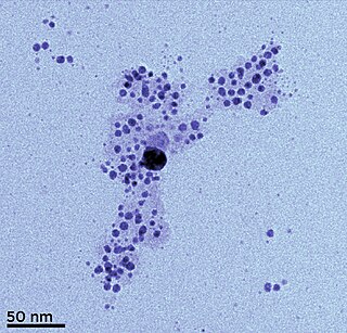
Silver nanoparticles are nanoparticles of silver of between 1 nm and 100 nm in size. While frequently described as being 'silver' some are composed of a large percentage of silver oxide due to their large ratio of surface to bulk silver atoms. Numerous shapes of nanoparticles can be constructed depending on the application at hand. Commonly used silver nanoparticles are spherical, but diamond, octagonal, and thin sheets are also common.

<span class="mw-page-title-main">Marine fungi</span> Species of fungi that live in marine or estuarine environments

Marine fungi are species of fungi that live in marine or estuarine environments. They are not a taxonomic group, but share a common habitat. Obligate marine fungi grow exclusively in the marine habitat while wholly or sporadically submerged in sea water. Facultative marine fungi normally occupy terrestrial or freshwater habitats, but are capable of living or even sporulating in a marine habitat. About 444 species of marine fungi have been described, including seven genera and ten species of basidiomycetes, and 177 genera and 360 species of ascomycetes. The remainder of the marine fungi are chytrids and mitosporic or asexual fungi. Many species of marine fungi are known only from spores and it is likely a large number of species have yet to be discovered. In fact, it is thought that less than 1% of all marine fungal species have been described, due to difficulty in targeting marine fungal DNA and difficulties that arise in attempting to grow cultures of marine fungi. It is impracticable to culture many of these fungi, but their nature can be investigated by examining seawater samples and undertaking rDNA analysis of the fungal material found.

The mycorrhizosphere is the region around a mycorrhizal fungus in which nutrients released from the fungus increase the microbial population and its activities. The roots of most terrestrial plants, including most crop plants and almost all woody plants, are colonized by mycorrhiza-forming symbiotic fungi. In this relationship, the plant roots are infected by a fungus, but the rest of the fungal mycelium continues to grow through the soil, digesting and absorbing nutrients and water and sharing these with its plant host. The fungus in turn benefits by receiving photosynthetic sugars from its host. The mycorrhizosphere consists of roots, hyphae of the directly connected mycorrhizal fungi, associated microorganisms, and the soil in their direct influence.

Myceliophthora thermophila is an ascomycete fungus that grows optimally at 45–50 °C (113–122 °F). It efficiently degrades cellulose and is of interest in the production of biofuels. The genome has recently been sequenced, revealing the full range of enzymes this organism uses for the degradation of plant cell wall material.

<i>Chrysosporium keratinophilum</i> Species of fungus

Chrysosporium keratinophilum is a mold that is closely related to the dermatophytic fungi and is mainly found in soil and the coats of wild animals to break down keratin. Chrysosporium keratinophilum is one of the more commonly occurring species of the genus Chrysosporium in nature. It is easily detected due to its characteristic "light-bulb" shape and flat base. Chrysosporium keratinophilum is most commonly found in keratin-rich, dead materials such as feathers, skin scales, hair, and hooves. Although not identified as pathogenic, it is a regular contaminant of cutaneous specimens which leads to the common misinterpretation that this fungus is pathogenic.

<i>Aspergillus clavatus</i> Species of fungus

Aspergillus clavatus is a species of fungus in the genus Aspergillus with conidia dimensions 3–4.5 x 2.5–4.5 μm. It is found in soil and animal manure. The fungus was first described scientifically in 1834 by the French mycologist John Baptiste Henri Joseph Desmazières.

Plant–fungus horizontal gene transfer is the movement of genetic material between individuals in the plant and fungus kingdoms. Horizontal gene transfer is universal in fungi, viruses, bacteria, and other eukaryotes. Horizontal gene transfer research often focuses on prokaryotes because of the abundant sequence data from diverse lineages, and because it is assumed not to play a significant role in eukaryotes.

Cladosporium oxysporum is an airborne fungus that is commonly found outdoors and is distributed throughout the tropical and subtropical region, it is mostly located In Asia and Africa. It spreads through airborne spores and is often extremely abundant in outdoor air during the spring and summer seasons. It mainly feeds on decomposing organic matter in warmer climates, but can also be parasitic and feed on living plants. The airborne spores can occasionally cause cutaneous infections in humans, and the high prevalence of C. oxysporum in outdoor air during warm seasons contributes to its importance as an etiological agent of allergic disease and possibly human cutaneous phaeohyphomycosis in tropical regions.

Aspergillus giganteus is a species of fungus in the genus Aspergillus that grows as a mold. It was first described in 1901 by Wehmer, and is one of six Aspergillus species from the Clavati section of the subgenus Fumigati. Its closest taxonomic relatives are Aspergillus rhizopodus and Aspergillus longivescia.

Aspergillus carneus is a fast-growing, filamentous fungus found on detritus and in fertile soil worldwide. It is characterized by its yellow, thick-walled hyphae and biseriate sterigmata. The fungus produces citrinin and 5 unique depsipeptides, Aspergillicins A-E.

Priyabrata Mukherjee is an American, academic researcher and professor. He is a Presbyterian Health Foundation presidential professor at the University of Oklahoma Health Sciences Center and associate director for translational research at Stephenson Cancer Center at the OU Health Sciences Center. He also holds the Peggy and Charles Stephenson endowed chair in cancer laboratory research at the OU Health Sciences Center.

Hemibiotrophs are the spectrum of plant pathogens, including bacteria, oomycete and a group of plant pathogenic fungi that keep its host alive while establishing itself within the host tissue, taking up the nutrients with brief biotrophic-like phase. It then, in later stages of infection switches to a necrotrophic life-style, where it rampantly kills the host cells, deriving its nutrients from the dead tissues.

References

  1. Barredo JL, ed. (2005). "Microbial cells and enzymes". Microbial enzymes and biotransformations. Humana Press. pp. 1–10. ISBN   978-1-58829-253-7.
  2. 1 2 3 Ghorbani, HR; Safekordi AA; Attar H; Rezayat Sorkhabadi SM (2011). "Biological and non-biological methods for silver nanoparticles synthesis". Chemical and Biochemical Engineering Quarterly. 25: 317–326.
  3. 1 2 Abou El-Nour, MM; Eftaiha A; Al-Warthan A; Ammar RAA (2010). "Synthesis and application of silver nanoparticles". Arabian Journal of Chemistry. 3 (3): 135–140. doi: 10.1016/j.arabjc.2010.04.008 .
  4. Popescu, M; Velea A; Lőrinczi A (2010). "Biogenic production of nanoparticles". Digest J of Nanomaterials and Biostructures. 5: 1035–1040.
  5. Sastry, M; Ahmad A; Khan MI; Kumar R (2003). "Biosynthesis of metal nanoparticles using fungi and actinomycete". Current Science. 85: 162–170.
  6. 1 2 3 Vahabi, K; Mansoori GA; Karimi S (2011). "Biosynthesis of silver nanoparticles by fungus Trichoderma reesei: a route for large-scale production of AgNPs". Insciences Journal. 1: 65–79. doi: 10.5640/insc.010165 .
  7. 1 2 Basavaraja, S; Balaji SD; Lagashetty A; Rajasab AH; Venkataraman A (2008). "Extracellular biosynthesis of silver nanoparticles using the fungus Fusarium semitectum". Materials Research Bulletin. 45 (5): 1164–1170. doi:10.1016/j.materresbull.2007.06.020.
  8. 1 2 Durán, N; Marcato PD; Alves OL; IH de Souza G; Esposito E (2005). "Mechanistic aspects of biosynthesis of silver nanoparticles by several Fusarium oxysporum strains". Journal of Nanobiotechnology. 3: 8. doi: 10.1186/1477-3155-3-8 . PMC   1180851 . PMID   16014167.
  9. 1 2 Naveen, H; Kumar G; Karthik L; Roa B (2010). "Extracellular biosynthesis of silver nanoparticles using the filamentous fungus Penicillium sp". Archives of Applied Science Research. 2: 161–167.
  10. Bhainsa, KC; D’Sousa SF (2006). "Extracellular biosynthesis of silver nanoparticles using the fungus Aspergillus fumigatas". Colloids and Surfaces B: Biointerfaces. 47 (2): 160–164. doi:10.1016/j.colsurfb.2005.11.026. PMID   16420977.
  11. 1 2 Jain, N; Jain, N., Bhargava A, Majumdar S, Tarafdar J, Panwar J; Majumdar, Sonali; Tarafdar, J. C.; Panwar, Jitendra (2011). "Extracellular biosynthesis and characterization of silver nanoparticles using Aspergillus flavus NJP08: a mechanism perspective". Nanoscale. 3 (2): 635–641. Bibcode:2011Nanos...3..635J. doi:10.1039/c0nr00656d. PMID   21088776.{{cite journal}}: CS1 maint: multiple names: authors list (link)
  12. Mukherjee, P; Ahmad A, Mandal D, Senapati S, Sainkar S, Khan M, Parishcha R, Ajaykumar P, Alam M, Kumar R, Sastry M; Mandal, Deendayal; Senapati, Satyajyoti; Sainkar, Sudhakar R.; Khan, Mohammad I.; Parishcha, Renu; Ajaykumar, P. V.; Alam, Mansoor; Kumar, Rajiv; Sastry, Murali (2001). "Fungus-mediated synthesis of silver nanoparticles and their immobilization in the mycelial matrix; a novel biological approach to nanoparticle synthesis". Nano Letters. 1 (10): 515–519. Bibcode:2001NanoL...1..515M. doi:10.1021/nl0155274.{{cite journal}}: CS1 maint: multiple names: authors list (link)
  13. 1 2 Mukherjee, P; Senapati S; Mandal D; Ahmad A; Khan M; Kumar R; Sastry M (2002). "Extracellular synthesis of gold nanoparticles by the fungus Fusarium oxysporum". ChemBioChem. 3 (5): 461–463. doi:10.1002/1439-7633(20020503)3:5<461::AID-CBIC461>3.0.CO;2-X. PMID   12007181. S2CID   34520744.
  14. Castro-Longoria, E; Vilchis-Nestor A,and Avales-Borja M; Avalos-Borja, M. (2011). "Biosynthesis of silver, gold, and bimetallic nanoparticles using the filamentous fungus Neurospora crassa". Colloids and Surface B:Biointerfaces. 83 (1): 42–48. doi:10.1016/j.colsurfb.2010.10.035. PMID   21087843.
  15. Agnihotri, M; Joshi S; Kumar A; Zinjarde S; Kulkarni S (2009). "Biosynthesis of gold nanoparticles by the tropical marine yeast Yarrowia lipolytica NCIM 3589". Materials Letters. 63 (15): 1231–1234. doi:10.1016/j.matlet.2009.02.042.
  16. Chauhan, A; Zubair S; Tufail S; Sherwani A; Sajid M; Raman S; Azam A; Owais M (2011). "Fungus-mediated biological synthesis of gold nanoparticles: potential in detection of liver cancer". International Journal of Nanomedicine. 6: 2305–2319. doi: 10.2147/ijn.s23195 . PMC   3205127 . PMID   22072868.
  17. 1 2 3 4 Gericke, M; Pinches A (2006). "Biological synthesis of metal nanoparticles". Hydrometallurgy. 83 (1–4): 132–140. Bibcode:2006HydMe..83..132G. doi:10.1016/j.hydromet.2006.03.019.
  18. Li, X; Xu H; Chen Z; Chen G (2011). "Biosynthesis of nanoparticles by microorganisms and their applications". Journal of Nanomaterials. 2011: 1–16. doi: 10.1155/2011/270974 .
  19. Espinosa-Ortiz, EJ; Gonzalez-Gil G; Saikaly PE; van Hullebusch ED; Lens PNL (2014). "Effects of selenium oxyanions on the white-rot fungus Phanerochaete chrysosporium". Appl Microbiol Biotechnol. 99 (5): 2405–2418. doi:10.1007/s00253-014-6127-3. PMID   25341399. S2CID   253768254.
  20. 1 2 3 4 5 Ahmad, A; Mukherjee P; Mandal D; Senapati S; Khan M; Kumar R; Sastry M (2002). "Enzyme mediated extracellular synthesis of CdS nanoparticles by the fungus, Fusarium oxysporum". Journal of the American Chemical Society. 124 (41): 12108–12109. doi:10.1021/ja027296o. PMID   12371846.
  21. Shankar, S; Ahmad A; Sastry M (2003). "Geranium leaf assisted biosynthesis of silver nanoparticles". Biotechnol. Prog. 19 (6): 1627–1631. doi:10.1021/bp034070w. PMID   14656132. S2CID   10120705.