T-even bacteriophages

Last updated

A typical T-even bacteriophage PhageExterior.svg
A typical T-even bacteriophage

T-even phages, also known as the E. coli phages, are a group of double-stranded DNA bacteriophages from the family Myoviridae. Bacteriophage means to "eat bacteria", and phages are well known for being obligate intracellular parasites that reproduce within the host cell and are released when the host is destroyed by lysis. Containing about 160 genes, these virulent viruses are among the largest, most complex viruses that are known and one of the best studied model organisms. They have played a key role in the development of virology and molecular biology. [1] [2]

Contents

Discovery

Bacteriophages were first discovered by the English scientist Frederick Twort in 1915 and Félix d'Hérelle in 1917. In the late 1930s, T.L. Rakieten proposed either a mixture of raw sewerage or a lysate from E.coli infected with raw sewerage to the two researchers Milislav Demerec and Ugo Fano. These two researchers isolated T3, T4, T5, and T6 from E.coli. Also, in 1932, the researcher J.Bronfenbrenner had studied and worked on the T2 phage, at which the T2 phage was isolated from the virus. [3] This isolation was made from a fecal material rather than from sewerage. At any rate, Delbruck was involved in the discovery of the T even phages. His part was naming the bacteriophages into Type 1(T1), Type 2 (T2), Type 3 (T3), etc.

Structure

Phages have multiple factors contributing to their structure. It consists of the head, collar, helical sheath, the core or tube, hexagonal base plate, tail fibers (not all) and finally tail pins. The head’s job is to enfold or surround nucleic acids. The tail fibers help in attaching the phage to a bacterial cell. The tail acts as a duct through which the nucleic acid goes through during an infection. The collar helps support the head (to stay in place). Bacteriophages in general (including T-even bacteriophages) contain a head structure, which can vary in size and shape. The head enfolds nucleic acid and acts as the protective covering. [4] Certain phages have tails attached to the phage head. The tail is a hollow duct through which the nucleic acid passes during infection. T-even Bacteriophages have genomes that code for phage-specific DNA replication, recombination, and DNA repair functions. Also, they offer well branded genes and proteins. Similar to all viruses, they depend on many of their hosts important makeups and roles or functions (transcription and translation, membranes, breakdown of energy etc.), for their reproduction.

Unique features of the T-even phages

Dating back to the 1940s till date, T-even phages are considered the best studied model organisms. Model organisms are usually required to be simple with as few as five genes. Yet, T-even phages are in fact among the largest and highest complexity virus, in which these phages genetic information is made up of around 160 genes. Coincident with their complexity, T-even viruses were found to have an unimaginable feature of no other, the presence of the unusual base hydroxymethylcytosine (HMC) in place of the nucleic acid base cytosine. In addition to this, the HMC residues on the T-even phage are glucosylated in a specific pattern. This unique feature allowed the formation of new enzymes that never existed in T-even infected cells or any other cell and modifying T-even phage DNA provided a basic underlying advancement in viral and molecular levels. Other unique features of the T-even virus is its regulated gene expression. [5] These unique features and other features gave significance of the T-even phages, this includes transduction which is responsible for transfer of drug resistant features, lysogenic conversion is responsible for acquisition of new characteristics such as the formation of new enzymes, random insertion into bacterial chromosome can induce insertional mutation, epidemiological typing of bacteria (phage typing), phages are used extensively in genetic engineering where they serve as cloning vectors. Libraries of genes and monoclonal antibodies are maintained in phages. In addition to all this they are responsible for natural removal of bacteria from water bodies. [6]

Life cycle

Structural overview of T2 phage Tevenphage.svg
Structural overview of T2 phage

Adsorption and penetration

Just like all other viruses, T-even phages don't just randomly attach to the surface of their host; instead they "search" and bind to receptors, specific protein structures, found on the surface of the host. These receptors vary with the phage; teichoic acid, cell wall proteins and lipopolysaccharides, flagella, and pili all can serve as receptors for the phage to bind to. In order for the T-even phage to infect its host and begin its life cycle it must enter the first process of infection, adsorption of the phage to the bacterial cell. Adsorption is a value characteristic of phage-host pair and the adsorption of the phage on host cell surface is illustrated as a 2-stage process: reversible and irreversible. It involves the phages tail structure that begins when the phages tail fibers helps bind the phage to the appropriate receptor of its host. This process is reversible. One or more of the components of the base plate mediates irreversible process of binding of the phage to a bacterium.

Diagram of the DNA injection process Phage injection.png
Diagram of the DNA injection process

Penetration is also a value characteristic of phage-host infection that involves the injection of the phages genetic material inside the bacterium. Penetration of nucleic acid takes place after the irreversible adsorption phase. Mechanisms involving penetration of the phages nucleic acid are specific for each phage. This penetration mechanism can involve electrochemical membrane potential, ATP molecules, enzymatic splitting of peptidoglycan layer, or all three of these factor can be vital for the penetration of the nucleic acid inside the bacterial cell. Studies have been done on the T2 bacteriophage (T4-like phage) mechanism of penetration and it has shown that the phages tail does not penetrate inside the bacterial cell wall and penetration of this phage involves electrochemical membrane potential on the inner membrane. [7] [8]

Lytic cycle


Virulent bacteriophages multiply in their bacterial host immediately after entry. After the number of progeny phages reach a certain amount, they cause the host to lyse or break down, therefore they would be released and infect new host cells. [9] The process of host lyses and release is called the lytic cycle. Lytic cycle is a cycle of viral reproduction that involves the destruction of the infected cell and its membrane. This cycle involves a virus that overtakes the host cell and its machinery to reproduce. Therefore, the virus must go through 5 stages in order to reproduce and infect the host cell. Such stages are Attachment, Penetration, Biosynthesis, Maturation, and then Release.

Attachment

Attachment occurs when the bacteria and the phage particles come in contact. This actually happens when the attachment site found on the viral surface attaches to the complementary receptor site on the bacteria. Attachment is supported by weak chemical bonds between the attachment and the receptor site. Also, the fibers at the end of the T even bacteriophage tail plays a role in the attachment site. Therefore, there must be specific receptor attachment as the receptor determines the host preference.

Penetration

This stage involves for the virus to penetrate itself in the host cell. It does so by injecting its DNA into the cell. The process of penetration happens when the bacteriophage's tail secretes the enzyme lysozyme, which breaks down the bacterial cell wall into segments. Therefore, the break down of the cell by the enzyme lysozyme forms a hole on the host cell;so, the bacteriophage inserts the DNA (genetic material) within the cell as the capsid is kept outside the cell.

Biosynthesis

After the phage DNA is inserted into the cell, the process of biosynthesis is initiated. Biosynthesis involves the utilization of the host cells' nucleotides and enzymes to make copies of the phage DNA. The mRNA in the cytoplasm are transcribed from the phage DNA being involved in the biosynthesis of phage enzymes and capsids proteins. Therefore, the ribosomes, enzymes, and amino acids of the host are involved in translation.

Maturation

Maturation is submitted by the assembly of bacteriophage DNA and capsids into virions. Late proteins are important in assembly. Assembly is complicated but varies in bacteriophage stages, some are assembled in nucleus and some are assembled in cytoplasm, and may be seen as paracrystalline structures in cell. The tails and heads of the T even bacteriophage are assembled from protein subunits, and the head is packed with DNA and attached to the tail.

Release

The final step in viral reproduction and multiplication is determined by the release of virions from the host cell. The release of the virions occurs after the breakage of the bacterial plasma membrane. Nonenveloped viruses lyse the host cell which is characterized by viral proteins attacking the peptidoglycan or membrane. The lysis of the bacteria occurs when the capsids inside the cell release the enzyme lysozyme which break down the cell wall. The released bacteriophages infect other cells, and the viral multiplication cycle is repeated within those cells.

Lysogenic cycle

Phage2 Phage2.JPG
Phage2

Lysogenic phages multiply in one of two ways; either by entering an inactive or latent state or by multiplying through the lytic phase. Through a process known as lysogeny, the phage DNA replicates with the replication of the host chromosome by assimilating into the host chromosome itself. It is then passed on to its daughter cells; this is why it usually isn’t recognized by the host. A process known as lysogenic or phage conversion changes the properties of the bacteria cell; this is possible because the prophage itself contains genes that can present new properties into the bacteria cell or host cell. These bacteria are considered lysogenized. Lysogenized bacteria are resistant to superinfection by same or related phages. This is known as superinfection immunity. [10]

T even phage genome

T even bacteriophages are genetically related. The genetic diversity of bacteriophage is extraordinary and phenomenal. The genetic elements of a bacteriophage is transferred from one bacterium to another through the process of transduction. The process of transduction can be specialized or generalized. The generalized transduction is found within the lytic cycle where units of the bacterial DNA are packaged in a phage capsid. [11] Therefore, when these type of phages infect other bacterial cells, the bacterial DNA is inserted inside. Previously mentioned, bacterial genes can be transferred in generalized transduction. Whereas, in specialized transduction, only the genes that are adjacent to the prophage are transferred. Bacteriophage genomes can be highly mosaic at which the phage is made up of individual modules which may be found in other phage species in different arrangements. Based on correlative sequence similarity regulatory patterns, genomic organization, and virion structure of their essential genes, other phages from various parts of the world belong to this family.

See also

Related Research Articles

Bacteriophage virus that infects and replicates within bacteria

A bacteriophage, also known informally as a phage, is a virus that infects and replicates within bacteria and archaea. The term was derived from "bacteria" and the Greek φαγεῖν, meaning "to devour". Bacteriophages are composed of proteins that encapsulate a DNA or RNA genome, and may have structures that are either simple or elaborate. Their genomes may encode as few as four genes and as many as hundreds of genes. Phages replicate within the bacterium following the injection of their genome into its cytoplasm.

Hershey–Chase experiment Experiment

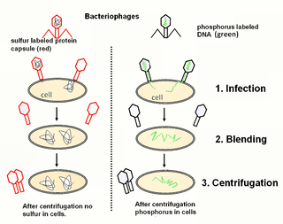
The Hershey–Chase experiments were a series of experiments conducted in 1952 by Alfred Hershey and Martha Chase that helped to confirm that DNA is genetic material. While DNA had been known to biologists since 1869, many scientists still assumed at the time that proteins carried the information for inheritance because DNA appeared to be an inert molecule, and, since it is located in the nucleus, its role was considered to be phosphorus storage. In their experiments, Hershey and Chase showed that when bacteriophages, which are composed of DNA and protein, infect bacteria, their DNA enters the host bacterial cell, but most of their protein does not. Hershey and Chase and subsequent discoveries all served to prove that DNA is the hereditary material.

Virology study of viruses

Virology is the study of viral – submicroscopic, parasitic particles of genetic material contained in a protein coat – and virus-like agents. It focuses on the following aspects of viruses: their structure, classification and evolution, their ways to infect and exploit host cells for reproduction, their interaction with host organism physiology and immunity, the diseases they cause, the techniques to isolate and culture them, and their use in research and therapy. Virology is considered to be a subfield of microbiology or of medicine.

Virulence is a pathogen's or microbe's ability to infect or damage a host.

Transduction (genetics) The transfer of genetic information to a bacterium from a bacteriophage or between bacterial or yeast cells mediated by a phage vector.

Transduction is the process by which foreign DNA is introduced into an eukaryotic cell by a virus or viral vector. An example is the viral transfer of DNA from one bacterium to another and hence an example of horizontal gene transfer. Transduction does not require physical contact between the cell donating the DNA and the cell receiving the DNA, and it is DNase resistant. Transduction is a common tool used by molecular biologists to stably introduce a foreign gene into a host cell's genome.

Lytic cycle Process of viral reproduction

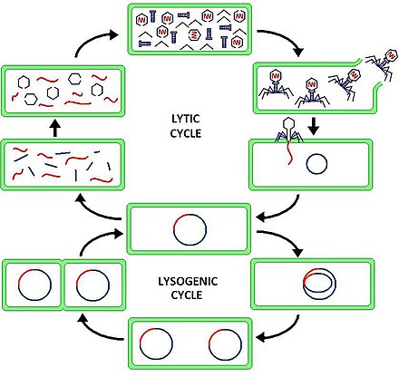
The lytic cycle is one of the two cycles of viral reproduction, the other being the lysogenic cycle. The lytic cycle results in the destruction of the infected cell and its membrane. Bacteriophages that only use the lytic cycle are called virulent phages.

Escherichia virus T4 is a species of bacteriophages that infect Escherichia coli bacteria. It is a member of virus subfamily Tevenvirinae and includes among other strains Enterobacteria phage T2, Enterobacteria phage T4 and Enterobacteria phage T6. T4 is capable of undergoing only a lytic lifecycle and not the lysogenic lifecycle.

Microviridae is a family of bacteriophages with a single-stranded DNA genome. The name of this family is derived from the ancient Greek word μικρός (mikrós), meaning "small". This refers to the size of their genomes, which are among the smallest of the DNA viruses. Enterobacteria, intracellular parasitic bacteria, and spiroplasma serve as natural hosts. There are currently 12 species in this family, divided among 7 genera and one subfamily.

Molecular virology

Molecular virology is the study of viruses on a molecular level. Viruses are submicroscopic parasites that replicate inside host cells. They are able to successfully infect and parasitize all kinds of life forms- from microorganisms to plants and animals- and as a result viruses have more biological diversity than the rest of the bacterial, plant, and animal kingdoms combined. Studying this diversity is the key to a better understanding of how viruses interact with their hosts, replicate inside them, and cause diseases.

<i>Caudovirales</i> order of viruses

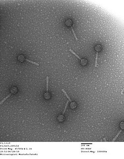
The Caudovirales are an order of viruses also known as the tailed bacteriophages. Under the Baltimore classification scheme, the Caudovirales are group I viruses as they have double stranded DNA (dsDNA) genomes, which can be anywhere from 18,000 base pairs to 500,000 base pairs in length. The virus particles have a distinct shape; each virion has an icosahedral head that contains the viral genome, and is attached to a flexible tail by a connector protein. The order encompasses a wide range of viruses, many containing genes of similar nucleotide sequence and function. However, some tailed bacteriophage genomes can vary quite significantly in nucleotide sequence, even among the same genus. Due to their characteristic structure and possession of potentially homologous genes, it is believed these bacteriophages possess a common origin.

Lysogenic cycle

Lysogeny, or the lysogenic cycle, is one of two cycles of viral reproduction. Lysogeny is characterized by integration of the bacteriophage nucleic acid into the host bacterium's genome or formation of a circular replicon in the bacterial cytoplasm. In this condition the bacterium continues to live and reproduce normally. The genetic material of the bacteriophage, called a prophage, can be transmitted to daughter cells at each subsequent cell division, and at later events can release it, causing proliferation of new phages via the lytic cycle. Lysogenic cycles can also occur in eukaryotes, although the method of DNA incorporation is not fully understood.

Bacteriophage T7 is a bacteriophage, a virus composed of DNA. It infects most strains of an Escherichia coli and relies on these hosts to propagate. Bacteriophage T7 has a lytic life cycle, meaning that it destroys the cell it infects. It also possesses several properties that make it an ideal phage for experimentation: its purification and concentration have produced consistent values in chemical analyses; it can be rendered noninfectious by exposure to UV light; and it can be used in phage display to clone RNA binding proteins.

Viral entry biological process

Viral entry is the earliest stage of infection in the viral life cycle, as the virus comes into contact with the host cell and introduces viral material into the cell. The major steps involved in viral entry are shown below. Despite the variation among viruses, there are several shared generalities concerning viral entry.

Enterobacteria phage P22 is a bacteriophage in the Podoviridae family that infects Salmonella typhimurium. Like many phages, it has been used in molecular biology to induce mutations in cultured bacteria and to introduce foreign genetic material. P22 has been used in generalized transduction and is an important tool for investigating Salmonella genetics.

Tectivirus family of viruses

Tectiviridae is a family of viruses with three genera. Gram-negative bacteria serve as natural hosts. There are currently four species in this genus including the type species Enterobacteria phage PRD1. Tectiviruses have no head-tail structure, but are capable of producing tail-like tubes of ~ 60×10 nm upon adsorption or after chloroform treatment. The name is derived from Latin tectus.

<i>Corticovirus</i> family of viruses

Corticovirus is a genus of viruses in the family Corticoviridae. Corticoviruses are bacteriophages; that is, their natural hosts are bacteria. The genus contains only one species, the type species Pseudoalteromonas virus PM2. The name is derived from Latin cortex, corticis. However, prophages closely related to PM2 are abundant in the genomes of aquatic bacteria, suggesting that the ecological importance of corticoviruses might be underestimated. Bacteriophage PM2 was first described in 1968 after isolation from seawater sampled from the coast of Chile.

Virus Small non-cellular infectious agent that only replicates in cells

A virus is a submicroscopic infectious agent that replicates only inside the living cells of an organism. Viruses can infect all types of life forms, from animals and plants to microorganisms, including bacteria and archaea. Since Dmitri Ivanovsky's 1892 article describing a non-bacterial pathogen infecting tobacco plants, and the discovery of the tobacco mosaic virus by Martinus Beijerinck in 1898, about 5,000 virus species have been described in detail, of the millions of types of viruses in the environment. Viruses are found in almost every ecosystem on Earth and are the most numerous type of biological entity. The study of viruses is known as virology, a subspeciality of microbiology.

Bacteriophage P2 species of virus

Bacteriophage P2, scientific name Escherichia virus P2, is a temperate phage that infects E. coli. It is a tailed virus with a contractile sheath and is thus classified in the genus P2virus, subfamily Peduovirinae, family Myoviridae within order Caudovirales. This genus of viruses includes many P2-like phages as well as the satellite phage P4.

<i>Spiroplasma phage 1-R8A2B</i> species of virus

Spiroplasma phage 1-R8A2B is a filamentous bacteriophage in the genus Plectrovirus of the family Inoviridae, part of the group of single-stranded DNA viruses. The virus has many synonyms, such as SpV1-R8A2 B, Spiroplasma phage 1, and Spiroplasma virus 1, SpV1. SpV1-R8A2 B infects Spiroplasma citri. Its host itself is a prokaryotic pathogen for citrus plants, causing Citrus stubborn disease.

Escherichia virus CC31, formerly known as Enterobacter virus CC31, is a dsDNA bacteriophage of the subfamily Tevenvirinae responsible for infecting the bacteria family of Enterobacteriaceae. It is one of two discovered viruses of the genus Karamvirus, diverging away from the previously discovered T4virus, as a clonal complex (CC). CC31 was first isolated from Escherichia coli B strain S/6/4 and is primarily associated with Escherichia, even though is named after Enterobacter.

References

  1. Norkin, Leonard C. (2010). Virology, Molecular Biology and Pathogenesis. Washington: American Society for Microbiology. p. 725. ISBN   978-1-55581-453-3.
  2. Prescott, Harley, and Klein (2008). Microbiology (seventh ed.). McGraw Hill. p. 1078. ISBN   978-007-126727-4.CS1 maint: multiple names: authors list (link)
  3. Willey, Joanne. Prescott's Microbiology (seventh ed.). McGraw-Hill.
  4. Prescott, Harley, and Klein (2008). Microbiology (seventh ed.). McGraw-Hill. ISBN   978-007-126727-4.
  5. Norkin, Leonard C. (2010). Virology, Molecular Biology and Pathogenesis. Washington: American Society for Microbiology. p. 725. ISBN   978-1-55581-453-3.
  6. Rao, Srdhar. "Bacteriophages" (PDF). microrao.
  7. Norkin, Leonard C. (2010). Virology, Molecular Biology and Pathogenesis. Washington: American Society for Microbiology. p. 31. ISBN   978-1-55581-453-3.
  8. Prescott, Harley, and Klein (2008). Microbiology (seventh ed.). McGraw Hill. p. 427. ISBN   978-007-126727-4.CS1 maint: multiple names: authors list (link)
  9. Sherwood, Linda (2011). Prescott's Microbiology (eighth ed.). McGraw-Hill.
  10. Willey, Joanne (2011). Prescott's Microbiology (eighth ed.). McGraw-Hill.
  11. Leonard C., Norkin (2010). Virology, Molecular Biology and Pathogenesis. Washington: American Society for Microbiology.