Transepidermal water loss

Last updated
Principle of an instrument measuring transepidermal water loss. Water vapor is diffusing through the transparently shown cylinder. The yellow arrow symbolizes the diffusion direction. The two dark red square elements are two pairs of sensors each measuring relative humidity and temperature. Tewameter Principle.png
Principle of an instrument measuring transepidermal water loss. Water vapor is diffusing through the transparently shown cylinder. The yellow arrow symbolizes the diffusion direction. The two dark red square elements are two pairs of sensors each measuring relative humidity and temperature.

Transepidermal water loss (TEWL or TWL) is the loss of water that passes from inside a body (animal or plant) through the epidermis (that is, either the epidermal layer of animal skin or the epidermal layer of plants) to the surrounding atmosphere via diffusion and evaporation processes. TEWL in mammals is also known as insensible water loss (IWL), as it is a process over which organisms have little physiologic control and of which they are usually mostly unaware. Insensible loss of body water can threaten fluid balance; in humans, substantial dehydration sometimes occurs before a person realizes what is happening.

Contents

Measurements of TEWL [1] may be useful for identifying skin damage caused by certain chemicals, physical insult (such as "tape stripping") or pathological conditions such as eczema, as rates of TEWL increase in proportion to the level of damage. However, TEWL is also affected by environmental factors such as humidity, temperature, the time of year (season variation) and the moisture content of the skin (hydration level). Therefore, care must be taken when interpreting the meaning of TEWL rates.

Implications

Human health

From a clinical standpoint, TEWL measurements are of great importance in evaluating skin barrier functionality. [2] Often normal rates of TEWL – from 2.3 g/(m2h) to 44 g/(m2h) [3] – are compromised due to injury, infection and/or severe damage as in the case of burns causing rates over 50 or even over 100 g/m2/h. [4] Damage to the stratum corneum and superficial skin layers not only results in physical vulnerability, but also results in an excess rate of water loss. Therefore, dehydration, metabolic acidosis, and conditions such as anhydremia or concentration of the blood are often critical issues for healthcare providers to consider in the treatment of burn patients. [5] [6]

TEWL is of major concern in public health, considering the relatively high rate of burn incidence among communities in the developing world due to poor quality cooking stoves. [7] Resources for burn care in local clinics are often scarce and depending on the affected surface area, TEWL is a major issue that can be overlooked. Furthermore, TEWL is also affected by variations in sweat gland activity, temperature, and metabolism. [8] Therefore, transepidermal water loss becomes a significant factor in dehydration associated with several major disease states.

Botany and earth science

The amount of water that trees and other plants move from the ground to the atmosphere with TEWL is of interest in botany and earth science, as transpiration by plants is a substantial component of the water cycle on Earth.

Acronym variation

Web corpus searches show that the acronym TEWL is about 40 times more common than TWL in reference to transepidermal water loss. A large advantage of TEWL is that it has higher specificity to that sense than does TWL, which has more alternative senses, including, most importantly, two other senses having to do with evaporation of body water: thermal work limit (TWL), which is the highest sustainable metabolic rate that well-hydrated, acclimatized individuals can maintain in a specific thermal environment, and total water loss (TWL), which in its physiologic sense is a superset of transepidermal water loss, because it also includes respiratory losses and urinary losses. Use of the acronym TEWL for transepidermal water loss avoids this ambiguity. As with many other acronyms in biology and medicine (for example, polymorphonuclear [PMN], bronchioloalveolar carcinoma [BAC], dolutegravir [DTG]), there is no law preventing the letters of the acronym from representing combining forms (trans- + epidermal) or other syllables irrespective of word boundaries.

For certain regions of human epidermis specific acronyms for TEWL are common. Water loss through finger and toe nails is called transonychial water loss, abbreviated TOWL. [9]

Related Research Articles

<span class="mw-page-title-main">Skin</span> Soft outer covering organ of vertebrates

Skin is the layer of usually soft, flexible outer tissue covering the body of a vertebrate animal, with three main functions: protection, regulation, and sensation.

<span class="mw-page-title-main">Integumentary system</span> Skin and other protective organs

The integumentary system is the set of organs forming the outermost layer of an animal's body. It comprises the skin and its appendages, which act as a physical barrier between the external environment and the internal environment that it serves to protect and maintain the body of the animal. Mainly it is the body's outer skin.

<span class="mw-page-title-main">Keratinocyte</span> Primary type of cell found in the epidermis

Keratinocytes are the primary type of cell found in the epidermis, the outermost layer of the skin. In humans, they constitute 90% of epidermal skin cells. Basal cells in the basal layer of the skin are sometimes referred to as basal keratinocytes. Keratinocytes form a barrier against environmental damage by heat, UV radiation, water loss, pathogenic bacteria, fungi, parasites, and viruses. A number of structural proteins, enzymes, lipids, and antimicrobial peptides contribute to maintain the important barrier function of the skin. Keratinocytes differentiate from epidermal stem cells in the lower part of the epidermis and migrate towards the surface, finally becoming corneocytes and eventually be shed off, which happens every 40 to 56 days in humans.

<span class="mw-page-title-main">Epidermis</span> Outermost of the three layers that make up the skin

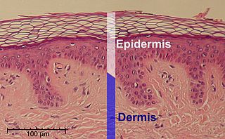
The epidermis is the outermost of the three layers that comprise the skin, the inner layers being the dermis and hypodermis. The epidermis layer provides a barrier to infection from environmental pathogens and regulates the amount of water released from the body into the atmosphere through transepidermal water loss.

<span class="mw-page-title-main">Skin condition</span> Any medical condition that affects the integumentary system

A skin condition, also known as cutaneous condition, is any medical condition that affects the integumentary system—the organ system that encloses the body and includes skin, nails, and related muscle and glands. The major function of this system is as a barrier against the external environment.

<span class="mw-page-title-main">Stratum corneum</span> Outermost layer of the epidermis

The stratum corneum is the outermost layer of the epidermis. The human stratum corneum comprises several levels of flattened corneocytes that are divided into two layers: the stratum disjunctum and stratum compactum. The skin's protective acid mantle and lipid barrier sit on top of the stratum disjunctum. The stratum disjunctum is the uppermost and loosest layer of skin. The stratum compactum is the comparatively deeper, more compacted and more cohesive part of the stratum corneum. The corneocytes of the stratum disjunctum are larger, more rigid and more hydrophobic than that of the stratum compactum.

<span class="mw-page-title-main">Crocodilian armor</span>

Crocodile armor consists of the protective dermal and epidermal components of the integumentary system in animals of the order Crocodilia.

<span class="mw-page-title-main">Moisturizer</span> Type of cosmetics

A moisturizer, or emollient, is a cosmetic preparation used for protecting, moisturizing, and lubricating the skin. These functions are normally performed by sebum produced by healthy skin. The word "emollient" is derived from the Latin verb mollire, to soften.

<span class="mw-page-title-main">Desquamation</span> Medical condition

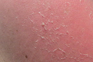
Desquamation occurs when the outermost layer of a tissue, such as the skin, is shed. The term is from Latin desquamare 'to scrape the scales off a fish'.

<span class="mw-page-title-main">Hyperkeratosis</span> Medical condition

Hyperkeratosis is thickening of the stratum corneum, often associated with the presence of an abnormal quantity of keratin, and also usually accompanied by an increase in the granular layer. As the corneum layer normally varies greatly in thickness in different sites, some experience is needed to assess minor degrees of hyperkeratosis.

Diprobase is an emollient, specifically targeted at eczema and dermatitis. It is an occlusive emollient, meaning that it restores the layer of oil on the surface of the skin to slow water loss. This gives its tendency to make the applied area sticky.

<span class="mw-page-title-main">Stratum lucidum</span>

The stratum lucidum is a thin, clear layer of dead skin cells in the epidermis named for its translucent appearance under a microscope. It is readily visible by light microscopy only in areas of thick skin, which are found on the palms of the hands and the soles of the feet.

<span class="mw-page-title-main">Stratified squamous epithelium</span>

A stratified squamous epithelium consists of squamous (flattened) epithelial cells arranged in layers upon a basal membrane. Only one layer is in contact with the basement membrane; the other layers adhere to one another to maintain structural integrity. Although this epithelium is referred to as squamous, many cells within the layers may not be flattened; this is due to the convention of naming epithelia according to the cell type at the surface. In the deeper layers, the cells may be columnar or cuboidal. There are no intercellular spaces. This type of epithelium is well suited to areas in the body subject to constant abrasion, as the thickest layers can be sequentially sloughed off and replaced before the basement membrane is exposed. It forms the outermost layer of the skin and the inner lining of the mouth, esophagus and vagina.

<span class="mw-page-title-main">Human skin</span> Outer covering of the body

The human skin is the outer covering of the body and is the largest organ of the integumentary system. The skin has up to seven layers of ectodermal tissue guarding muscles, bones, ligaments and internal organs. Human skin is similar to most of the other mammals' skin, and it is very similar to pig skin. Though nearly all human skin is covered with hair follicles, it can appear hairless. There are two general types of skin, hairy and glabrous skin (hairless). The adjective cutaneous literally means "of the skin".

<span class="mw-page-title-main">Transdermal</span>

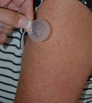
Transdermal is a route of administration wherein active ingredients are delivered across the skin for systemic distribution. Examples include transdermal patches used for medicine delivery. The drug is administered in the form of a patch or ointment that delivers the drug into the circulation for systemic effect.

Corneocytes are terminally differentiated keratinocytes and compose most of the stratum corneum, the outermost layer of the epidermis. They are regularly replaced through desquamation and renewal from lower epidermal layers and are essential for its function as a skin barrier.

Keratohyalin is a protein structure found in cytoplasmic granules of the keratinocytes in the stratum granulosum of the epidermis. Keratohyalin granules (KHG) mainly consist of keratin, profilaggrin, loricrin and trichohyalin proteins which contribute to cornification or keratinization, the process of the formation of epidermal cornified cell envelope. During the keratinocyte differentiation, these granules maturate and expand in size, which leads to the conversion of keratin tonofilaments into a homogenous keratin matrix, an important step in cornification.

<span class="mw-page-title-main">Bullous impetigo</span> Medical condition

Bullous impetigo is a bacterial skin infection caused by Staphylococcus aureus that results in the formation of large blisters called bullae, usually in areas with skin folds like the armpit, groin, between the fingers or toes, beneath the breast, and between the buttocks. It accounts for 30% of cases of impetigo, the other 70% being non-bullous impetigo.

Skin sloughing is the process of shedding dead surface cells from the skin. It is most associated with cosmetic skin maintenance via exfoliation, but can also occur biologically or for medical reasons.

TGM5 is a transglutaminase enzyme.

References

  1. Fluhr, Joachim; Berardesca, Enzo; Elsner, Peter; Maibach, Howard I. (2004). Bioengineering of the skin: water and stratum corneum (2 ed.). CRC Press. ISBN   0-8493-1443-7.
  2. Gioia, Francesco and Leonardo Celleno (2002) The dynamics of TWL from hydrated skin. Skin Research and Technology Vol. 8 pp. 178–186
  3. Jan Kottner, Andrea Lichterfeld, Ulrike Blume-Peytavi (2013) Transepidermal water loss in young and aged healthy humans: a systematic review and meta-analysis, Arch Dermatol Res Vol. 305 pp. 315–323, doi : 10.1007/s00403-012-1313-6
  4. K.L. Gardien, D.C. Baas, H.C. de Vet, E. Middelkoop (2016) Transepidermal water loss measured with the Tewameter TM300 in burn scars. Burns Vol. 42(7) pp. 1455-1462, doi : 10.1016/j.burns.2016.04.018
  5. Underhill, Frank P. (1927) The Significance of Anhydremia in Extensive Superficial Burns Journal of the American Medical Association Vol. 95 No. 12.
  6. Artz, Lieut. Col. Curtis P. and Capt. Harry S. Soroff (MC) (1955) Modern Concepts in the Treatment of Burns Journal of the American Medical Association Vol. 159 No. 5
  7. Ahuja, Rajeev B. and Sameek Bhattacharya (2004) Burns in the developing world and burn disasters British Medical Journal Vol. 329.
  8. Chilcott, Robert et al. (2002) Transepidermal water loss does not correlate with skin barrier function in vitro The Journal of Investigative Dermatology Vol. 118 pp. 871–875
  9. Krönauer C., Gfesser M., Ring J., Abeck D.: Transonychial water loss in healthy and diseased nails. Acta Derm Venereol. (2001) 81(3):175–177. doi : 10.1080/000155501750376249