Related Research Articles

Infrared spectroscopy is the measurement of the interaction of infrared radiation with matter by absorption, emission, or reflection. It is used to study and identify chemical substances or functional groups in solid, liquid, or gaseous forms. It can be used to characterize new materials or identify and verify known and unknown samples. The method or technique of infrared spectroscopy is conducted with an instrument called an infrared spectrometer which produces an infrared spectrum. An IR spectrum can be visualized in a graph of infrared light absorbance on the vertical axis vs. frequency, wavenumber or wavelength on the horizontal axis. Typical units of wavenumber used in IR spectra are reciprocal centimeters, with the symbol cm−1. Units of IR wavelength are commonly given in micrometers, symbol μm, which are related to the wavenumber in a reciprocal way. A common laboratory instrument that uses this technique is a Fourier transform infrared (FTIR) spectrometer. Two-dimensional IR is also possible as discussed below.

Raman spectroscopy is a spectroscopic technique typically used to determine vibrational modes of molecules, although rotational and other low-frequency modes of systems may also be observed. Raman spectroscopy is commonly used in chemistry to provide a structural fingerprint by which molecules can be identified.

Resonance Raman spectroscopy is a variant of Raman spectroscopy in which the incident photon energy is close in energy to an electronic transition of a compound or material under examination. This similarity in energy (resonance) leads to greatly increased intensity of the Raman scattering of certain vibrational modes, compared to ordinary Raman spectroscopy.

Laser-induced breakdown spectroscopy (LIBS) is a type of atomic emission spectroscopy which uses a highly energetic laser pulse as the excitation source. The laser is focused to form a plasma, which atomizes and excites samples. The formation of the plasma only begins when the focused laser achieves a certain threshold for optical breakdown, which generally depends on the environment and the target material.
Coherent anti-Stokes Raman spectroscopy, also called Coherent anti-Stokes Raman scattering spectroscopy (CARS), is a form of spectroscopy used primarily in chemistry, physics and related fields. It is sensitive to the same vibrational signatures of molecules as seen in Raman spectroscopy, typically the nuclear vibrations of chemical bonds. Unlike Raman spectroscopy, CARS employs multiple photons to address the molecular vibrations, and produces a coherent signal. As a result, CARS is orders of magnitude stronger than spontaneous Raman emission. CARS is a third-order nonlinear optical process involving three laser beams: a pump beam of frequency ωp, a Stokes beam of frequency ωS and a probe beam at frequency ωpr. These beams interact with the sample and generate a coherent optical signal at the anti-Stokes frequency (ωpr+ωp-ωS). The latter is resonantly enhanced when the frequency difference between the pump and the Stokes beams (ωp-ωS) coincides with the frequency of a Raman resonance, which is the basis of the technique's intrinsic vibrational contrast mechanism.

Surface-enhanced Raman spectroscopy or surface-enhanced Raman scattering (SERS) is a surface-sensitive technique that enhances Raman scattering by molecules adsorbed on rough metal surfaces or by nanostructures such as plasmonic-magnetic silica nanotubes. The enhancement factor can be as much as 1010 to 1011, which means the technique may detect single molecules.

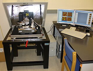
Near-field scanning optical microscopy (NSOM) or scanning near-field optical microscopy (SNOM) is a microscopy technique for nanostructure investigation that breaks the far field resolution limit by exploiting the properties of evanescent waves. In SNOM, the excitation laser light is focused through an aperture with a diameter smaller than the excitation wavelength, resulting in an evanescent field on the far side of the aperture. When the sample is scanned at a small distance below the aperture, the optical resolution of transmitted or reflected light is limited only by the diameter of the aperture. In particular, lateral resolution of 6 nm and vertical resolution of 2–5 nm have been demonstrated.
Chemical imaging is the analytical capability to create a visual image of components distribution from simultaneous measurement of spectra and spatial, time information. Hyperspectral imaging measures contiguous spectral bands, as opposed to multispectral imaging which measures spaced spectral bands.
Multivariate optical computing, also known as molecular factor computing, is an approach to the development of compressed sensing spectroscopic instruments, particularly for industrial applications such as process analytical support. "Conventional" spectroscopic methods often employ multivariate and chemometric methods, such as multivariate calibration, pattern recognition, and classification, to extract analytical information from data collected at many different wavelengths. Multivariate optical computing uses an optical computer to analyze the data as it is collected. The goal of this approach is to produce instruments which are simple and rugged, yet retain the benefits of multivariate techniques for the accuracy and precision of the result.
Photothermal microspectroscopy (PTMS), alternatively known as photothermal temperature fluctuation (PTTF), is derived from two parent instrumental techniques: infrared spectroscopy and atomic force microscopy (AFM). In one particular type of AFM, known as scanning thermal microscopy (SThM), the imaging probe is a sub-miniature temperature sensor, which may be a thermocouple or a resistance thermometer. This same type of detector is employed in a PTMS instrument, enabling it to provide AFM/SThM images: However, the chief additional use of PTMS is to yield infrared spectra from sample regions below a micrometer, as outlined below.

Scanning thermal microscopy (SThM) is a type of scanning probe microscopy that maps the local temperature and thermal conductivity of an interface. The probe in a scanning thermal microscope is sensitive to local temperatures – providing a nano-scale thermometer. Thermal measurements at the nanometer scale are of both scientific and industrial interest. The technique was invented by Clayton C. Williams and H. Kumar Wickramasinghe in 1986.

Spatially offset Raman spectroscopy (SORS) is a variant of Raman spectroscopy that allows highly accurate chemical analysis of objects beneath obscuring surfaces, such as tissue, coatings and bottles. Examples of uses include analysis of: bone beneath skin, tablets inside plastic bottles, explosives inside containers and counterfeit tablets inside blister packs. There have also been advancements in the development of deep non-invasive medical diagnosis using SORS with the hopes of being able to detect breast tumors.
Acoustic resonance spectroscopy (ARS) is a method of spectroscopy in the acoustic region, primarily the sonic and ultrasonic regions. ARS is typically much more rapid than HPLC and NIR. It is non destructive and requires no sample preparation as the sampling waveguide can simply be pushed into a sample powder/liquid or in contact with a solid sample. To date, the AR spectrometer has successfully differentiated and quantified sample analytes in various forms;. It has been used to measure and monitor the progression of chemical reactions, such as the setting and hardening of concrete from cement paste to solid. Acoustic spectrometry has also been used to measure the volume fraction of colloids in a dispersion medium, as well as for the investigation of physical properties of colloidal dispersions, such as aggregation and particle size distribution. Typically, these experiments are carried out with sinusoidal excitation signals and the experimental observation of signal attenuation. From a comparison of theoretical attenuation to experimental observation, the particle size distribution and aggregation phenomena are inferred.

The Raman microscope is a laser-based microscopic device used to perform Raman spectroscopy. The term MOLE is used to refer to the Raman-based microprobe. The technique used is named after C. V. Raman, who discovered the scattering properties in liquids.
Gas in scattering media absorption spectroscopy (GASMAS) is an optical technique for sensing and analysis of gas located within porous and highly scattering solids, e.g. powders, ceramics, wood, fruit, translucent packages, pharmaceutical tablets, foams, human paranasal sinuses etc. It was introduced in 2001 by Prof. Sune Svanberg and co-workers at Lund University (Sweden). The technique is related to conventional high-resolution laser spectroscopy for sensing and spectroscopy of gas, but the fact that the gas here is "hidden" inside solid materials give rise to important differences.
In statistics, the bootstrap error-adjusted single-sample technique is a non-parametric method that is intended to allow an assessment to be made of the validity of a single sample. It is based on estimating a probability distribution representing what can be expected from valid samples. This is done use a statistical method called bootstrapping, applied to previous samples that are known to be valid.
The technique of vibrational analysis with scanning probe microscopy allows probing vibrational properties of materials at the submicrometer scale, and even of individual molecules. This is accomplished by integrating scanning probe microscopy (SPM) and vibrational spectroscopy. This combination allows for much higher spatial resolution than can be achieved with conventional Raman/FTIR instrumentation. The technique is also nondestructive, requires non-extensive sample preparation, and provides more contrast such as intensity contrast, polarization contrast and wavelength contrast, as well as providing specific chemical information and topography images simultaneously.
AFM-IR or infrared nanospectroscopy is one of a family of techniques that are derived from a combination of two parent instrumental techniques. AFM-IR combines the chemical analysis power of infrared spectroscopy and the high-spatial resolution of scanning probe microscopy (SPM). The term was first used to denote a method that combined a tuneable free electron laser with an atomic force microscope equipped with a sharp probe that measured the local absorption of infrared light by a sample with nanoscale spatial resolution.
Tip-enhanced Raman spectroscopy (TERS) is a variant of surface-enhanced Raman spectroscopy (SERS) that combines scanning probe microscopy with Raman spectroscopy. High spatial resolution chemical imaging is possible via TERS, with routine demonstrations of nanometer spatial resolution under ambient laboratory conditions, or better at ultralow temperatures and high pressure.
Micro-spatially offset Raman spectroscopy (micro-SORS) is an analytical technique developed in 2014 that combines SORS with microscopy. The technique derives its sublayer‐resolving properties from its parent technique SORS. The main difference between SORS and micro-SORS is the spatial resolution: while SORS is suited to the analysis of millimetric layers, micro-SORS is able to resolve thin, micrometric-scale layers. Similarly to SORS technique, micro-SORS is able to preferentially collect the Raman photons generated under the surface in turbid media. In this way, it is possible to reconstruct the chemical makeup of micrometric multi-layered turbid system in a non destructive way. Micro-SORS is particularly useful when dealing with precious or unique objects as for Cultural Heritage field and Forensic Science or in biomedical applications, where a non-destructive molecular characterization constitute a great advantage.
References
- ↑ B. Schrader; G. Bergmann (1967). "Die Intensität des Ramanspektrums polykristalliner Substanzen". Fresenius' Z. Anal. Chem. 225 (2): 225–230. doi:10.1007/bf00983673. S2CID 94487523.
- ↑ P. Matousek; A. W. Parker (2006). "Bulk Raman Analysis of Pharmaceutical Tablets". Applied Spectroscopy. 60 (12): 1353–1357. Bibcode:2006ApSpe..60.1353M. doi:10.1366/000370206779321463. PMID 17217583. S2CID 32218439.
- ↑ H. Wang; C.K. Mann; T.J. Vickers (2002). "Effect of Powder Properties on the Intensity of Raman Scattering by Crystalline Solids". Appl. Spectrosc. 56 (12): 1538–1544. Bibcode:2002ApSpe..56.1538W. doi:10.1366/000370202321115779. S2CID 96236279.
- ↑ J. Johansson; S. Pettersson; S. Folestad (2005). "Characterization of different laser irradiation methods for quantitative Raman tablet assessment". Journal of Pharmaceutical and Biomedical Analysis. 39 (3–4): 516. doi:10.1016/j.jpba.2005.04.029. PMID 15950422.
- ↑ P. Matousek; A. W. Parker (2006). "Bulk Raman Analysis of Pharmaceutical Tablets". Applied Spectroscopy. 60 (12): 1353–7. Bibcode:2006ApSpe..60.1353M. doi:10.1366/000370206779321463. PMID 17217583. S2CID 32218439.
- ↑ P. Matousek; A. W. Parker (2007). "Non-invasive probing of pharmaceutical capsules using transmission Raman spectroscopy". J. Raman Spectrosc. 38. 38 (5): 563–567. Bibcode:2007JRSp...38..563M. doi:10.1002/jrs.1688.
- ↑ J. Johansson; A. Sparen; O. Svensson; S. Folestad; et al. (2007). "Quantitative transmission Raman spectroscopy of pharmaceutical tablets and capsules". Appl. Spectrosc. 61 (11): 1211–8. Bibcode:2007ApSpe..61.1211J. doi:10.1366/000370207782597085. PMID 18028700. S2CID 6622891.
- ↑ Charlotte Eliasson; Neil A. Macleod; Linda C. Jayes; Fiona C. Clarke; et al. (2008). "Non-invasive quantitative assessment of the content of pharmaceutical capsules using transmission Raman spectroscopy". Journal of Pharmaceutical and Biomedical Analysis. 47 (2): 221–229. doi:10.1016/j.jpba.2008.01.013. PMID 18296001.
- ↑ Vigh, T.; et al. (2013). "Polymer-free and polyvinylpirrolidone-based electrospun solid dosage forms for drug dissolution enhancement". European Journal of Pharmaceutical Sciences. 49 (4): 595–602. doi:10.1016/j.ejps.2013.04.034. PMID 23684933.
- ↑ P. Matousek; N. Stone (2007). "Prospects for the diagnosis of breast cancer by noninvasive probing of calcifications using transmission Raman spectroscopy". Journal of Biomedical Optics. 12 (2): 024008. Bibcode:2007JBO....12b4008M. doi: 10.1117/1.2718934 . PMID 17477723.
- ↑ Ellis, David I.; Cowcher, David P.; Ashton, Lorna; O'Hagan, Steve; Goodacre, Royston (2013). "Illuminating disease and enlightening biomedicine: Raman spectroscopy as a diagnostic tool". The Analyst. 138 (14): 3871–3884. Bibcode:2013Ana...138.3871E. doi: 10.1039/c3an00698k . ISSN 0003-2654. PMID 23722248.