Triple test score

Last updated
Triple Test Score components: physical exam, imaging, and needle biopsy TTS components diagram.png
Triple Test Score components: physical exam, imaging, and needle biopsy

The triple test score is a diagnostic tool for examining potentially cancerous breasts. Diagnostic accuracy of the triple test score is nearly 100%. Scoring includes using the procedures of physical examination, mammography and needle biopsy. If the results of a triple test score are greater than five, an excisional biopsy is indicated. [1]

Contents

The term triple test scoring (TSS) was first noted in 1975 as a means of rapidly diagnosing and examining breast malignancies. [2] TSS developed as a useful and accurate clinical tool for breast masses because it was cheaper and it cut down on the diagnosis time.

Scoring

To obtain the triple test score, a number from 1 through 3 is assigned to each one of the procedures. A score of 1 is assigned to a benign test result, 2 applies to a suspicious test result, and 3 applies to a malignant result. The sum of the scores of all three procedures is the triple test score.

If the total summed score from the three tests is 3 to 4 then the diagnosis is most likely benign. A total summed score of 5 is considered suspicious. A score of 6 or greater is possibly malignant. There have been different inclusions for the components of the triple test score in the past, such as using the procedures of physical examination, mammography, and cytology. Other versions of the triple test score have included mammography, ultrasound, and magnetic resonance imaging (MRI). [3]

Summed total scoreDiagnosis
3-4Benign
5Suspicious
6 <Malignant

TTS vs. Modified TTS (mTTS)

The TTS was first implemented and then changed to create a modified TTS. The main difference between the two diagnostic tools is the substitution of the mammogram for the ultrasound in persons under the age of 40. [4] [5] This is because ultrasound has been found to be more effecting at early detection of breast cancer and masses for persons with denser breast tissues. These individuals with denser breast tissues have also been found to be at an increased risk of developing breast cancer. [6] Most modified TTS exams contain a combination of physical examination, ultrasound, and needle biopsy. [7] There are no changes to the scoring system of the mTSS.

TTS vs. BI-RADS

Like the triple test score, BI-RADS (breast imaging-reporting and data system) uses a similar method of scoring breast imaging reports to help with evaluating and determining treatment for breast masses. [8] Like the triple test score, BI-RADS employs a numerical scoring system to determine whether a mass is benign or malignant. The triple test score assigns a numerical indicator of 1 to 3 while BI-RADS assigns a numerical indicator of 1 to 6. The BI-RADS scoring for mammograms can be comparable to the triple test score's scoring for mammograms. [2] For instance, a BI-RADS of 1 or 2 is equivalent to a triple test score of 1. Similar to the triple test score, a lower scoring on BI-RADS (i.e. 1 or 2) is indicative of a benign screening while a high scoring (i.e. 5 or 6) is indicative of malignancy. [2]

Unlike the triple test score which scores three different exams, BI-RADS focuses on evaluating findings from one exam: mammograms.

Cost

The triple test score reduces cost for evaluating breast masses compared to traditional methods due to reducing the likelihood of people undergoing an excisional biopsy while still providing effective diagnoses. [2] An excisional biopsy can be performed to remove a palpable breast mass. Because of the triple test score's high accuracy, it can be used to replace excisional biopsy if all three portions of the triple test score are scored a 1 (benign), indicating that the mass does not necessarily need to be removed. [9] Cost differences between the traditional methods and the triple test score varied based on the stages of breast mass evaluation. Traditional methods of evaluating breast masses include radiological assessments (e.g. mammography, ultrasound, MRI) and pathologic analyses (e.g. fine-needle aspiration cytology, core biopsy). [10] During early work-up stages to evaluate suspicion of a breast mass—such as mammography imaging due to a palpable mass—triple test score was found to cost more than traditional methods. During diagnosis of malignancy, triple test score was found to cost less than traditional methods. [11]

Components of TTS

Physical exam

A physical exam of the breast is one of the three tests that is scored that is a part of a triple test score. [12] A clinical breast examination (CBE) is different from a breast self-exam because a CBE is conducted by a physician during an appointment whereas a self-exam is recommend to be conducted monthly by patients at home. The physician will ask the patient to stand in various poses during a CBE because this will allow them to look for abnormalities that may be present in a patient's breast. [13] An overview of 11 different systemic reviews summarized the effectiveness of a clinical breast examination as the sole method of screening for breast cancer by using sensitivity, specificity and positive predictive value as measures of accuracy. A total of 8 out of 11 reviews reported a sensitivity range between 40% and 69% which when averaged gave a result of 54.1%. [14]

Imaging

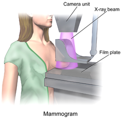
Imaging example of a mammogram Blausen 0628 Mammogram.png
Imaging example of a mammogram

Various imaging tests can be conducted, such as magnetic resonance imaging (MRI), ultrasound, or mammogram, as one of the three tests that is scored that is a part of a triple test score. [15]

Needle breast biopsy of a lump Needle Breast Biopsy.png
Needle breast biopsy of a lump

Fine-needle aspiration biopsy

Over a review of 46 studies using sensitivity, specificity, and other measures of accuracy, fine-needle aspiration biopsy proved to be a very accurate yet minimally invasive diagnostic method for evaluating breast malignancy. With the exclusion of unsatisfactory samples, fine-needle aspiration biopsy sensitivity proportion was 0.927 and the specificity proportion was 0.948. In the unsatisfactory samples, the pooled sensitivity proportion was 0.920, and the pooled specificity proportion was 0.768. [19]

One way that fine-needle aspiration biopsy cytology is reported is via the International Academy of Cytology (IAC) Yokohama System, which "defines five categories for reporting breast cytology, each with a clear descriptive term for the category, a definition, a risk of malignancy (ROM) and a suggested management algorithm." [20] This suggested management algorithm may be particularly useful in countries utilizing the triple test score, as it can provide various management strategies based on the breast lesions from fine-needle aspiration biopsy. [20]

In another review of 22 studies with over 10,000 subjects, using the IAC Yokohama Reporting System, fine-needle aspiration showed strong overall accuracy. "Sensitivity and specificity, with 95% confidence intervals, were 0.978 [0.968, 0.985] and 0.832 [0.76, 0.886] for the diagnostic cut-off of "Atypical considered positive for malignancy," 0.916 [0.892, 0.935] and 0.983 [0.97, 0.99] for the cut-off of "Suspicious of Malignancy considered positive," and 0.763 [0.706, 0.812] and 0.999 [0.994, 1] for the cut-off of "Malignant considered positive." [21]

The IAC Yokohama Reporting System was also evaluated on the pooled risk of malignancy in a meta analysis of 18 different studies with a total of 7,969 cases. They found that when considering both "suspicious" and "malignant" as positive results, the sensitivity was 91%, and the false positive rate was 2.33%. [22]

Overall, fine-needle aspiration cytopathology can greatly benefit low medical infrastructure communities as it is "minimally invasive and well-tolerated by patients, inexpensive, and requires minimal laboratory infrastructure and proceduralist costs." However, two major requirements that may slow the integration of fine-needle aspiration cytology is actually attaining or training pathologists and to encourage the use cytopathology in the education of local clinicians. [23]

Use today

Studies investigating the applicability and the effectiveness of the triple test score in the United States have ranged from the 1991 to 2010. [1] [24] [9] The current prevalence and usage of the triple test score in the United States are not well understood.

In the United Kingdom, the triple test score is usually referred to as the "triple assessment". The majority of hospitals in the UK have implemented rapid-access breast cancer screening clinics where the triple test score is used as a clinical diagnostic tool. [25] [26]

Ongoing research efforts are essential for the long-term effectiveness and applicability of the triple test score in healthcare settings.

Reframing guidelines

Breast cancer is not a gender-specific disease; anyone who has breast tissue has a risk of getting breast cancer. Gendering guidelines for breast cancer excludes individuals who do not identify as female, which can potentially lead to late detection of breast cancer in those individuals. There has been a push by various healthcare providers to make guidelines more inclusive when it comes to breast cancer screenings and awareness. [27] [ relevant? ]

Related Research Articles

<span class="mw-page-title-main">Cytopathology</span> A branch of pathology that studies and diagnoses diseases on the cellular level

Cytopathology is a branch of pathology that studies and diagnoses diseases on the cellular level. The discipline was founded by George Nicolas Papanicolaou in 1928. Cytopathology is generally used on samples of free cells or tissue fragments, in contrast to histopathology, which studies whole tissues. Cytopathology is frequently, less precisely, called "cytology", which means "the study of cells".

<span class="mw-page-title-main">Mammography</span> Process of using low-energy X-rays to examine the human breast for diagnosis and screening

Mammography is the process of using low-energy X-rays to examine the human breast for diagnosis and screening. The goal of mammography is the early detection of breast cancer, typically through detection of characteristic masses or microcalcifications.

<span class="mw-page-title-main">Fibroadenoma</span> Medical condition

Fibroadenomas are benign breast tumours characterized by an admixture of stromal and epithelial tissue. Breasts are made of lobules and ducts. These are surrounded by glandular, fibrous and fatty tissues. Fibroadenomas develop from the lobules. The glandular tissue and ducts grow over the lobule to form a solid lump.

<span class="mw-page-title-main">Fine-needle aspiration</span> Diagnostic medical procedure

Fine-needle aspiration (FNA) is a diagnostic procedure used to investigate lumps or masses. In this technique, a thin, hollow needle is inserted into the mass for sampling of cells that, after being stained, are examined under a microscope (biopsy). The sampling and biopsy considered together are called fine-needle aspiration biopsy (FNAB) or fine-needle aspiration cytology (FNAC). Fine-needle aspiration biopsies are very safe minor surgical procedures. Often, a major surgical biopsy can be avoided by performing a needle aspiration biopsy instead, eliminating the need for hospitalization. In 1981, the first fine-needle aspiration biopsy in the United States was done at Maimonides Medical Center. Today, this procedure is widely used in the diagnosis of cancer and inflammatory conditions. Fine needle aspiration is generally considered a safe procedure. Complications are infrequent.

<span class="mw-page-title-main">Phyllodes tumor</span> Medical condition

Phyllodes tumors, are a rare type of biphasic fibroepithelial mass that form from the periductal stromal and epithelial cells of the breast. They account for less than 1% of all breast neoplasms. They were previously termed cystosarcoma phyllodes, coined by Johannes Müller in 1838, before being renamed to phyllodes tumor by the World Health Organization in 2003. Phullon, which means 'leaf' in Greek, describes the unique papillary projections characteristic of phyllodes tumors on histology. Diagnosis is made via a core-needle biopsy and treatment is typically surgical resection with wide margins (>1 cm), due to their propensity to recur.

<span class="mw-page-title-main">Endoscopic ultrasound</span> Medical imaging procedure

Endoscopic ultrasound (EUS) or echo-endoscopy is a medical procedure in which endoscopy is combined with ultrasound to obtain images of the internal organs in the chest, abdomen and colon. It can be used to visualize the walls of these organs, or to look at adjacent structures. Combined with Doppler imaging, nearby blood vessels can also be evaluated.

<span class="mw-page-title-main">Fibrocystic breast changes</span> Medical condition

Fibrocystic breast changes is a condition of the breasts where there may be pain, breast cysts, and breast masses. The breasts may be described as "lumpy" or "doughy". Symptoms may worsen during certain parts of the menstrual cycle due to hormonal stimulation. These are normal breast changes, not associated with cancer.

The Breast Imaging-Reporting and Data System (BI-RADS) is a quality assurance tool originally designed for use with mammography. The system is a collaborative effort of many health groups but is published and trademarked by the American College of Radiology (ACR).

<span class="mw-page-title-main">Breast cyst</span> Medical condition

A breast cyst is a cyst, a fluid-filled sac, within the breast. One breast can have one or more cysts. They are often described as round or oval lumps with distinct edges. In texture, a breast cyst usually feels like a soft grape or a water-filled balloon, but sometimes a breast cyst feels firm.

<span class="mw-page-title-main">Thyroid nodule</span> Medical condition

Thyroid nodules are nodules which commonly arise within an otherwise normal thyroid gland. They may be hyperplastic or tumorous, but only a small percentage of thyroid tumors are malignant. Small, asymptomatic nodules are common, and often go unnoticed. Nodules that grow larger or produce symptoms may eventually need medical care. A goitre may have one nodule – uninodular, multiple nodules – multinodular, or be diffuse.

<span class="mw-page-title-main">Breast cancer screening</span> Medical screening of asymptomatic, healthy women for breast cancer

Breast cancer screening is the medical screening of asymptomatic, apparently healthy women for breast cancer in an attempt to achieve an earlier diagnosis. The assumption is that early detection will improve outcomes. A number of screening tests have been employed, including clinical and self breast exams, mammography, genetic screening, ultrasound, and magnetic resonance imaging.

Stereotactic biopsy, also known as stereotactic core biopsy, is a biopsy procedure that uses a computer and imaging performed in at least two planes to localize a target lesion in three-dimensional space and guide the removal of tissue for examination by a pathologist under a microscope. Stereotactic core biopsy makes use of the underlying principle of parallax to determine the depth or "Z-dimension" of the target lesion.

Malignant pleural effusion is a condition in which cancer causes an abnormal amount of fluid to collect between the thin layers of tissue (pleura) lining the outside of the lung and the wall of the chest cavity. Lung cancer and breast cancer account for about 50-65% of malignant pleural effusions. Other common causes include pleural mesothelioma and lymphoma.

<span class="mw-page-title-main">Molecular breast imaging</span>

Molecular breast imaging (MBI), also known as scintimammography, is a type of breast imaging test that is used to detect cancer cells in breast tissue of individuals who have had abnormal mammograms, especially for those who have dense breast tissue, post-operative scar tissue or breast implants.

Daniel B. Kopans, MD, FACR is a radiologist specializing in mammography and other forms of breast imaging.

<span class="mw-page-title-main">Breast mass</span> Localized swellings that feel different from the surrounding tissue

A breast mass, also known as a breast lump, is a localized swelling that feels different from the surrounding tissue. Breast pain, nipple discharge, or skin changes may be present. Concerning findings include masses that are hard, do not move easily, are of an irregular shape, or are firmly attached to surrounding tissue.

<span class="mw-page-title-main">Cervical lymphadenopathy</span> Disease of the lymph nodes

Cervical lymphadenopathy refers to lymphadenopathy of the cervical lymph nodes. The term lymphadenopathy strictly speaking refers to disease of the lymph nodes, though it is often used to describe the enlargement of the lymph nodes. Similarly, the term lymphadenitis refers to inflammation of a lymph node, but often it is used as a synonym of lymphadenopathy.

<span class="mw-page-title-main">Breast ultrasound</span> Type of medical imaging

Breast ultrasound is a medical imaging technique that uses medical ultrasonography to perform imaging of the breast. It can be performed for either diagnostic or screening purposes and can be used with or without a mammogram. In particular, breast ultrasound may be useful for younger women who have denser fibrous breast tissue that may make mammograms more challenging to interpret.

<span class="mw-page-title-main">Breast biopsy</span> Surgical diagnostic procedure for breast tumours

A breast biopsy is usually done after a suspicious lesion is discovered on either mammography or ultrasound to get tissue for pathological diagnosis. Several methods for a breast biopsy now exist. The most appropriate method of biopsy for a patient depends upon a variety of factors, including the size, location, appearance and characteristics of the abnormality. The different types of breast biopsies include fine-needle aspiration (FNA), vacuum-assisted biopsy, core needle biopsy, and surgical excision biopsy. Breast biopsies can be done utilizing ultrasound, MRI or a stereotactic biopsy imaging guidance. Vacuum assisted biopsies are typically done using stereotactic techniques when the suspicious lesion can only be seen on mammography. On average, 5–10 biopsies of a suspicious breast lesion will lead to the diagnosis of one case of breast cancer. Needle biopsies have largely replaced open surgical biopsies in the initial assessment of imaging as well as palpable abnormalities in the breast.

<span class="mw-page-title-main">Breast imaging</span>

In medicine, breast imaging is a sub-speciality of diagnostic radiology that involves imaging of the breasts for screening or diagnostic purposes. There are various methods of breast imaging using a variety of technologies as described in detail below. Traditional screening and diagnostic mammography uses x-ray technology and has been the mainstay of breast imaging for many decades. Breast tomosynthesis is a relatively new digital x-ray mammography technique that produces multiple image slices of the breast similar to, but distinct from, computed tomography (CT). Xeromammography and galactography are somewhat outdated technologies that also use x-ray technology and are now used infrequently in the detection of breast cancer. Breast ultrasound is another technology employed in diagnosis and screening that can help differentiate between fluid filled and solid lesions, an important factor to determine if a lesion may be cancerous. Breast MRI is a technology typically reserved for high-risk patients and patients recently diagnosed with breast cancer. Lastly, scintimammography is used in a subgroup of patients who have abnormal mammograms or whose screening is not reliable on the basis of using traditional mammography or ultrasound.

References

  1. 1 2 Morris A, Pommier RF, Schmidt WA, Shih RL, Alexander PW, Vetto JT (September 1998). "Accurate Evaluation of Palpable Breast Masses by the Triple Test Score". Archives of Surgery. 133 (9): 930–934. doi:10.1001/archsurg.133.9.930. PMID   9749842.
  2. 1 2 3 4 Johnsén C (1975-01-01). "Breast disease. A clinical study with special reference to diagnostic procedures". Acta Chirurgica Scandinavica. Supplementum. 454: 1–108. PMID   1059305.
  3. Egyed Z, Járay B, Kulka J, Péntek Z (28 August 2008). "Triple test score for the evaluation of invasive ductal and lobular breast cancer". Pathology & Oncology Research. 15 (2): 159–166. doi:10.1007/s12253-008-9083-3. PMID   18752055. S2CID   6523239.
  4. Harvey JA (August 2006). "Sonography of Palpable Breast Masses". Seminars in Ultrasound, CT, and MR. Breast Imaging Update. 27 (4): 284–297. doi:10.1053/j.sult.2006.05.003. PMID   16915997.
  5. Morris KT, Vetto JT, Petty JK, Lum SS, Schmidt WA, Toth-Fejel S, Pommier RF (October 2002). "A new score for the evaluation of palpable breast masses in women under age 40". American Journal of Surgery. 184 (4): 346–347. doi:10.1016/S0002-9610(02)00947-9. PMID   12383898.
  6. Thigpen D, Kappler A, Brem R (March 2018). "The Role of Ultrasound in Screening Dense Breasts-A Review of the Literature and Practical Solutions for Implementation". Diagnostics. 8 (1): 20. doi: 10.3390/diagnostics8010020 . PMC   5872003 . PMID   29547532.
  7. Kachewar SS, Dongre SD (2015). "Role of triple test score in the evaluation of palpable breast lump". Indian Journal of Medical and Paediatric Oncology. 36 (2): 123–127. doi: 10.4103/0971-5851.158846 . PMC   4477375 . PMID   26157290.
  8. Magny SJ, Shikhman R, Keppke AL (August 29, 2022). "Breast Imaging Reporting and Data System". StatPearls. Treasure Island (FL): StatPearls Publishing. PMID   29083600 . Retrieved 2023-07-30.
  9. 1 2 Morris KT, Pommier RF, Morris A, Schmidt WA, Beagle G, Alexander PW, et al. (September 2001). "Usefulness of the triple test score for palpable breast masses; discussion 1012-3". Archives of Surgery. 136 (9): 1008–1012. doi:10.1001/archsurg.136.9.1008. PMID   11529822. S2CID   22625052.
  10. Daly C, Puckett Y (August 1, 2022). "New Breast Mass". StatPearls. Treasure Island (FL): StatPearls Publishing. PMID   32809592 . Retrieved 2023-07-29.
  11. Morris AM, Flowers CR, Morris KT, Schmidt WA, Pommier RF, Vetto JT (August 2003). "Comparing the cost-effectiveness of the triple test score to traditional methods for evaluating palpable breast masses". Medical Care. 41 (8): 962–971. doi:10.1097/00005650-200308000-00009. PMID   12886175. S2CID   41315487.
  12. 1 2 Klein S (May 2005). "Evaluation of palpable breast masses". American Family Physician. 71 (9): 1731–1738. PMID   15887452.
  13. "Clinical Breast Exam". National Breast Cancer Foundation. Retrieved 2023-07-30.
  14. Ngan TT, Nguyen NT, Van Minh H, Donnelly M, O'Neill C (November 2020). "Effectiveness of clinical breast examination as a 'stand-alone' screening modality: an overview of systematic reviews". BMC Cancer. 20 (1): 1070. doi: 10.1186/s12885-020-07521-w . PMC   7653771 . PMID   33167942.
  15. "The Triple Test". www.breastcancerfoundation.org.nz. Retrieved 2023-07-30.
  16. Sood R, Rositch AF, Shakoor D, Ambinder E, Pool KL, Pollack E, et al. (August 2019). "Ultrasound for Breast Cancer Detection Globally: A Systematic Review and Meta-Analysis". Journal of Global Oncology. 5 (5): 1–17. doi:10.1200/JGO.19.00127. PMC   6733207 . PMID   31454282.
  17. 1 2 Chen HL, Zhou JQ, Chen Q, Deng YC (July 2021). "Comparison of the sensitivity of mammography, ultrasound, magnetic resonance imaging and combinations of these imaging modalities for the detection of small (≤2cm) breast cancer". Medicine. 100 (26): e26531. doi:10.1097/MD.0000000000026531. PMC   8257894 . PMID   34190189.
  18. "What Is a Mammogram?". Centers for Disease Control and Prevention. 2022-09-26. Retrieved 2023-07-30.
  19. Yu YH, Wei W, Liu JL (January 2012). "Diagnostic value of fine-needle aspiration biopsy for breast mass: a systematic review and meta-analysis". BMC Cancer. 12: 41. doi: 10.1186/1471-2407-12-41 . PMC   3283452 . PMID   22277164.
  20. 1 2 Field AS, Raymond WA, Rickard M, Arnold L, Brachtel EF, Chaiwun B, et al. (2019). "The International Academy of Cytology Yokohama System for Reporting Breast Fine-Needle Aspiration Biopsy Cytopathology". Acta Cytologica. 63 (4): 257–273. doi:10.1159/000499509. PMID   31112942. S2CID   263539317.
  21. Paul P, Azad S, Agrawal S, Rao S, Chowdhury N (2023). "Systematic Review and Meta-Analysis of the Diagnostic Accuracy of the International Academy of Cytology Yokohama System for Reporting Breast Fine-Needle Aspiration Biopsy in Diagnosing Breast Cancer". Acta Cytologica. 67 (1): 1–16. doi: 10.1159/000527346 . PMID   36412573. S2CID   253760182.
  22. Nikas IP, Vey JA, Proctor T, AlRawashdeh MM, Ishak A, Ko HM, Ryu HS (February 2023). "The Use of the International Academy of Cytology Yokohama System for Reporting Breast Fine-Needle Aspiration Biopsy". American Journal of Clinical Pathology. 159 (2): 138–145. doi:10.1093/ajcp/aqac132. PMC   9891409 . PMID   36370120.
  23. Field AS (March 2018). "Cytopathology in Low Medical Infrastructure Countries: Why and How to Integrate to Capacitate Health Care". Clinics in Laboratory Medicine. Global Health and Pathology. 38 (1): 175–182. doi:10.1016/j.cll.2017.10.014. PMID   29412881.
  24. Vetto J, Pommier R, Schmidt W, Wachtel M, DuBois P, Jones M, Thurmond A (May 1995). "Use of the "triple test" for palpable breast lesions yields high diagnostic accuracy and cost savings". American Journal of Surgery. 169 (5): 519–522. doi:10.1016/S0002-9610(99)80209-8. PMID   7747833.
  25. "Overview | Early and locally advanced breast cancer: diagnosis and management | Guidance". www.nice.org.uk. 2018-07-18. Retrieved 2023-07-26.
  26. Chalasani P, Stopeck AT, Thompson PA (May 2023). Kiluk JV (ed.). "Breast Cancer Workup: Approach Considerations, Breast Cancer Screening, Ultrasonography". emedicine.medscape.com. Retrieved 2023-07-30.
  27. "Breast Cancer Has No Gender". Cedars-Sinai. October 18, 2021. Retrieved 2023-08-02.