X-ray machine

Last updated
A radiology room table. The X-ray housing is turned by 90deg for a chest radiograph X-ray table.JPG
A radiology room table. The X-ray housing is turned by 90° for a chest radiograph

An X-ray machine is a device that uses X-rays for a variety of applications including medicine, X-ray fluorescence, electronic assembly inspection, and measurement of material thickness in manufacturing operations. In medical applications, X-ray machines are used by radiographers to acquire x-ray images of the internal structures (e.g., bones) of living organisms, and also in sterilization.

Contents

Structure

GemX-160 - Portable Wireless Controlled Battery Powered X-ray Generator for use in Non Destructive Testing and Security. GemX2.png
GemX-160 - Portable Wireless Controlled Battery Powered X-ray Generator for use in Non Destructive Testing and Security.
XR150 - Portable pulsed X-ray Battery Powered X-ray Generator used in Security. Portable x-ray device.jpg
XR150 - Portable pulsed X-ray Battery Powered X-ray Generator used in Security.

An X-ray generator generally contains an X-ray tube to produce the X-rays. Possibly, radioisotopes can also be used to generate X-rays. [1]

An X-ray tube is a simple vacuum tube that contains a cathode, which directs a stream of electrons into a vacuum, and an anode, which collects the electrons and is made of tungsten to evacuate the heat generated by the collision. When the electrons collide with the target, about 1% of the resulting energy is emitted as X-rays, with the remaining 99% released as heat. Due to the high energy of the electrons that reach relativistic speeds the target is usually made of tungsten even if other material can be used particularly in XRF applications.[ citation needed ]

An X-ray generator also needs to contain a cooling system to cool the anode; many X-ray generators use water or oil recirculating systems. [2]

Medical imaging

Acquisition of projectional radiography, with an X-ray generator and a detector. Projectional radiography components.jpg
Acquisition of projectional radiography, with an X-ray generator and a detector.

In medical imaging applications, an X-ray machine has a control console that is used by a radiologic technologist to select X-ray techniques suitable for the specific exam, a power supply that creates and produces the desired kVp (peak kilovoltage), mA (milliamperes, sometimes referred to as mAs which is actually mA multiplied by the desired exposure length) for the X-ray tube, and the X-ray tube itself.

History

The discovery of X-rays came from experimenting with Crookes tubes, an early experimental electrical discharge tube invented by English physicist William Crookes around 1869–1875. In 1895, Wilhelm Röntgen discovered X-rays emanating from Crookes tubes and the many uses for X-rays were immediately apparent. One of the first X-ray photographs was made of the hand of Röntgen's wife. The image displayed both her wedding ring and bones. On January 18, 1896 an X-ray machine was formally displayed by Henry Louis Smith. A fully functioning unit was introduced to the public at the 1904 World's Fair by Clarence Dally. [3] The technology developed quickly: In 1909 Mónico Sánchez Moreno had produced the first portable medical device and during World War I Marie Curie led the development of X-ray machines mounted in "radiological cars" to provide mobile X-ray services for military field hospitals.

In the 1940s and 1950s, X-ray machines were used in stores to help sell footwear. These were known as Shoe-fitting fluoroscopes. However, as the harmful effects of X-ray radiation were properly considered, they finally fell out of use. Shoe-fitting use of the device was first banned by the state of Pennsylvania in 1957. (They were more a clever marketing tool to attract customers, rather than a fitting aid.) Together with Robert J. Van de Graaff, John G. Trump developed one of the first million-volt X-ray generators.

Overview

An X-ray imaging system consists of a generator control console where the operator selects desired techniques to obtain a quality readable image(kVp, mA and exposure time), an x-ray generator which controls the x-ray tube current, x-ray tube kilovoltage and x-ray emitting exposure time, an X-ray tube that converts the kilovoltage and mA into actual x-rays and an image detection system which can be either a film (analog technology) or a digital capture system and a PACS.

Applications

X-ray machines are used in health care for visualising bone structures, during surgeries (especially orthopedic) to assist surgeons in reattaching broken bones with screws or structural plates, assisting cardiologists in locating blocked arteries and guiding stent placements or performing angioplasties and for other dense tissues such as tumours. Non-medicinal applications include security and material analysis.

Medicine

Mobile fluoroscopy units can produce images continuously. Mobile X-ray machine.jpg
Mobile fluoroscopy units can produce images continuously.

The main fields in which x-ray machines are used in medicine are radiography, radiotherapy, and fluoroscopic-type procedures. Radiography is generally used for fast, highly penetrating images, and is usually used in areas with a high bone content but can also be used to look for tumors such as with mammography imaging. Some forms of radiography include:

In fluoroscopy, imaging of the digestive tract is done with the help of a radiocontrast agent such as barium sulfate, which is opaque to X-rays.

Radiotherapy — the use of x-ray radiation to treat malignant and benign cancer cells, a non-imaging application

Fluoroscopy is used in cases where real-time visualization is necessary (and is most commonly encountered in everyday life at airport security). Some medical applications of fluoroscopy include:

X-rays are highly penetrating, ionizing radiation, therefore X-ray machines are used to take pictures of dense tissues such as bones and teeth. This is because bones absorb the radiation more than the less dense soft tissue. X-rays from a source pass through the body and onto a photographic cassette. Areas where radiation is absorbed show up as lighter shades of grey (closer to white). This can be used to diagnose broken or fractured bones.

In 2012, European Commission of Radiation Protection set leakage radiation limit from X-ray generators such as X-ray tubes and CT machines as one mGy/hour at one metre distance from the machine. [4]

Security

Hand-luggage inspection machine at Berlin Schonefeld Airport. Flughafenkontrolle.jpg
Hand-luggage inspection machine at Berlin Schönefeld Airport.

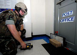
X-ray machines are used to screen objects non-invasively. Luggage at airports and student baggage at some schools are examined for possible weapons, including bombs. Prices of these Luggage X-rays vary from $50,000 to $300,000. The main parts of an X-ray Baggage Inspection System are the generator used to generate x-rays, the detector to detect radiation after passing through the baggage, signal processor unit (usually a PC) to process the incoming signal from the detector, and a conveyor system for moving baggage into the system. Portable pulsed X-ray Battery Powered X-ray Generator used in Security as shown in the figure provides EOD responders safer analysis of any possible target hazard.

Operation

When baggage is placed on the conveyor, it is moved into the machine by the operator. There is an infrared transmitter and receiver assembly to detect the baggage when it enters the tunnel. This assembly gives the signal to switch on the generator and signal processing system. The signal processing system processes incoming signals from the detector and reproduce an image based upon the type of material and material density inside the baggage. This image is then sent to the display unit.

Color classification

X-ray image of a backpack. Organic and inorganic materials are discriminated in using dual energy techniques. Xray-verkehrshaus.jpg
X-ray image of a backpack. Organic and inorganic materials are discriminated in using dual energy techniques.

The colour of the image displayed depends upon the material and material density : organic material such as paper, clothes and most explosives are displayed in orange. Mixed materials such as aluminum are displayed in green. Inorganic materials such as copper are displayed in blue and non-penetrable items are displayed in black (some machines display this as a yellowish green or red). The darkness of the color depends upon the density or thickness of the material.

The material density determination is achieved by two-layer detector. The layers of the detector pixels are separated with a strip of metal. The metal absorbs soft rays, letting the shorter, more penetrating wavelengths through to the bottom layer of detectors, turning the detector to a crude two-band spectrometer.

Advances in X-ray technology

5.5-pound (2.5 kg) dental digital X-ray system under testing in 2011 NomadPortableDentalXRayCropped.jpg
5.5-pound (2.5 kg) dental digital X-ray system under testing in 2011

A film of carbon nanotubes (as a cathode) that emits electrons at room temperature when exposed to an electrical field has been fashioned into an X-ray device. An array of these emitters can be placed around a target item to be scanned and the images from each emitter can be assembled by computer software to provide a 3-dimensional image of the target in a fraction of the time it takes using a conventional X-ray device. The system also allows rapid, precise control, enabling prospective physiological gated imaging. [6]

Engineers at the University of Missouri (MU), Columbia, have invented a compact source of x-rays and other forms of radiation. The radiation source is the size of a stick of gum and could be used to create portable x-ray scanners. A prototype handheld x-ray scanner using the source could be manufactured in as soon as three years. [7]

See also

Notes

  1. Page 442 in: Rene Van Grieken, A. Markowicz (2001). Handbook of X-Ray Spectrometry, Second Edition, Practical Spectroscopy. CRC Press. ISBN   9780203908709.
  2. "X-ray Generators" Archived 2011-04-23 at the Wayback Machine , NDT Resource Center. Page fetched April 21, 2011.
  3. King, Gilbert (14 March 2012). "Clarence Dally - The Man Who Gave Thomas Edison X-Ray Vision". smithsonianmag.com. Retrieved 13 November 2016.
  4. Criteria for Acceptability of Medical Radiological Equipment used in Diagnostic Radiology, Nuclear Medicine and Radiotherapy (PDF). Luxembourg: European Union. 2012. p. 26. Archived from the original (PDF) on 9 March 2021. Retrieved 12 February 2022.
  5. "Deployed dentists test lightweight mobile X-ray system", Spc. Jonathan W. Thomas, 16th Mobile Public Affairs Detachment, April 21, 2011, www.army.mil. Fetched from URL on April 25, 2011.
  6. Zhang; et al. "UNC News release -- New method of using nanotube x-rays creates CT images faster than traditional scanners". Archived from the original on 2014-08-24. Retrieved 2012-08-20.
  7. Editorial Staff. "MU researchers develop super compact x-ray source" . Retrieved 2013-01-19.

Related Research Articles

<span class="mw-page-title-main">X-ray</span> Form of short-wavelength electromagnetic radiation

X-ray is a high-energy electromagnetic radiation. In many languages, it is referred to as Röntgen radiation, after the German scientist Wilhelm Conrad Röntgen, who discovered it in 1895 and named it X-radiation to signify an unknown type of radiation.

<span class="mw-page-title-main">CT scan</span> Medical imaging procedure using X-rays to produce cross-sectional images

A computed tomography scan is a medical imaging technique used to obtain detailed internal images of the body. The personnel that perform CT scans are called radiographers or radiology technologists.

<span class="mw-page-title-main">Radiography</span> Imaging technique using ionizing and non-ionizing radiation

Radiography is an imaging technique using X-rays, gamma rays, or similar ionizing radiation and non-ionizing radiation to view the internal form of an object. Applications of radiography include medical and industrial radiography. Similar techniques are used in airport security,. To create an image in conventional radiography, a beam of X-rays is produced by an X-ray generator and it is projected towards the object. A certain amount of the X-rays or other radiation are absorbed by the object, dependent on the object's density and structural composition. The X-rays that pass through the object are captured behind the object by a detector. The generation of flat two-dimensional images by this technique is called projectional radiography. In computed tomography, an X-ray source and its associated detectors rotate around the subject, which itself moves through the conical X-ray beam produced. Any given point within the subject is crossed from many directions by many different beams at different times. Information regarding the attenuation of these beams is collated and subjected to computation to generate two-dimensional images on three planes which can be further processed to produce a three-dimensional image.

<span class="mw-page-title-main">Radiology</span> Branch of Medicine

Radiology is the medical discipline that uses medical imaging to diagnose diseases and guide their treatment, within the bodies of humans and other animals. It began with radiography, but today it includes all imaging modalities, including those that use no electromagnetic radiation, as well as others that do, such as computed tomography (CT), fluoroscopy, and nuclear medicine including positron emission tomography (PET). Interventional radiology is the performance of usually minimally invasive medical procedures with the guidance of imaging technologies such as those mentioned above.

<span class="mw-page-title-main">Medical imaging</span> Technique and process of creating visual representations of the interior of a body

Medical imaging is the technique and process of imaging the interior of a body for clinical analysis and medical intervention, as well as visual representation of the function of some organs or tissues (physiology). Medical imaging seeks to reveal internal structures hidden by the skin and bones, as well as to diagnose and treat disease. Medical imaging also establishes a database of normal anatomy and physiology to make it possible to identify abnormalities. Although imaging of removed organs and tissues can be performed for medical reasons, such procedures are usually considered part of pathology instead of medical imaging.

<span class="mw-page-title-main">External beam radiotherapy</span> Treatment of cancer with ionized radiation

External beam radiation therapy (EBRT) is a form of radiotherapy that utilizes a high-energy collimated beam of ionizing radiation, from a source outside the body, to target and kill cancer cells. A radiotherapy beam is composed of particles which travel in a consistent direction; each radiotherapy beam consists of one type of particle intended for use in treatment, though most beams contain some contamination by other particle types.

<span class="mw-page-title-main">Fluoroscopy</span> Production of an image when X-rays strike a fluorescent screen

Fluoroscopy is an imaging technique that uses X-rays to obtain real-time moving images of the interior of an object. In its primary application of medical imaging, a fluoroscope allows a surgeon to see the internal structure and function of a patient, so that the pumping action of the heart or the motion of swallowing, for example, can be watched. This is useful for both diagnosis and therapy and occurs in general radiology, interventional radiology, and image-guided surgery.

Radiocontrast agents are substances used to enhance the visibility of internal structures in X-ray-based imaging techniques such as computed tomography, projectional radiography, and fluoroscopy. Radiocontrast agents are typically iodine, or more rarely barium sulfate. The contrast agents absorb external X-rays, resulting in decreased exposure on the X-ray detector. This is different from radiopharmaceuticals used in nuclear medicine which emit radiation.

Radiodensity is opacity to the radio wave and X-ray portion of the electromagnetic spectrum: that is, the relative inability of those kinds of electromagnetic radiation to pass through a particular material. Radiolucency or hypodensity indicates greater passage to X-ray photons and is the analogue of transparency and translucency with visible light. Materials that inhibit the passage of electromagnetic radiation are called radiodense or radiopaque, while those that allow radiation to pass more freely are referred to as radiolucent. Radiopaque volumes of material have white appearance on radiographs, compared with the relatively darker appearance of radiolucent volumes. For example, on typical radiographs, bones look white or light gray (radiopaque), whereas muscle and skin look black or dark gray, being mostly invisible (radiolucent).

An X-ray image intensifier (XRII) is an image intensifier that converts X-rays into visible light at higher intensity than the more traditional fluorescent screens can. Such intensifiers are used in X-ray imaging systems to allow low-intensity X-rays to be converted to a conveniently bright visible light output. The device contains a low absorbency/scatter input window, typically aluminum, input fluorescent screen, photocathode, electron optics, output fluorescent screen and output window. These parts are all mounted in a high vacuum environment within glass or, more recently, metal/ceramic. By its intensifying effect, It allows the viewer to more easily see the structure of the object being imaged than fluorescent screens alone, whose images are dim. The XRII requires lower absorbed doses due to more efficient conversion of X-ray quanta to visible light. This device was originally introduced in 1948.

A contrast agent is a substance used to increase the contrast of structures or fluids within the body in medical imaging. Contrast agents absorb or alter external electromagnetism or ultrasound, which is different from radiopharmaceuticals, which emit radiation themselves. In x-ray imaging, contrast agents enhance the radiodensity in a target tissue or structure. In magnetic resonance imaging, contrast agents shorten the relaxation times of nuclei within body tissues in order to alter the contrast in the image.

Digital radiography is a form of radiography that uses x-ray–sensitive plates to directly capture data during the patient examination, immediately transferring it to a computer system without the use of an intermediate cassette. Advantages include time efficiency through bypassing chemical processing and the ability to digitally transfer and enhance images. Also, less radiation can be used to produce an image of similar contrast to conventional radiography.

Peak kilovoltage (kVp) refers to the maximum high voltage applied across an X-ray tube to produce the X-rays. During X-ray generation, surface electrons are released from a heated cathode by thermionic emission. The applied voltage (kV) accelerates these electrons toward an anode target, ultimately producing X-rays when the electrons are stopped in the anode. Thus, the kVp corresponds to the highest kinetic energy of the electrons striking the target, and is proportional to the maximum photon energy of the resulting X-ray emission spectrum. In early and basic X-ray equipment, the applied voltage varies cyclically, with one, two, or more pulses per mains AC power cycle. One standard way to measure pulsating DC is its peak amplitude, hence kVp. Most modern X-ray generators apply a constant potential across the X-ray tube; in such systems, the kVp and the steady-state kV are identical.

<span class="mw-page-title-main">Industrial radiography</span> Type of non-destructive testing

Industrial radiography is a modality of non-destructive testing that uses ionizing radiation to inspect materials and components with the objective of locating and quantifying defects and degradation in material properties that would lead to the failure of engineering structures. It plays an important role in the science and technology needed to ensure product quality and reliability. In Australia, industrial radiographic non-destructive testing is colloquially referred to as "bombing" a component with a "bomb".

<span class="mw-page-title-main">Projectional radiography</span> Formation of 2D images using X-rays

Projectional radiography, also known as conventional radiography, is a form of radiography and medical imaging that produces two-dimensional images by X-ray radiation. The image acquisition is generally performed by radiographers, and the images are often examined by radiologists. Both the procedure and any resultant images are often simply called 'X-ray'. Plain radiography or roentgenography generally refers to projectional radiography. Plain radiography can also refer to radiography without a radiocontrast agent or radiography that generates single static images, as contrasted to fluoroscopy, which are technically also projectional.

Photostimulated luminescence (PSL) is the release of stored energy within a phosphor by stimulation with visible light, to produce a luminescent signal. X-rays may induce such an energy storage. A plate based on this mechanism is called a photostimulable phosphor (PSP) plate and is one type of X-ray detector used in projectional radiography. Creating an image requires illuminating the plate twice: the first exposure, to the radiation of interest, "writes" the image, and a later, second illumination "reads" the image. The device to read such a plate is known as a phosphorimager.

<span class="mw-page-title-main">Automatic exposure control</span>

Automatic Exposure Control (AEC) is an X-ray exposure termination device. A medical radiographic exposure is always initiated by a human operator but an AEC detector system may be used to terminate the exposure when a predetermined amount of radiation has been received. The intention of AEC is to provide consistent x-ray image exposure, whether to film, a digital detector or a CT scanner. AEC systems may also automatically set exposure factors such as the X-ray tube current and voltage in a CT.

<span class="mw-page-title-main">Flat-panel detector</span> Class of solid-state x-ray digital radiography devices

Flat-panel detectors are a class of solid-state x-ray digital radiography devices similar in principle to the image sensors used in digital photography and video. They are used in both projectional radiography and as an alternative to x-ray image intensifiers (IIs) in fluoroscopy equipment.

<span class="mw-page-title-main">Cone beam computed tomography</span> Medical imaging technique

Cone beam computed tomography is a medical imaging technique consisting of X-ray computed tomography where the X-rays are divergent, forming a cone.

<span class="mw-page-title-main">X-ray detector</span> Instrument that can measure properties of X-rays

X-ray detectors are devices used to measure the flux, spatial distribution, spectrum, and/or other properties of X-rays.

References

  1. Zhang, J; Yang, G; Cheng, Y; Gao, B Qiu, Q; Lee, YZ; Lu, JP & Zhou, O (2005). "Stationary scanning X-ray source based on carbon nanotube field emitters". Applied Physics Letters. 86 (May 2): 184104. Bibcode:2005ApPhL..86r4104Z. doi:10.1063/1.1923750. S2CID   120546441.{{cite journal}}: CS1 maint: multiple names: authors list (link)