Shoe-fitting fluoroscope

Last updated
A shoe fluoroscope displayed at the US National Museum of Health and Medicine. This machine was manufactured by Adrian Shoe Fitter, Inc. circa 1938 and used in a Washington, D.C., shoe store NMHM ShoeFluorscope.jpg
A shoe fluoroscope displayed at the US National Museum of Health and Medicine. This machine was manufactured by Adrian Shoe Fitter, Inc. circa 1938 and used in a Washington, D.C., shoe store

Shoe-fitting fluoroscopes, also sold under the names X-ray Shoe Fitter, Pedoscope and Foot-o-scope, were X-ray fluoroscope machines installed in shoe stores from the 1920s until about the 1970s in the United States, Canada, United Kingdom, Australia, South Africa, Germany and Switzerland. [1] In the UK, they were known as Pedoscopes, after the company based in St. Albans that manufactured them. [2] An example can be seen at the Science Museum, London. [3] At the beginning of the 1930s, Bally was the first company to import pedoscopes into Switzerland from the UK. In the second half of the 20th century, growing awareness of radiation hazards and increasingly stringent regulations forced their gradual phasing out. They were widely used particularly when buying shoes for children, whose shoe size continually changed until adulthood.

Contents

A shoe-fitting fluoroscope was a metal construction covered in finished wood, approximately 4 feet (1.2 m) high in the shape of short column, with a ledge with an opening through which the standing customer (adult or child) would put their feet and look through a viewing porthole at the top of the fluoroscope down at the X-ray view of the feet and shoes. Two other viewing portholes on either side enabled the parent and a sales assistant to observe the toes being wiggled to show how much room for the toes there was inside the shoe. The bones of the feet were clearly visible, as was the outline of the shoe, including the stitching around the edges.

Invention

There are multiple claims for the invention of the shoe-fitting fluoroscope. The most likely is Jacob Lowe, who demonstrated a modified medical device at shoe retailer conventions in 1920 in Boston and in 1921 in Milwaukee. Lowe filed a US patent application in 1919, granted in 1927, and assigned it to the Adrian Company of Milwaukee for US$15,000. Syl Adrian claims that his brother, Matthew Adrian, invented and built the first machine in Milwaukee; his name is featured in a 1922 advertisement for an X-ray shoe fitter. Clarence Karrer, the son of an X-ray equipment distributor, claims to have built the first unit in 1924 in Milwaukee, but had his idea stolen and patented by one of his father's employees. In the meantime, the British company Pedoscope filed a British patent application in 1924, granted in 1926, and claimed to have been building these machines since 1920. [4]

The X-ray Shoe Fitter Corporation of Milwaukee and Pedoscope Company became the largest manufacturers of shoe-fitting fluoroscopes in the world.

Health concerns

An Adrian Fluoroscope at the Dufferin County Museum, Ontario, Canada (2012). The x-ray tube was removed to render the apparatus completely harmless before being put on public display due to the possible risk of radiation burn or other health problems If it was switched on. Adrian Fluoroscope operating controls.jg.jpg
An Adrian Fluoroscope at the Dufferin County Museum, Ontario, Canada (2012). The x-ray tube was removed to render the apparatus completely harmless before being put on public display due to the possible risk of radiation burn or other health problems If it was switched on.

The risk of radiation burns to extremities was known since Wilhelm Röntgen's 1895 experiment, but this was a short-term effect with early warning from reddening of the skin (erythema). The long-term risks from chronic exposure to radiation began to emerge with Hermann Joseph Muller's 1927 paper showing genetic effects, [5] and the incidence of bone cancer in radium dial painters of the same time period. However, there was not enough data to quantify the level of risk until atomic bomb survivors began to experience the long-term effects of radiation in the late 1940s. The first scientific evaluations of these machines in 1948 immediately sparked concern for radiation protection and electrical safety reasons, and found them ineffective at shoe fitting. [6]

Large variations in dose were possible depending on the machine design, displacement of the shielding materials, and the duration and frequency of use. Radiation surveys showed that American machines delivered an average of 13 roentgen (r) (roughly 0.13 sievert (Sv) of equivalent dose in modern units) to the customer's feet during a typical 20-second viewing, with one capable of delivering 116 r (~1 Sv) in 20 seconds. [6] British Pedoscopes produced about ten times less radiation. [7]

A customer might try several shoes in a day, or return several times in a year, and radiation dose effects may be cumulative. [7] A dose of 300 r can cause growth disturbance in a child, [6] and 600 r can cause erythema in an adult. Hands and feet are relatively resistant to other forms of radiation damage, such as carcinogenesis.

Although most of the dose was directed at the feet, a substantial amount would scatter or leak in all directions. Shielding materials were sometimes displaced to improve image quality, to make the machine lighter, or out of carelessness, and this aggravated the leakage. The resulting whole-body dose may have been hazardous to the salesmen, who were chronically exposed, and to children, who are about twice as radiosensitive as adults. [8] Monitoring of American salespersons found dose rates at pelvis height of up to 95 mr/week, with an average of 7.1 mr/week (up to ~50 mSv/a, avg ~3.7 mSv/an effective dose). [6] A 2007 paper suggested that even higher doses of 0.5 Sv/a were plausible. [9] The most widely accepted model of radiation-induced cancer posits that the incidence of cancers due to ionizing radiation increases linearly with effective (i.e., whole-body) dose. [10]

Years or decades may elapse between radiation exposure and a related occurrence of cancer, and no follow-up studies of customers can be performed for lack of records. According to a 1950 medical article on the machines: "Present evidence indicates that at least some radiation injuries are statistical processes that do not have a threshold. If this evidence is valid, there is no exposure which is absolutely safe and which produces no effect." [6] Three shoe salespersons were identified with rare conditions that might have been associated with their chronic occupational exposure: a severe radiation burn requiring amputation in 1950, [11] a case of dermatitis with ulceration in 1957, [12] and a case of basal-cell carcinoma of the sole in 2004. [9]

Shoe industry response

Representatives of the shoe retail industry denied claims of potential harm in newspaper articles and opinion pieces. They argued that use of the devices prevented harm to customers' feet from poorly-fitted shoes. [13] [14] [15]

Regulation

There were no applicable regulations when shoe-fitting fluoroscopes were introduced. An estimated 10,000 machines were sold in the US, 3,000 in the UK, 1,500 in Switzerland, and 1,000 in Canada before authorities began discouraging their use. [9] As understanding grew of the long-term health effects of radiation, a variety of bodies began speaking out and regulating the machines.

1931 ACXRP recommends limiting dose to 0.1 r per day (~0.5 r/week) in all applications. [16]
1934 IXRPC recommends limiting dose to 0.2 r per day (~1 r/week) in all applications. [17]
1946 ASA recommends limiting foot dose to 2 r per 5 second exposure. [4]
Children to be limited to 12 exposures per year. [4]
1948Warnings specific to the shoe-fitting fluoroscope start appearing in US journals. [18]
1949 Tripartite Conference on Radiation Protection recommends lowering the dose limits: [19]
0.3  rep/week (~0.3 r/week) for whole body bone marrow
1.5 rep/week (~1.5 r/week) for the hands
1950Warnings start appearing in UK journals. [7]
A public inquiry was held in Queensland, Australia and warned against uncontrolled use [20]
ICRP adopts the Tripartite recommendations, with some lack of clarity about units. [21]
1953A definitive recommendation against use on children was published in the journal Pediatrics [9]
US Food and Drug Administration bans the machines. [22]
1954 NCRP recommends reducing dose limits by a factor of 10 for children, and other changes: [23]
15.6 mSv/a (~0.03 r/week) for whole body bone marrow
78 mSv/a (~0.15 r/week) for the hands
1956UK Ministry of Health considers regulating the machines. [7]
1957Pennsylvania is first US state to ban use of these machines. [4]
ICRP recommends limiting occupational whole body dose to 50 mSv/a (~0.1 r/week)[ citation needed ]
1958The UK Government required all machines be fitted with a warning sign advising customers of possible health risks, and that they should not use a machine more than 12 times a year. [24]
1958NCRP recommends limiting public whole body dose to 5 mSv/a (~0.01 r/week) [25]
1960160 devices still in use in the Canton of Zürich. [26]
1970sBy 1970, 33 US states have banned the machine. [9]
Late 1970s: Last recorded sighting of a shoe-fitting fluoroscope in service in Boston. [11]
1973The last devices still in use in Germany are banned. [27]
1989Switzerland prohibits the machines. [27]
1990ICRP recommends reducing limits on exposure and other changes: [28]
occupational foot dose to 500 mSv/a (~1 r/week)
occupational whole body dose to 20 mSv/a (~0.04 r/week)
public whole body dose to 1 mSv/a (~0.002 r/week)

Related Research Articles

Background radiation is a measure of the level of ionizing radiation present in the environment at a particular location which is not due to deliberate introduction of radiation sources.

<span class="mw-page-title-main">Sievert</span> SI unit of equivalent dose of ionizing radiation

The sievert is a unit in the International System of Units (SI) intended to represent the stochastic health risk of ionizing radiation, which is defined as the probability of causing radiation-induced cancer and genetic damage. The sievert is important in dosimetry and radiation protection. It is named after Rolf Maximilian Sievert, a Swedish medical physicist renowned for work on radiation dose measurement and research into the biological effects of radiation.

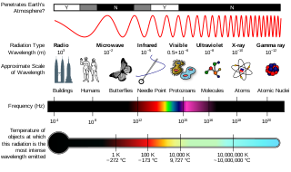
Ionizing radiation (US) (or ionising radiation [UK]), including nuclear radiation, consists of subatomic particles or electromagnetic waves that have sufficient energy to ionize atoms or molecules by detaching electrons from them. Some particles can travel up to 99% of the speed of light, and the electromagnetic waves are on the high-energy portion of the electromagnetic spectrum.

The gray is the unit of ionizing radiation dose in the International System of Units (SI), defined as the absorption of one joule of radiation energy per kilogram of matter.

Radiation dosimetry in the fields of health physics and radiation protection is the measurement, calculation and assessment of the ionizing radiation dose absorbed by an object, usually the human body. This applies both internally, due to ingested or inhaled radioactive substances, or externally due to irradiation by sources of radiation.

Radiation protection, also known as radiological protection, is defined by the International Atomic Energy Agency (IAEA) as "The protection of people from harmful effects of exposure to ionizing radiation, and the means for achieving this". Exposure can be from a source of radiation external to the human body or due to internal irradiation caused by the ingestion of radioactive contamination.
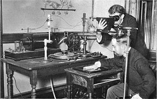
<span class="mw-page-title-main">Fluoroscopy</span> Production of an image when X-rays strike a fluorescent screen

Fluoroscopy is an imaging technique that uses X-rays to obtain real-time moving images of the interior of an object. In its primary application of medical imaging, a fluoroscope allows a surgeon to see the internal structure and function of a patient, so that the pumping action of the heart or the motion of swallowing, for example, can be watched. This is useful for both diagnosis and therapy and occurs in general radiology, interventional radiology, and image-guided surgery.

Equivalent dose is a dose quantity H representing the stochastic health effects of low levels of ionizing radiation on the human body which represents the probability of radiation-induced cancer and genetic damage. It is derived from the physical quantity absorbed dose, but also takes into account the biological effectiveness of the radiation, which is dependent on the radiation type and energy. In the SI system of units, the unit of measure is the sievert (Sv).

<span class="mw-page-title-main">Health physics</span>

Health physics, also referred to as the science of radiation protection, is the profession devoted to protecting people and their environment from potential radiation hazards, while making it possible to enjoy the beneficial uses of radiation. Health physicists normally require a four-year bachelor’s degree and qualifying experience that demonstrates a professional knowledge of the theory and application of radiation protection principles and closely related sciences. Health physicists principally work at facilities where radionuclides or other sources of ionizing radiation are used or produced; these include research, industry, education, medical facilities, nuclear power, military, environmental protection, enforcement of government regulations, and decontamination and decommissioning—the combination of education and experience for health physicists depends on the specific field in which the health physicist is engaged.

The roentgen equivalent man (rem) is a CGS unit of equivalent dose, effective dose, and committed dose, which are dose measures used to estimate potential health effects of low levels of ionizing radiation on the human body.

Absorbed dose is a dose quantity which is the measure of the energy deposited in matter by ionizing radiation per unit mass. Absorbed dose is used in the calculation of dose uptake in living tissue in both radiation protection, and radiology. It is also used to directly compare the effect of radiation on inanimate matter such as in radiation hardening.

In radiation physics, kerma is an acronym for "kinetic energy released per unit mass", defined as the sum of the initial kinetic energies of all the charged particles liberated by uncharged ionizing radiation in a sample of matter, divided by the mass of the sample. It is defined by the quotient .

<span class="mw-page-title-main">Rolf Maximilian Sievert</span> Swedish medical physicist, professor

Rolf Maximilian Sievert was a Swedish medical physicist whose major contribution was in the study of the biological effects of ionizing radiation.

The Ionising Radiations Regulations (IRR) are statutory instruments which form the main legal requirements for the use and control of ionising radiation in the United Kingdom. There have been several versions of the regulations, the current legislation was introduced in 2017 (IRR17), repealing the 1999 regulations and implementing the 2013/59/Euratom European Union directive.

The International Commission on Radiological Protection (ICRP) is an independent, international, non-governmental organization, with the mission to protect people, animals, and the environment from the harmful effects of ionising radiation. Its recommendations form the basis of radiological protection policy, regulations, guidelines and practice worldwide.

<span class="mw-page-title-main">Roentgen (unit)</span> Measurement of radiation exposure

The roentgen or röntgen is a legacy unit of measurement for the exposure of X-rays and gamma rays, and is defined as the electric charge freed by such radiation in a specified volume of air divided by the mass of that air . In 1928, it was adopted as the first international measurement quantity for ionizing radiation to be defined for radiation protection, as it was then the most easily replicated method of measuring air ionization by using ion chambers. It is named after the German physicist Wilhelm Röntgen, who discovered X-rays and was awarded the first Nobel Prize in Physics for the discovery.

Effective dose is a dose quantity in the International Commission on Radiological Protection (ICRP) system of radiological protection.

The committed dose in radiological protection is a measure of the stochastic health risk due to an intake of radioactive material into the human body. Stochastic in this context is defined as the probability of cancer induction and genetic damage, due to low levels of radiation. The SI unit of measure is the sievert.

<span class="mw-page-title-main">Radiation exposure</span> Measure of ionization of air by ionizing radiation

Radiation exposure is a measure of the ionization of air due to ionizing radiation from photons. It is defined as the electric charge freed by such radiation in a specified volume of air divided by the mass of that air. As of 2007, "medical radiation exposure" was defined by the International Commission on Radiological Protection as exposure incurred by people as part of their own medical or dental diagnosis or treatment; by persons, other than those occupationally exposed, knowingly, while voluntarily helping in the support and comfort of patients; and by volunteers in a programme of biomedical research involving their exposure. Common medical tests and treatments involving radiation include X-rays, CT scans, mammography, lung ventilation and perfusion scans, bone scans, cardiac perfusion scan, angiography, radiation therapy, and more. Each type of test carries its own amount of radiation exposure. There are two general categories of adverse health effects caused by radiation exposure: deterministic effects and stochastic effects. Deterministic effects are due to the killing/malfunction of cells following high doses; and stochastic effects involve either cancer development in exposed individuals caused by mutation of somatic cells, or heritable disease in their offspring from mutation of reproductive (germ) cells.

<span class="mw-page-title-main">Flight-time equivalent dose</span> Dose measurement of radiation

Flight-time equivalent dose (FED) is an informal unit of measurement of ionizing radiation exposure. Expressed in units of flight-time, one unit of flight-time is approximately equivalent to the radiological dose received during the same unit of time spent in an airliner at cruising altitude. FED is intended as a general educational unit to enable a better understanding of radiological dose by converting dose typically presented in sieverts into units of time. FED is only meant as an educational exercise and is not a formally adopted dose measurement.
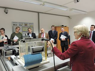
References

  1. Shoe-Fitting Fluoroscope (ca. 1930-1940) Archived 2008-12-17 at the Wayback Machine Oak Ridge Associated Universities.
  2. "Pedoscope X-ray apparatus, St Albans, England 1930-1955". Archived from the original on 2010-05-22. Retrieved 2010-06-03.
  3. "Pedoscope X-ray apparatus". Science Museum Group Collection. 2022. Archived from the original on 2020-11-11. Retrieved 2022-04-11.
  4. 1 2 3 4 Frame, Paul. "Shoe-Fitting Fluoroscope". Health Physics Historical Instrument Collection. Oak Ridge Associated Universities. Archived from the original on 26 September 2021. Retrieved 14 November 2012.
  5. Muller, Hermann Joseph (22 July 1927). "Artificial Mutation of the Gene" (PDF). Science. LXVI (1699): 84–87. Bibcode:1927Sci....66...84M. doi:10.1126/science.66.1699.84. PMID   17802387. Archived (PDF) from the original on 10 November 2019. Retrieved 13 November 2012.
  6. 1 2 3 4 5 Lewis, Leon; Paul E. Caplan (January 1950). "The Shoe-Fitting Fluoroscope as a Radiation Hazard". California Medicine. 72 (1): 26–30. PMC   1520288 . PMID   15408494.
  7. 1 2 3 4 Dyson, E. D. (4 August 1956). "Shoe Fitting X-Ray Fluoroscopes: Radiation Measurements and Hazards". British Medical Journal. 2 (4987): 269–272. doi:10.1136/bmj.2.4987.269. PMC   2034993 . PMID   13342467.
  8. Little JB (2000). "Chapter 14: Ionizing Radiation". In Kufe DW, Pollock RE, Weichselbaum RR, Bast RC Jr, Gansler TS, Holland JF, Frei E III (eds.). Cancer medicine (6th ed.). Hamilton, Ont: B.C. Decker. ISBN   1-55009-113-1. Archived from the original on 2016-01-02. Retrieved 2017-09-11.
  9. 1 2 3 4 5 Smullen, Michael J.; David E. Bertler (2007). "Basal Cell Carcinoma of the Sole: Possible Association with the Shoe-Fitting Fluoroscope" (PDF). Wisconsin Medical Journal. 106 (5): 275–278. PMID   17874675. Archived (PDF) from the original on 2 February 2017. Retrieved 13 November 2012.
  10. "The 2007 Recommendations of the International Commission on Radiological Protection". Annals of the ICRP. ICRP publication 103. 37 (2–4). 2007. ISBN   978-0-7020-3048-2. Archived from the original on 16 November 2012.
  11. 1 2 Lapp, David R. (1 January 2004). "The X-ray Shoe Fitter — An Early Application of Roentgen's 'New Kind of Ray'" (PDF). The Physics Teacher. 42 (6): 354–358. Bibcode:2004PhTea..42..354L. doi:10.1119/1.1790343. Archived from the original (PDF) on March 26, 2010. Retrieved 13 November 2012.
  12. Kopp, H. (7 December 1957). "Radiation Damage Caused by Shoe-Fitting Fluoroscope". British Medical Journal. 2 (5057): 1344–1345. doi:10.1136/bmj.2.5057.1344. PMC   1963031 . PMID   13479715.
  13. "Good shoe X-ray can't hurt'". Argus (Melbourne, Vic. : 1848 - 1957). 1951-03-01. p. 5. Archived from the original on 2022-05-09. Retrieved 2020-11-27.
  14. "Ban On Shoe X-ray Machines Resented". Canberra Times (ACT : 1926 - 1995). 1957-06-26. p. 4. Archived from the original on 2022-05-09. Retrieved 2020-11-27.
  15. "Shoe Men Say X-ray Harmless". The Daily News. Perth, Western Australia. 1949-11-26. p. 10. Archived from the original on 2022-05-09.
  16. Meinhold, C. B. (April 1996). One Hundred Years of X Rays and Radioactivity—Radiation Protection: Then and Now (PDF). Ninth International Congress. Vienna: International Radiation Protection Association. Archived (PDF) from the original on 3 March 2016.
  17. Clarke, R. H.; Valentin, J. (2009). "The History of ICRP and the Evolution of its Policies" (PDF). Annals of the ICRP. 39 (1): 75–110. doi:10.1016/j.icrp.2009.07.009. S2CID   71278114. Archived (PDF) from the original on 2012-05-08. Retrieved 2012-11-14.
  18. Fredrick, W. G.; Smith, R. G. (1948). "Health Problems of X-Ray Shoe Fitting". American Industrial Hygiene Association Quarterly. 9 (4): 89–93. doi:10.1080/00968204809344081.
  19. Lauriston S. Taylor, ed. (September 1949). "Appendix 16". The Tripartite Conferences on Radiation Protection. US Department of Energy. Archived from the original on 9 May 2022. Retrieved 14 November 2012.
  20. "Inquiry in Queensland". Sunday Mail (Brisbane, Qld. : 1926 - 1954). 1950-04-16. p. 4. Archived from the original on 2022-05-09. Retrieved 2020-11-27.
  21. Recommendations of the International Commission on Radiological Protection and of the International Commission on Radiological Units (PDF). National Bureau of Standards Handbook. Vol. 47. US Department of Commerce. 1950. Archived (PDF) from the original on 1 July 2015. Retrieved 14 November 2012.
  22. Nedd, C. A. II (1992). "When the Solution is the Problem: A Brief History of the Shoe Fluoroscope". American Journal of Roentgenology. 158 (6): 1270. doi: 10.2214/ajr.158.6.1590121 . PMID   1590121.
  23. Permissible Doses from External Sources of Radiation (PDF). National Bureau of Standards Handbook. Vol. 59. US Department of Commerce. 24 September 1954. p. 56. Archived (PDF) from the original on 8 September 2015. Retrieved 14 November 2012.
  24. Charlton, Amy (11 September 2012). "If the shoe fits…". Science Museum, London. Archived from the original on 21 June 2019. Retrieved 21 June 2019.
  25. Inkret, W. C.; Meinhold, C. B.; Taschner, J. C. (1995). "A Brief History of Radiation Protection Standards" (PDF). Los Alamos Science. 23: 116–123. Archived (PDF) from the original on 29 October 2012. Retrieved 12 November 2012.
  26. Dommann, Monika (2003). Durchsicht, Einsicht, Vorsicht, Eine Geschichte der Röntgenstrahlen 1896 bis 1963 (in German). Zürich: Chronos Verlag. ISBN   3-0340-0587-3.
  27. 1 2 Bauer, Elke (2019). Strahlenschutzunterweisung - Arbeitsbereich Isotopenanwendung (PDF) (Report) (in German). University of Ulm.
  28. Vennart, J. (1991). "1990 Recommendations of the International Commission on Radiological Protection". Journal of Radiological Protection. ICRP publication 60. 21 (1–3): 199–203. Bibcode:1991JRP....11..199V. doi:10.1088/0952-4746/11/3/006. ISBN   978-0-08-041144-6. S2CID   250822587 . Retrieved 17 May 2012.
  29. August, Melissa; Barovick, Harriet; Derrow, Michelle; Gray, Tam; Levy, Daniel S.; Lofaro, Lina; Spitz, David; Stein, Joel; Taylor, Chris (June 14, 1999). "The 100 Worst Ideas Of The Century". Time . Archived from the original on October 21, 2018. Retrieved April 3, 2020.(subscription required)
  30. "100 Worst Ideas of the Century". Archived from the original on 2020-07-14. Retrieved 2020-07-14.
  31. "American Restoration" Apples and X-Rays (TV Episode 2011) - IMDb , retrieved 2023-02-22
  32. "Readings Are Off the Scale". TV Tropes. Retrieved 2023-02-22.

Patents