
A body cavity is any space or compartment, or potential space, in an animal body. Cavities accommodate organs and other structures; cavities as potential spaces contain fluid.

A hernia is the abnormal exit of tissue or an organ, such as the bowel, through the wall of the cavity in which it normally resides. The term is also used for the normal development of the intestinal tract, referring to the retraction of the intestine from the extra-embryonal navel coelom into the abdomen in the healthy embryo at about 7½ weeks.
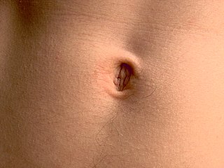
The navel is a protruding, flat, or hollowed area on the abdomen at the attachment site of the umbilical cord. All placental mammals have a navel, although it is generally more conspicuous in humans.

The umbilical vein is a vein present during fetal development that carries oxygenated blood from the placenta into the growing fetus. The umbilical vein provides convenient access to the central circulation of a neonate for restoration of blood volume and for administration of glucose and drugs.
The umbilical artery is a paired artery that is found in the abdominal and pelvic regions. In the fetus, it extends into the umbilical cord.

A Meckel's diverticulum, a true congenital diverticulum, is a slight bulge in the small intestine present at birth and a vestigial remnant of the vitelline duct. It is the most common malformation of the gastrointestinal tract and is present in approximately 2% of the population, with males more frequently experiencing symptoms.

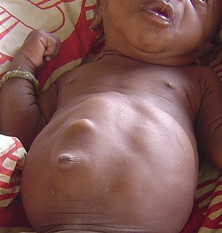
Omphalocele or omphalocoele also called exomphalos, is a rare abdominal wall defect. Beginning at the 6th week of development, rapid elongation of the gut and increased liver size reduces intra abdominal space, which pushes intestinal loops out of the abdominal cavity. Around 10th week, the intestine returns to the abdominal cavity and the process is completed by the 12th week. Persistence of intestine or the presence of other abdominal viscera in the umbilical cord results in an omphalocele.
An inguinal hernia or groin hernia is a hernia (protrusion) of abdominal cavity contents through the inguinal canal. Symptoms, which may include pain or discomfort especially with or following coughing, exercise, or bowel movements, are absent in about a third of patients. Symptoms often get worse throughout the day and improve when lying down. A bulging area may occur that becomes larger when bearing down. Inguinal hernias occur more often on the right than left side. The main concern is strangulation, where the blood supply to part of the intestine is blocked. This usually produces severe pain and tenderness of the area.

Gastroschisis is a birth defect in which the baby's intestines extend outside of the abdomen through a hole next to the belly button. The size of the hole is variable, and other organs including the stomach and liver may also occur outside the baby's body. Complications may include feeding problems, prematurity, intestinal atresia, and intrauterine growth restriction.
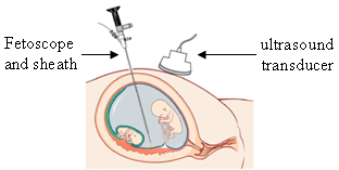
Fetoscopy is an endoscopic procedure during pregnancy to allow surgical access to the fetus, the amniotic cavity, the umbilical cord, and the fetal side of the placenta. A small (3–4 mm) incision is made in the abdomen, and an endoscope is inserted through the abdominal wall and uterus into the amniotic cavity. Fetoscopy allows for medical interventions such as a biopsy or a laser occlusion of abnormal blood vessels or the treatment of spina bifida.

An umbilical hernia is a health condition where the abdominal wall behind the navel is damaged. It may cause the navel to bulge outwards—the bulge consisting of abdominal fat from the greater omentum or occasionally parts of the small intestine. The bulge can often be pressed back through the hole in the abdominal wall, and may "pop out" when coughing or otherwise acting to increase intra-abdominal pressure. Treatment is surgical, and surgery may be performed for cosmetic as well as health-related reasons.
An epigastric hernia is a type of hernia that causes fat to push through a weakened area in the walls of the abdomen. It may develop in the epigastrium. Epigastric hernias are more common in adults and usually appear above the umbilical region of the abdomen. It is a common condition that is usually asymptomatic although sometimes their unusual clinical presentation can present a diagnostic dilemma for the clinician. Unlike the benign diastasis recti, epigastric hernia may trap fat and other tissues inside the opening of the hernia, causing pain and tissue damage. It is usually present at birth and may appear and disappear only when the patient is doing an activity that creates abdominal pressure, pushing to have bowel movements, or crying.

A Spigelian is the type of ventral hernia where aponeurotic fascia pushes through a hole in the junction of the linea semilunaris and the arcuate line, creating a bulge. It appears in the lower quadrant of the abdomen between an area of dense fibrous tissue and abdominal wall muscles causing a.
Femoral hernias occur just below the inguinal ligament, when abdominal contents pass through a naturally occurring weakness in the abdominal wall called the femoral canal. Femoral hernias are a relatively uncommon type, accounting for only 3% of all hernias. While femoral hernias can occur in both males and females, almost all develop in women due to the increased width of the female pelvis. Femoral hernias are more common in adults than in children. Those that do occur in children are more likely to be associated with a connective tissue disorder or with conditions that increase intra-abdominal pressure. Seventy percent of pediatric cases of femoral hernias occur in infants under the age of one.
An incisional hernia is a type of hernia caused by an incompletely-healed surgical wound. Since median incisions in the abdomen are frequent for abdominal exploratory surgery, ventral incisional hernias are often also classified as ventral hernias due to their location. Not all ventral hernias are from incisions, as some may be caused by other trauma or congenital problems.

The midgut is the portion of the human embryo from which most of the intestines develop. After it bends around the superior mesenteric artery, it is called the "midgut loop". It comprises the portion of the alimentary canal from the end of the foregut at the opening of the bile duct to the hindgut, about two-thirds of the way through the transverse colon.
Diastasis recti, or rectus abdominis diastasis, is defined as a gap of about 2.7 cm or greater between the two sides of the rectus abdominis muscle. The distance between the right and left rectus abdominis muscles is created by the stretching of the linea alba, a connective collagen sheath created by the aponeurosis insertions of the transverse abdominis, internal oblique, and external oblique. This condition has no associated morbidity or mortality. Physical therapy is often required to repair this separation and surgery is an option for more severe cases. Standard exercise rarely results in complete healing of the separated muscles.
The development of the reproductive system is the part of embryonic growth that results in the sex organs and contributes to sexual differentiation. Due to its large overlap with development of the urinary system, the two systems are typically described together as the genitourinary system.
Abdominal wall defects are a type of congenital defect that allows the stomach, the intestines, or other organs to protrude through an unusual opening that forms on the abdomen.
The development of the digestive system in the human embryo concerns the epithelium of the digestive system and the parenchyma of its derivatives, which originate from the endoderm. Connective tissue, muscular components, and peritoneal components originate in the mesoderm. Different regions of the gut tube such as the esophagus, stomach, duodenum, etc. are specified by a retinoic acid gradient that causes transcription factors unique to each region to be expressed. Differentiation of the gut and its derivatives depends upon reciprocal interactions between the gut endoderm and its surrounding mesoderm. Hox genes in the mesoderm are induced by a Hedgehog signaling pathway secreted by gut endoderm and regulate the craniocaudal organization of the gut and its derivatives. The gut system extends from the oropharyngeal membrane to the cloacal membrane and is divided into the foregut, midgut, and hindgut.