Vagal maneuver

Last updated

A vagal maneuver is an action used to stimulate the parasympathetic nervous system by activating the vagus nerve. The vagus nerve is the longest nerve of the autonomic nervous system and helps regulate many critical aspects of human physiology, including heart rate, blood pressure, sweating, and digestion through the release of acetylcholine. Common maneuvers that activate the vagus nerve include the Valsalva maneuver and carotid sinus massage, which can serve diagnostic or therapeutic functions.

Contents

Clinical indications

There are both diagnostic and therapeutic indications for the use of vagal maneuvers in clinical practice.

Diagnostic:

Therapeutic:

Types of vagal maneuvers

While many physical maneuvers can elicit autonomic responses, only some are appropriate for use in a clinical setting. The vagal maneuvers most often used for diagnostic or therapeutic purposes are those that can be reliably performed at bedside or in an office setting with minimal risk. A list of vagal maneuvers are listed below: [12] [4]

Other less clinically useful physical maneuvers that elicit a similar autonomic response through stimulation of the vagus nerve include:

Physiology

Parasympathetic nervous system mediated by vagal innervation Blausen 0703 Parasympathetic Innervation.png
Parasympathetic nervous system mediated by vagal innervation

Vagal maneuvers serve to stimulate the vagus nerve (cranial nerve X) through various mechanisms. The longest nerve in the body, the vagus nerve serves both motor and sensory functions through afferent and efferent signaling to and from the brain. The vagus nerve releases the neurotransmitter acetylcholine, [13] and is a main mediator for the parasympathetic nervous system.

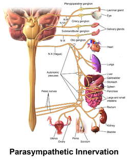
The vagus nerve exits the skull through the jugular foramen, moving down through the carotid sheath and dividing many times to influence multiple organ systems and directly innervating the pharynx, larynx, esophagus, heart, lung, and GI tract. Due to this wide nerve distribution, many physiologic process may be influenced through its stimulation, including heart rate and blood pressure. [14]

Stimulation of the vagus nerve through vagal maneuvers is thought to effect afferent fibers that carry sensory information from its distribution throughout the body to the nucleus tractus solitarii (NTS) in the dorsal medullary complex, where it is then relayed to other areas of the brain. This stimulation can also be done more directly through a therapy called Vagus Nerve Stimulation (VNS), which utilizes an implanted neuro-stimulator device and is approved clinically for controlling seizures in epilepsy patients and drug resistant depression. [15]

Vagal maneuvers make use of the vagus nerve's afferent and efferent bifunctional role, triggering reflexes (like the baroreceptor reflex, chemoreceptor reflex) and utilizing those afferent nerve fibers to increase nerve activity. This results in increased parasympathetic signaling through its efferent distribution and is mediated by the chemical messenger acetylcholine. [4]

Related Research Articles

<span class="mw-page-title-main">Vagus nerve</span> Cranial nerve X, for visceral innervation

The vagus nerve, also known as the tenth cranial nerve, cranial nerve X, or simply CN X, is a cranial nerve that carries sensory fibers that create a pathway that interfaces with the parasympathetic control of the heart, lungs, and digestive tract. It comprises two nerves—the left and right vagus nerves—but they are typically referred to collectively as a single subsystem. The vagus is the longest nerve of the autonomic nervous system in the human body and comprises both sensory and motor fibers. The sensory fibers originate from neurons of the nodose ganglion, whereas the motor fibers come from neurons of the dorsal motor nucleus of the vagus and the nucleus ambiguus. The vagus was also historically called the pneumogastric nerve.

<span class="mw-page-title-main">Parasympathetic nervous system</span> Division of the autonomic nervous system

The parasympathetic nervous system (PSNS) is one of the three divisions of the autonomic nervous system, the others being the sympathetic nervous system and the enteric nervous system. The enteric nervous system is sometimes considered part of the autonomic nervous system, and sometimes considered an independent system.

<span class="mw-page-title-main">Glossopharyngeal nerve</span> Cranial nerve IX, for the tongue and pharynx

The glossopharyngeal nerve, also known as the ninth cranial nerve, cranial nerve IX, or simply CN IX, is a cranial nerve that exits the brainstem from the sides of the upper medulla, just anterior to the vagus nerve. Being a mixed nerve (sensorimotor), it carries afferent sensory and efferent motor information. The motor division of the glossopharyngeal nerve is derived from the basal plate of the embryonic medulla oblongata, whereas the sensory division originates from the cranial neural crest.

<span class="mw-page-title-main">Valsalva maneuver</span> Technique for equalising pressure in the middle ears

The Valsalva maneuver is performed by a forceful attempt of exhalation against a closed airway, usually done by closing one's mouth and pinching one's nose shut while expelling air out as if blowing up a balloon. Variations of the maneuver can be used either in medical examination as a test of cardiac function and autonomic nervous control of the heart, or to clear the ears and sinuses when ambient pressure changes, as in scuba diving, hyperbaric oxygen therapy, or air travel.

<span class="mw-page-title-main">Palpitations</span> Perceived cardiac abnormality in which ones heartbeat can be felt

Palpitations are perceived abnormalities of the heartbeat characterized by awareness of cardiac muscle contractions in the chest, which is further characterized by the hard, fast and/or irregular beatings of the heart.

<span class="mw-page-title-main">Solitary nucleus</span> Sensory nuclei in medulla oblongata

The solitary nucleus is a series of sensory nuclei forming a vertical column of grey matter in the medulla oblongata of the brainstem. It receives general visceral and/or special visceral inputs from the facial nerve, glossopharyngeal nerve and vagus nerve ; it receives and relays stimuli related to taste and visceral sensation. It sends outputs to various parts of the brain. Neuron cell bodies of the SN are roughly somatotopically arranged along its length according to function.

<span class="mw-page-title-main">Supraventricular tachycardia</span> Abnormally fast heart rhythm

Supraventricular tachycardia (SVT) is an umbrella term for fast heart rhythms arising from the upper part of the heart. This is in contrast to the other group of fast heart rhythms – ventricular tachycardia, which start within the lower chambers of the heart. There are four main types of SVT: atrial fibrillation, atrial flutter, paroxysmal supraventricular tachycardia (PSVT), and Wolff–Parkinson–White syndrome. The symptoms of SVT include palpitations, feeling of faintness, sweating, shortness of breath, and/or chest pain.

<span class="mw-page-title-main">AV nodal reentrant tachycardia</span> Medical condition

AV-nodal reentrant tachycardia (AVNRT) is a type of abnormal fast heart rhythm. It is a type of supraventricular tachycardia (SVT), meaning that it originates from a location within the heart above the bundle of His. AV nodal reentrant tachycardia is the most common regular supraventricular tachycardia. It is more common in women than men. The main symptom is palpitations. Treatment may be with specific physical maneuvers, medications, or, rarely, synchronized cardioversion. Frequent attacks may require radiofrequency ablation, in which the abnormally conducting tissue in the heart is destroyed.

<span class="mw-page-title-main">Baroreflex</span> Homeostatic mechanism in the body

The baroreflex or baroreceptor reflex is one of the body's homeostatic mechanisms that helps to maintain blood pressure at nearly constant levels. The baroreflex provides a rapid negative feedback loop in which an elevated blood pressure causes the heart rate to decrease. Decreased blood pressure decreases baroreflex activation and causes heart rate to increase and to restore blood pressure levels. Their function is to sense pressure changes by responding to change in the tension of the arterial wall The baroreflex can begin to act in less than the duration of a cardiac cycle and thus baroreflex adjustments are key factors in dealing with postural hypotension, the tendency for blood pressure to decrease on standing due to gravity.

<span class="mw-page-title-main">Carotid sinus</span> Dilated area near internal carotid artery above bifurcation

In human anatomy, the carotid sinus is a dilated area at the base of the internal carotid artery just superior to the bifurcation of the internal carotid and external carotid at the level of the superior border of thyroid cartilage. The carotid sinus extends from the bifurcation to the "true" internal carotid artery. The carotid sinus is sensitive to pressure changes in the arterial blood at this level. It is the major baroreception site in humans and most mammals.

<span class="mw-page-title-main">Paroxysmal supraventricular tachycardia</span> Medical condition

Paroxysmal supraventricular tachycardia (PSVT) is a type of supraventricular tachycardia, named for its intermittent episodes of abrupt onset and termination. Often people have no symptoms. Otherwise symptoms may include palpitations, feeling lightheaded, sweating, shortness of breath, and chest pain.

The oculocardiac reflex, also known as Aschner phenomenon, Aschner reflex, or Aschner–Dagnini reflex, is a decrease in pulse rate associated with traction applied to extraocular muscles and/or compression of the eyeball. The reflex is mediated by nerve connections between the ophthalmic branch of the trigeminal cranial nerve via the ciliary ganglion, and the vagus nerve of the parasympathetic nervous system. Nerve fibres from the maxillary and mandibular divisions of the trigeminal nerve have also been documented. These afferents synapse with the visceral motor nucleus of the vagus nerve, located in the reticular formation of the brain stem. The efferent portion is carried by the vagus nerve from the cardiovascular center of the medulla to the heart, of which increased stimulation leads to decreased output of the sinoatrial node. This reflex is especially sensitive in neonates and children, particularly during strabismus correction surgery. Oculocardiac reflex can be profound during eye examination for retinopathy of prematurity. However, this reflex may also occur with adults. Bradycardia, junctional rhythm and asystole, all of which may be life-threatening, can be induced through this reflex. This reflex has been seen to occur during many pan facial trauma surgeries due to stimulation of any of the three branches of trigeminal nerve.

<span class="mw-page-title-main">Vagovagal reflex</span>

Vagovagal reflex refers to gastrointestinal tract reflex circuits where afferent and efferent fibers of the vagus nerve coordinate responses to gut stimuli via the dorsal vagal complex in the brain. The vagovagal reflex controls contraction of the gastrointestinal muscle layers in response to distension of the tract by food. This reflex also allows for the accommodation of large amounts of food in the gastrointestinal tracts.

The Bainbridge reflex or Bainbridge effect, also called the atrial reflex, is an increase in heart rate due to an increase in central venous pressure. Increased blood volume is detected by stretch receptors located in both sides of atria at the venoatrial junctions.

Reflex bradycardia is a bradycardia in response to the baroreceptor reflex, one of the body's homeostatic mechanisms for preventing abnormal increases in blood pressure. In the presence of high mean arterial pressure, the baroreceptor reflex produces a reflex bradycardia as a method of decreasing blood pressure by decreasing cardiac output.

The Bezold–Jarisch reflex involves a variety of cardiovascular and neurological processes which cause hypopnea, hypotension and bradycardia in response to noxious stimuli detected in the cardiac ventricles. The reflex is named after Albert von Bezold and Adolf Jarisch Junior. The significance of the discovery is that it was the first recognition of a chemical (non-mechanical) reflex.

The nervous system, and endocrine system collaborate in the digestive system to control gastric secretions, and motility associated with the movement of food throughout the gastrointestinal tract, including peristalsis, and segmentation contractions.

The inflammatory reflex is a neural circuit that regulates the immune response to injury and invasion. All reflexes have an afferent and efferent arc. The Inflammatory reflex has a sensory afferent arc, which is activated by cytokines, and a motor or efferent arc, which transmits action potentials in the vagus nerve to suppress cytokine production. Increased signaling in the efferent arc inhibits inflammation and prevents organ damage.

The Czermak–Hering test is a vagal maneuver consisting of the application of external digital pressure to the carotid sinus. The test is performed at the patient's bedside by imposing moderate pressure with the fingers, repeatedly massaging the left or the right carotid arteries.

<span class="mw-page-title-main">Automatic tachycardia</span> Medical condition

An automatic tachycardia is a cardiac arrhythmia which involves an area of the heart generating an abnormally fast rhythm, sometimes also called enhanced automaticity. These tachycardias, or fast heart rhythms, differ from reentrant tachycardias in which there is an abnormal electrical pathway which gives rise to the pathology. Most automatic tachycardias are supraventricular tachycardias (SVT). It is important to recognize an automatic tachycardia because the treatment will be different to that for a reentrant tachycardia. The most useful clue will be the presence of 'warm up' and 'cool down'. This means that whereas a reentrant tachycardia will both begin and end abruptly as cardiac conduction uses then ceases to use the accessory pathway, an automatic tachycardia will rise and fall gradually in rate as the automatic focus increases and decreases its automatic rate of electrical discharge.

References

  1. Rivasi, Giulia; Rafanelli, Martina; Ungar, Andrea (1 August 2018). "Usefulness of Tilt Testing and Carotid Sinus Massage for Evaluating Reflex Syncope". The American Journal of Cardiology. 122 (3): 517–520. doi:10.1016/j.amjcard.2018.04.033. ISSN   0002-9149. PMID   29954601. S2CID   49597948.
  2. Pasquier, Mathieu; Clair, Mathieu; Pruvot, Etienne; Hugli, Olivier; Carron, Pierre-Nicolas (12 October 2017). "Carotid Sinus Massage". The New England Journal of Medicine. 377 (15): e21. doi:10.1056/NEJMvcm1313338. ISSN   1533-4406. PMID   29020587.
  3. Page, Richard L.; Joglar, José A.; Caldwell, Mary A.; Calkins, Hugh; Conti, Jamie B.; Deal, Barbara J.; Estes, N. A. Mark; Field, Michael E.; Goldberger, Zachary D.; Hammill, Stephen C.; Indik, Julia H. (5 April 2016). "2015 ACC/AHA/HRS Guideline for the Management of Adult Patients With Supraventricular Tachycardia: A Report of the American College of Cardiology/American Heart Association Task Force on Clinical Practice Guidelines and the Heart Rhythm Society". Circulation. 133 (14): e506–574. doi: 10.1161/CIR.0000000000000311 . ISSN   1524-4539. PMID   26399663.
  4. 1 2 3 Niehues, Logan J.; Klovenski, Victoria (9 July 2021). Vagal Maneuver. StatPearls Publishing. PMID   31855402.
  5. Ceylan, Ezgi; Ozpolat, Cigdem; Onur, Ozge; Akoglu, Haldun; Denizbasi, Arzu (1 September 2019). "Initial and Sustained Response Effects of 3 Vagal Maneuvers in Supraventricular Tachycardia: A Randomized, Clinical Trial". Journal of Emergency Medicine. 57 (3): 299–305. doi:10.1016/j.jemermed.2019.06.008. ISSN   0736-4679. PMID   31443919. S2CID   201631721.
  6. Campbell, Marion; Buitrago, Silvia Ruiz (January 2017). "BET 2: Ice water immersion, other vagal manoeuvres or adenosine for SVT in children: Table 2". Emergency Medicine Journal. 34 (1): 58–60. doi:10.1136/emermed-2016-206487.2. ISSN   1472-0205. PMID   27974431. S2CID   29012408.
  7. Wei, J. Y.; Greene, H. L.; Weisfeldt, M. L. (January 1980). "Cough-facilitated conversion of ventricular tachycardia". The American Journal of Cardiology. 45 (1): 174–176. doi:10.1016/0002-9149(80)90235-0. ISSN   0002-9149. PMID   7350763.
  8. Hess, D. S.; Hanlon, T.; Scheinman, M.; Budge, R.; Desai, J. (March 1982). "Termination of ventricular tachycardia by carotid sinus massage". Circulation. 65 (3): 627–633. doi: 10.1161/01.cir.65.3.627 . ISSN   0009-7322. PMID   7055883.
  9. "UpToDate". www.uptodate.com. Retrieved 11 November 2021.
  10. Srivastava, Anubha; Kumar, Sanjay; Agarwal, Anil; Khetan, Dheeraj; Katharia, Rahul; Mishra, Prabhaker; Khati, Shikha; Gautam, Sujeet; Sandeep, Khuba (1 January 2021). "Evaluation of efficacy of Valsalva for attenuating needle puncture pain in first time nonremunerated voluntary plateletpheresis donors: A prospective, randomized controlled trial". Asian Journal of Transfusion Science. 15 (1): 68–74. doi: 10.4103/ajts.AJTS_95_20 . ISSN   0973-6247. PMC   8294442 . PMID   34349460.
  11. Kumar, Sanjay; Khuba, Sandeep; Agarwal, Anil; Gautam, Sujeet; Yadav, Madhulika; Dixit, Aanchal (December 2018). "Evaluation of efficacy of Valsalva maneuver for attenuating propofol injection pain: a prospective, randomized, single blind, placebo controlled study". Korean Journal of Anesthesiology. 71 (6): 453–458. doi:10.4097/kja.d.18.00017. ISSN   2005-6419. PMC   6283717 . PMID   29843507.
  12. "UpToDate". www.uptodate.com. Retrieved 11 November 2021.
  13. Lund, D D; Oda, R P; Pardini, B J; Schmid, P G (1 March 1986). "Vagus nerve stimulation alters regional acetylcholine turnover in rat heart". Circulation Research. 58 (3): 372–377. doi: 10.1161/01.RES.58.3.372 . PMID   3719926. S2CID   17017695.
  14. Kenny, Brian J.; Bordoni, Bruno (2021), "Neuroanatomy, Cranial Nerve 10 (Vagus Nerve)", StatPearls, Treasure Island (FL): StatPearls Publishing, PMID   30725856 , retrieved 11 November 2021
  15. Nemeroff, Charles B.; Mayberg, Helen S.; Krahl, Scott E.; McNamara, James; Frazer, Alan; Henry, Thomas R.; George, Mark S.; Charney, Dennis S.; Brannan, Stephen K. (July 2006). "VNS Therapy in Treatment-Resistant Depression: Clinical Evidence and Putative Neurobiological Mechanisms". Neuropsychopharmacology. 31 (7): 1345–1355. doi: 10.1038/sj.npp.1301082 . ISSN   1740-634X. PMID   16641939. S2CID   11455427.