Related Research Articles
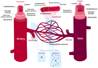
Blood vessels are the tubular structures of a circulatory system that transport blood throughout a vertebrate's body. Blood vessels transport blood cells, nutrients, and oxygen to most of the tissues of a body. They also take waste and carbon dioxide away from the tissues. Some tissues such as cartilage, epithelium, and the lens and cornea of the eye are not supplied with blood vessels and are termed avascular.

The sympathetic nervous system is one of the three divisions of the autonomic nervous system, the others being the parasympathetic nervous system and the enteric nervous system. The enteric nervous system is sometimes considered part of the autonomic nervous system, and sometimes considered an independent system.

The adrenergic receptors or adrenoceptors are a class of G protein-coupled receptors that are targets of many catecholamines like norepinephrine (noradrenaline) and epinephrine (adrenaline) produced by the body, but also many medications like beta blockers, beta-2 (β2) agonists and alpha-2 (α2) agonists, which are used to treat high blood pressure and asthma, for example.

Vasoconstriction is the narrowing of the blood vessels resulting from contraction of the muscular wall of the vessels, in particular the large arteries and small arterioles. The process is the opposite of vasodilation, the widening of blood vessels. The process is particularly important in controlling hemorrhage and reducing acute blood loss. When blood vessels constrict, the flow of blood is restricted or decreased, thus retaining body heat or increasing vascular resistance. This makes the skin turn paler because less blood reaches the surface, reducing the radiation of heat. On a larger level, vasoconstriction is one mechanism by which the body regulates and maintains mean arterial pressure.

The microcirculation is the circulation of the blood in the smallest blood vessels, the microvessels of the microvasculature present within organ tissues. The microvessels include terminal arterioles, metarterioles, capillaries, and venules. Arterioles carry oxygenated blood to the capillaries, and blood flows out of the capillaries through venules into veins.

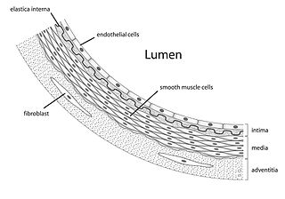
Vasodilation, also known as vasorelaxation, is the widening of blood vessels. It results from relaxation of smooth muscle cells within the vessel walls, in particular in the large veins, large arteries, and smaller arterioles. Blood vessel walls are composed of endothelial tissue and a basal membrane lining the lumen of the vessel, concentric smooth muscle layers on top of endothelial tissue, and an adventitia over the smooth muscle layers. Relaxation of the smooth muscle layer allows the blood vessel to dilate, as it is held in a semi-constricted state by sympathetic nervous system activity. Vasodilation is the opposite of vasoconstriction, which is the narrowing of blood vessels.
A neuroeffector junction is a site where a motor neuron releases a neurotransmitter to affect a target—non-neuronal—cell. This junction functions like a synapse. However, unlike most neurons, somatic efferent motor neurons innervate skeletal muscle, and are always excitatory. Visceral efferent neurons innervate smooth muscle, cardiac muscle, and glands, and have the ability to be either excitatory or inhibitory in function. Neuroeffector junctions are known as neuromuscular junctions when the target cell is a muscle fiber.
Vascular resistance is the resistance that must be overcome for blood to flow through the circulatory system. The resistance offered by the systemic circulation is known as the systemic vascular resistance or may sometimes be called by another term total peripheral resistance, while the resistance caused by the pulmonary circulation is known as the pulmonary vascular resistance. Vasoconstriction increases resistance, whereas vasodilation decreases resistance. Blood flow and cardiac output are related to blood pressure and inversely related to vascular resistance.
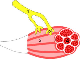
Vascular smooth muscle is the type of smooth muscle that makes up most of the walls of blood vessels.
An adrenergic nerve fibre is a neuron for which the neurotransmitter is either adrenaline (epinephrine), noradrenaline or dopamine. These neurotransmitters are released at a location known as the synapse, which is a junction point between the axon of one nerve cell and the dendrite of another. The neurotransmitters are first released from the axon and then bind to the receptor site on the dendrite. Adrenergic nerve terminals are found in the secondary neurons of the sympathetic nervous system, one of two divisions of the autonomic nervous system which is responsible for the fight-or-flight response. This system increases heart rate, slows digestion, dilates pupils, and also controls the secretion of apocrine sweat glands in the dermal layer of skin, in addition to other responses.

Phentolamine, sold under the brand name Regitine among others, is a reversible nonselective α-adrenergic antagonist.
alpha-1 (α1) adrenergic receptors are G protein-coupled receptors (GPCRs) associated with the Gq heterotrimeric G protein. α1-adrenergic receptors are subdivided into three highly homologous subtypes, i.e., α1A-, α1B-, and α1D-adrenergic receptor subtypes. There is no α1C receptor. At one time, there was a subtype known as α1C, but it was found to be identical to the previously discovered α1A receptor subtype. To avoid confusion, naming was continued with the letter D. Catecholamines like norepinephrine (noradrenaline) and epinephrine (adrenaline) signal through the α1-adrenergic receptors in the central and peripheral nervous systems. The crystal structure of the α1B-adrenergic receptor subtype has been determined in complex with the inverse agonist (+)-cyclazosin.
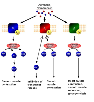
The alpha-2 (α2) adrenergic receptor is a G protein-coupled receptor (GPCR) associated with the Gi heterotrimeric G-protein. It consists of three highly homologous subtypes, including α2A-, α2B-, and α2C-adrenergic. Some species other than humans express a fourth α2D-adrenergic receptor as well. Catecholamines like norepinephrine (noradrenaline) and epinephrine (adrenaline) signal through the α2-adrenergic receptor in the central and peripheral nervous systems.
There are at least four known endothelin receptors, ETA, ETB1, ETB2 and ETC, all of which are G protein-coupled receptors whose activation result in elevation of intracellular-free calcium, which constricts the smooth muscles of the blood vessels, raising blood pressure, or relaxes the smooth muscles of the blood vessels, lowering blood pressure, among other functions.

Alpha blockers, also known as α-blockers or α-adrenoreceptor antagonists, are a class of pharmacological agents that act as antagonists on α-adrenergic receptors (α-adrenoceptors).
The vasomotor center (VMC) is a portion of the medulla oblongata. Together with the cardiovascular center and respiratory center, it regulates blood pressure. It also has a more minor role in other homeostatic processes. Upon increase in carbon dioxide level at central chemoreceptors, it stimulates the sympathetic system to constrict vessels. This is opposite to carbon dioxide in tissues causing vasodilatation, especially in the brain. Cranial nerves IX and X both feed into the vasomotor centre and are themselves involved in the regulation of blood pressure.
The catecholamines are a group of neurotransmitters composed of the endogenous substances dopamine, noradrenaline (norepinephrine), and adrenaline (epinephrine), as well as numerous artificially synthesized compounds such as isoprenaline - an anti-bradycardiac medication. Their investigation constitutes a major chapter in the history of physiology, biochemistry, and pharmacology. Adrenaline was the first hormone extracted from an endocrine gland and obtained in pure form, before the word hormone was coined. Adrenaline was also the first hormone whose structure and biosynthesis was discovered. Second to acetylcholine, adrenaline and noradrenaline were some of the first neurotransmitters discovered, and the first intercellular biochemical signals to be found in intracellular vesicles. The β-adrenoceptor gene was the first G protein-coupled receptor to be cloned.
Autonomic drugs are substances that can either inhibit or enhance the functions of the parasympathetic and sympathetic nervous systems. This type of drug can be used to treat a wide range of diseases an disorders, including glaucoma, asthma, and disorders of the urinary, gastrointestinal and circulatory systems.
Adrenergic neurone blockers, commonly known as adrenergic antagonists, are a group of drugs that inhibit the sympathetic nervous system by blocking the activity of adrenergic neurones. They prevent the action or release of catecholamines such as norepinephrine and epinephrine. They are located throughout the body, causing various physiological reactions including bronchodilation, accelerated heartbeat, and vasoconstriction. They work by inhibiting the synthesis, release, or reuptake of the neurotransmitters or by antagonising the receptors on postsynaptic neurones. Their medical uses, mechanisms of action, adverse effects, and contraindications depend on the specific types of adrenergic blockers used, including alpha 1, alpha 2, beta 1, and beta 2.

Pulmonary Arterial Hypertension (PAH) is a syndrome in which the blood pressure in the pulmonary arteries and pulmonary arterioles is elevated. This pre-capillary pulmonary artery pressure being elevated is essential, and by definition a mean pulmonary artery pressure greater than 20 mmHg as measured by a right heart catheterization is required for the diagnosis. This pre-capillary pulmonary hypertension is confirmed with measuring pulmonary vascular resistance being greater than 3 Woods Units. A pulmonary artery wedge pressure being less than 15 mmHg excludes post-capillary bed pulmonary hypertension. Pulmonary arterial hypertension is a subgroup of pulmonary hypertension and is categorized as World Health Organization as group 1. PAH is further subdivided into various categories based on the cause, including idiopathic, heritable, drug and toxin induced, PAH associated with specific diseases, PAH that is responsive to vasodilators, PAH with venous or capillary involvement, and persistent PAH in the newborn period.
References
- ↑ "Vasomotor" at Dorland's Medical Dictionary
- ↑ WebMD (2009). "vasomotor". Webster's New World Medical Dictionary (3rd ed.). Houghton Mifflin Harcourt. p. 447. ISBN 978-0-544-18897-6.
- 1 2 Robert C. Ward et al. (2002). In Foundations for osteopathic medicine . Lippincott Williams & Wilkins. 2nd edition. p. 98. ISBN 978-0-7817-3497-4. Google Book Search. Retrieved on 5 December 2010.
- ↑ Cater, D B; Adair, H M; Grove, C A (1966). "Effects of vasomotor drugs and 'mediators' of the inflammatory reaction upon the oxygen tension of tumours and tumour blood-flow". British Journal of Cancer. 20 (3): 504–16. doi:10.1038/bjc.1966.62. PMC 2007997 . PMID 4288476.
- ↑ B.D Cahurasia (2010). Human Anatomy: Regional & Applied (Dissection & Clinical) 5e (in 3 Vols.) Vol. 1: Upper Limb & Thorax With CD. New Delhi: CBS Publishers & Distributors. pp. 59, 133, 296. ISBN 978-81-239-1863-1.
- ↑ Guyton, Arthur C.; Hall, John Edward (2006). Textbook of Medical Physiology . Pennsylvania, United States: Elsevier Saunders. pp. 285, 1116. ISBN 978-0-7216-0240-0.