Vitreous hemorrhage

Last updated
Vitreous hemorrhage
Slit lamp photograph showing retinal detachment in Von Hippel-Lindau disease EDA08.JPG
Slit lamp photograph showing retinal detachment with visible vitreous hemorrhage.
Specialty Ophthalmology

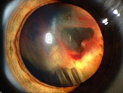
Vitreous hemorrhage is the extravasation, or leakage, of blood into the areas in and around the vitreous humor of the eye. [1] The vitreous humor is the clear gel that fills the space between the lens and the retina of the eye. A variety of conditions can result in blood leaking into the vitreous humor, which can cause impaired vision, floaters, and photopsia. [2]

Contents

Symptoms

Common symptoms of vitreous hemorrhage include:

Small vitreous hemorrhage often manifests itself as "floaters." A moderate case will often result in dark streaks in the vision, while dense vitreous hemorrhage can significantly inhibit vision. [3]

Causes

There are many factors known to cause vitreous hemorrhage.

Diabetic retinopathy

The most common cause found in adults is diabetic retinopathy. Abnormal blood vessels can form in the back of the eye of a person with diabetes. These new blood vessels are weaker and prone to breaking and causing hemorrhage. [2] Diabetic retinopathy accounts for 31.5–54% of all cases of vitreous hemorrhage in adults in the United States. [1]

Trauma

Some injuries can cause blood vessels in the back of the eye to bleed. Trauma is the leading cause of vitreous hemorrhage in young people, and accounts for 12–18.8% of cases in adults. [1]

Retinal tear or detachment

A tear in the retina can allow fluids from the eye to leak in behind the retina, which causes retinal detachment. When this occurs, blood from the retinal blood vessels can bleed into the vitreous. [4] Retinal tear accounts for 11.4–44% of vitreous hemorrhage cases. [1]

Posterior vitreous detachment

As one gets older, pockets of fluid can develop in the vitreous. When these pockets develop near the back of the eye, the vitreous can pull away from the retina and possibly tear it. [2] Posterior vitreous detachment accounts for 3.7–11.7% of vitreous hemorrhage cases. [1]

Other causes

Less common causes of vitreous hemorrhage make up 6.4–18% of cases, and include:

Diagnosis

Vitreous hemorrhage is diagnosed by identifying symptoms, examining the eye, and performing tests to identify the cause. Some common tests include:

Treatments

The treatment method used depends on the cause of the hemorrhage. In most cases, the patient is advised to rest with the head elevated 30–45°, and sometimes to put patches over the eyes to limit movement prior to treatment in order to allow the blood to settle. The patient is also advised to avoid taking medications that cause blood thinning (such as aspirin or similar medications).

The goal of the treatment is to fix the cause of the hemorrhage as quickly as possible. Retinal tears are closed by laser treatment or cryotherapy, and detached retinas are reattached surgically. [5]

Even after treatment, it can take months for the body to clear all of the blood from the vitreous. [2] In cases of vitreous hemorrhage due to detached retina, long-standing vitreous hemorrhage with a duration of more than 2–3 months, or cases associated with rubeosis iridis or glaucoma, a vitrectomy may be necessary to remove the standing blood in the vitreous.

Related Research Articles

Retinopathy

Retinopathy is any damage to the retina of the eyes, which may cause vision impairment. Retinopathy often refers to retinal vascular disease, or damage to the retina caused by abnormal blood flow. Age-related macular degeneration is technically included under the umbrella term retinopathy but is often discussed as a separate entity. Retinopathy, or retinal vascular disease, can be broadly categorized into proliferative and non-proliferative types. Frequently, retinopathy is an ocular manifestation of systemic disease as seen in diabetes or hypertension. Diabetes is the most common cause of retinopathy in the U.S. as of 2008. Diabetic retinopathy is the leading cause of blindness in working-aged people. It accounts for about 5% of blindness worldwide and is designated a priority eye disease by the World Health Organization.

Diabetic retinopathy

Diabetic retinopathy, also known as diabetic eye disease (DED), is a medical condition in which damage occurs to the retina due to diabetes mellitus. It is a leading cause of blindness in developed countries.

Floater Deposits within the eyes vitreous humour

Floaters or eye floaters are sometimes visible deposits within the eye's vitreous humour, which is normally transparent, or between the vitreous and retina. Each floater can be measured by its size, shape, consistency, refractive index, and motility. They are also called muscae volitantes, or mouches volantes. The vitreous usually starts out transparent, but imperfections may gradually develop as one ages. The common type of floater, present in most people's eyes, is due to these degenerative changes of the vitreous. The perception of floaters, which may be annoying or problematic to some people, is known as myodesopsia, or, less commonly, as myodaeopsia, myiodeopsia, or myiodesopsia. It is not often treated, except in severe cases, where vitrectomy (surgery), laser vitreolysis, and medication may be effective.

Vitrectomy Type of eye surgery

Vitrectomy is a surgery to remove some or all of the vitreous humor from the eye.

Retinoschisis Eye disease involving splitting of the retina

Retinoschisis is an eye disease characterized by the abnormal splitting of the retina's neurosensory layers, usually in the outer plexiform layer. Retinoschisis can be divided into degenerative forms which are very common and almost exclusively involve the peripheral retina and hereditary forms which are rare and involve the central retina as sometimes the peripheral retina. The degenerative forms are asymptomatic and involve the peripheral retina only and do not affect the visual acuity., Some rarer forms result in a loss of vision in the corresponding visual field.

Retinopathy of prematurity (ROP), also called retrolental fibroplasia (RLF) and Terry syndrome, is a disease of the eye affecting prematurely born babies generally having received neonatal intensive care, in which oxygen therapy is used due to the premature development of their lungs. It is thought to be caused by disorganized growth of retinal blood vessels which may result in scarring and retinal detachment. ROP can be mild and may resolve spontaneously, but it may lead to blindness in serious cases. Thus, all preterm babies are at risk for ROP, and very low birth-weight is an additional risk factor. Both oxygen toxicity and relative hypoxia can contribute to the development of ROP.

Macular edema

Macular edema occurs when fluid and protein deposits collect on or under the macula of the eye and causes it to thicken and swell (edema). The swelling may distort a person's central vision, because the macula holds tightly packed cones that provide sharp, clear, central vision to enable a person to see detail, form, and color that is directly in the centre of the field of view.

Retinal detachment Eye disorder

Retinal detachment is a disorder of the eye in which the retina separates from the layer underneath. Symptoms include an increase in the number of floaters, flashes of light, and worsening of the outer part of the visual field. This may be described as a curtain over part of the field of vision. In about 7% of cases both eyes are affected. Without treatment permanent loss of vision may occur.

Retinal haemorrhage is a disorder of the eye in which bleeding occurs in the retina, the light sensitive tissue, located on the back wall of the eye. There are photoreceptor cells in the retina called rods and cones, which transduce light energy into nerve signals that can be processed by the brain to form visual images. Retinal hemorrhage can affect adults, and newborn babies and infants may also suffer from this disorder.

Posterior vitreous detachment Eye condition

A posterior vitreous detachment (PVD) is a condition of the eye in which the vitreous membrane separates from the retina. It refers to the separation of the posterior hyaloid membrane from the retina anywhere posterior to the vitreous base.

Photopsia

Photopsia is the presence of perceived flashes of light in the field of vision.

Neovascularization is the natural formation of new blood vessels, usually in the form of functional microvascular networks, capable of perfusion by red blood cells, that form to serve as collateral circulation in response to local poor perfusion or ischemia.

Eales disease is a type of obliterative vasculopathy, also known as angiopathia retinae juvenilis, periphlebitis retinae, primary perivasculitis of the retina. It was first described by the British ophthalmologist Henry Eales (1852–1913) in 1880 and is a rare ocular disease characterized by inflammation and possible blockage of retinal blood vessels, abnormal growth of new blood vessels (neovascularization), and recurrent retinal and vitreal hemorrhages. This disease is identified by its three characteristic steps: vasculitis, occlusion, and retinal neovascularization, leading to recurrent vitreous hemorrhages and vision loss. Eales Disease with a characteristic clinical picture, fluorescein angiographic finding, and natural course is considered a specific disease entity. The exact cause of this disease is unknown but it appears to affect individuals that are from Asian subcontinents. This disease tends to begin between the ages of 20-30 years and begins with unilateral peripheral retinal perivasculitis and peripheral retinal capillary non-perfusion. These are not normally recognized until the inflammation results in vitreous hemorrhage. This disease has been found to affect the second eye 50-90% of the time so there is a large chance that both eyes will begin to show signs of the disease.

Coats disease Human eye disease causing full or partial blindness

Coats' disease, is a rare congenital, nonhereditary eye disorder, causing full or partial blindness, characterized by abnormal development of blood vessels behind the retina. Coats' disease can also fall under glaucoma.

Ocular ischemic syndrome is the constellation of ocular signs and symptoms secondary to severe, chronic arterial hypoperfusion to the eye. Amaurosis fugax is a form of acute vision loss caused by reduced blood flow to the eye; it may be a warning sign of an impending stroke, as both stroke and retinal artery occlusion can be caused by thromboembolism due to atherosclerosis elsewhere in the body. Consequently, those with transient blurring of vision are advised to urgently seek medical attention for a thorough evaluation of the carotid artery. Anterior segment ischemic syndrome is a similar ischemic condition of anterior segment usually seen in post-surgical cases. Retinal artery occlusion leads to rapid death of retinal cells, thereby resulting in severe loss of vision.

Epiretinal membrane Eye disease

Epiretinal membrane or macular pucker is a disease of the eye in response to changes in the vitreous humor or more rarely, diabetes. Sometimes, as a result of immune system response to protect the retina, cells converge in the macular area as the vitreous ages and pulls away in posterior vitreous detachment (PVD). PVD can create minor damage to the retina, stimulating exudate, inflammation, and leucocyte response. These cells can form a transparent layer gradually and, like all scar tissue, tighten to create tension on the retina which may bulge and pucker, or even cause swelling or macular edema. Often this results in distortions of vision that are clearly visible as bowing and blurring when looking at lines on chart paper within the macular area, or central 1.0 degree of visual arc. Usually it occurs in one eye first, and may cause binocular diplopia or double vision if the image from one eye is too different from the image of the other eye. The distortions can make objects look different in size, especially in the central portion of the visual field, creating a localized or field dependent aniseikonia that cannot be fully corrected optically with glasses. Partial correction often improves the binocular vision considerably though. In the young, these cells occasionally pull free and disintegrate on their own; but in the majority of sufferers the condition is permanent. The underlying photoreceptor cells, rod cells and cone cells, are usually not damaged unless the membrane becomes quite thick and hard; so usually there is no macular degeneration.

Laser coagulation or laser photocoagulation surgery is used to treat a number of eye diseases and has become widely used in recent decades. During the procedure, a laser is used to finely cauterize ocular blood vessels to attempt to bring about various therapeutic benefits.

Vitreomacular adhesion Human medical condition

Vitreomacular adhesion (VMA) is a human medical condition where the vitreous gel of the human eye adheres to the retina in an abnormally strong manner. As the eye ages, it is common for the vitreous to separate from the retina. But if this separation is not complete, i.e. there is still an adhesion, this can create pulling forces on the retina that may result in subsequent loss or distortion of vision. The adhesion in of itself is not dangerous, but the resulting pathological vitreomacular traction (VMT) can cause severe ocular damage.

Intravitreal injection

Intravitreal injection is the method of administration of drugs into the eye by injection with a fine needle. The medication will be directly applied into the vitreous humor. It is used to treat various eye diseases, such as age-related macular degeneration (AMD), diabetic retinopathy, and infections inside the eye such as endophthalmitis. As compared to topical administration, this method is beneficial for a more localized delivery of medications to the targeted site, as the needle can directly pass through the anatomical eye barrier and dynamic barrier. It could also minimize adverse drug effect to other body tissues via the systemic circulation, which could be a possible risk for intravenous injection of medications. Although there are risks of infections or other complications, with suitable precautions throughout the injection process, chances for these complications could be lowered.

Sickle cell retinopathy is a major ocular complication of the sickle cell disease (SCD) which causes permanent loss of vision. Retinopathy can occur in sickling hemoglobinopathies like sickle cell disease, sickle cell C disease, and sickle cell thalassaemia disease.

References

  1. 1 2 3 4 5 "Vitreous Hemorrhage: Background, Pathophysiology, Epidemiology". 20 October 2019. Retrieved 5 November 2019 via eMedicine.Cite journal requires |journal= (help)
  2. 1 2 3 4 5 6 Garibaldi, Daniel. "Vitreous Hemorrhage." CRS – Eye Advisor (2010): 1. Health Source – Consumer Edition. Web. 29 November 2011.
  3. "Vitreous Hemorrhage Clinical Presentation: History, Physical, Causes". emedicine.medscape.com. Retrieved 5 November 2019.
  4. "Retinal detachment: MedlinePlus Medical Encyclopedia". medlineplus.gov. Retrieved 5 November 2019.
  5. "Vitreous Hemorrhage Treatment & Management: Medical Care, Surgical Care, Consultations". 20 October 2019. Retrieved 5 November 2019 via eMedicine.Cite journal requires |journal= (help)
Classification
D