Related Research Articles

Coagulation, also known as clotting, is the process by which blood changes from a liquid to a gel, forming a blood clot. It potentially results in hemostasis, the cessation of blood loss from a damaged vessel, followed by repair. The mechanism of coagulation involves activation, adhesion and aggregation of platelets, as well as deposition and maturation of fibrin.

Von Willebrand disease (VWD) is the most common hereditary blood-clotting disorder in humans. An acquired form can sometimes result from other medical conditions. It arises from a deficiency in the quality or quantity of von Willebrand factor (VWF), a multimeric protein that is required for platelet adhesion. It is known to affect several breeds of dogs as well as humans. The three forms of VWD are hereditary, acquired, and pseudo or platelet type. The three types of hereditary VWD are VWD type 1, VWD type 2, and VWD type 3. Type 2 contains various subtypes. Platelet type VWD is also an inherited condition.

Factor VIII (FVIII) is an essential blood-clotting protein, also known as anti-hemophilic factor (AHF). In humans, factor VIII is encoded by the F8 gene. Defects in this gene result in hemophilia A, a recessive X-linked coagulation disorder. Factor VIII is produced in liver sinusoidal cells and endothelial cells outside the liver throughout the body. This protein circulates in the bloodstream in an inactive form, bound to another molecule called von Willebrand factor, until an injury that damages blood vessels occurs. In response to injury, coagulation factor VIII is activated and separates from von Willebrand factor. The active protein interacts with another coagulation factor called factor IX. This interaction sets off a chain of additional chemical reactions that form a blood clot.

von Willebrand factor (VWF) is a blood glycoprotein involved in hemostasis. It is deficient and/or defective in von Willebrand disease and is involved in many other diseases, including thrombotic thrombocytopenic purpura, Heyde's syndrome, and possibly hemolytic–uremic syndrome. Increased plasma levels in many cardiovascular, neoplastic, and connective tissue diseases are presumed to arise from adverse changes to the endothelium, and may predict an increased risk of thrombosis.
Weibel–Palade bodies (WPBs) are the storage granules of endothelial cells, the cells that form the inner lining of the blood vessels and heart. They store and release two principal molecules, von Willebrand factor and P-selectin, and thus play a dual role in hemostasis and inflammation.

ADAMTS13 —also known as von Willebrand factor-cleaving protease (VWFCP)—is a zinc-containing metalloprotease enzyme that cleaves von Willebrand factor (vWf), a large protein involved in blood clotting. It is secreted into the blood and degrades large vWf multimers, decreasing their activity.

Vitamin K epoxide reductase (VKOR) is an enzyme that reduces vitamin K after it has been oxidised in the carboxylation of glutamic acid residues in blood coagulation enzymes. VKOR is a member of a large family of predicted enzymes that are present in vertebrates, Drosophila, plants, bacteria and archaea. In some plant and bacterial homologues, the VKOR domain is fused with domains of the thioredoxin family of oxidoreductases.

Fibulin (FY-beau-lin) is the prototypic member of a multigene family, currently with seven members. Fibulin-1 is a calcium-binding glycoprotein. In vertebrates, fibulin-1 is found in blood and extracellular matrices. In the extracellular matrix, fibulin-1 associates with basement membranes and elastic fibers. The association with these matrix structures is mediated by its ability to interact with numerous extracellular matrix constituents including fibronectin, proteoglycans, laminins and tropoelastin. In blood, fibulin-1 binds to fibrinogen and incorporates into clots.
Platelet membrane glycoproteins are surface glycoproteins found on platelets (thrombocytes) which play a key role in hemostasis. When the blood vessel wall is damaged, platelet membrane glycoproteins interact with the extracellular matrix.

Platelet glycoprotein Ib alpha chain also known as glycoprotein Ib (platelet), alpha polypeptide or CD42b, is a protein that in humans is encoded by the GP1BA gene.

Discoidin domain is major protein domain of many blood coagulation factors.

Glycoprotein Ib (platelet), beta polypeptide (GP1BB) also known as CD42c, is a protein that in humans is encoded by the GP1BB gene.

Glycoprotein V (platelet) (GP5) also known as CD42d, is a human gene.
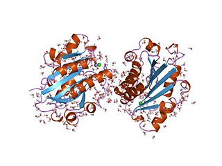
The von Willebrand factor type A (vWA) domain is a protein domain named after its occurrence in von Willebrand factor (vWF), a large multimeric glycoprotein found in blood plasma. Mutant forms of vWF are involved in the aetiology of bleeding disorders. This type A domain is the prototype for a protein superfamily.
Fibronectin, type I repeats are one of the three repeats found in the fibronectin protein. Fibronectin is a plasma protein that binds cell surfaces and various compounds including collagen, fibrin, heparin, DNA, and actin. Type I domain (FN1) is approximately 40 residues in length. Four conserved cysteines are involved in disulfide bonds. The 3D structure of the FN1 domain has been determined. It consists of two antiparallel beta-sheets, first a double-stranded one, that is linked by a disulfide bond to a triple-stranded beta-sheet. The second conserved disulfide bridge links the C-terminal adjacent strands of the domain.
Von Willebrand factor, type C is a protein domain is found in various blood plasma proteins: complement factors B, C2, CR3 and CR4; the integrins (I-domains); collagen types VI, VII, XII and XIV; and other extracellular proteins.

In molecular biology, the ars operon is an operon found in several bacterial taxon. It is required for the detoxification of arsenate, arsenite, and antimonite. This system transports arsenite and antimonite out of the cell. The pump is composed of two polypeptides, the products of the arsA and arsB genes. This two-subunit enzyme produces resistance to arsenite and antimonite. Arsenate, however, must first be reduced to arsenite before it is extruded. A third gene, arsC, expands the substrate specificity to allow for arsenate pumping and resistance. ArsC is an approximately 150-residue arsenate reductase that uses reduced glutathione (GSH) to convert arsenate to arsenite with a redox active cysteine residue in the active site. ArsC forms an active quaternary complex with GSH, arsenate, and glutaredoxin 1 (Grx1). The three ligands must be present simultaneously for reduction to occur.

A Link domain or Link module, also known as Xlink domain, is a protein domain that binds to hyaluronic acid. It is important in blood cell migration and apoptosis. The link domain is found in some extracellular proteins in vertebrates such as the hyalectans. It appears to be involved in extracellular matrix assembly and stability, cell adhesion, and migration.
SNED1 is a human protein expressed at low levels in a wide range of tissues. The protein is soluble and found in circulating blood and the conceptually translated protein has four domains of interest. These domains include a nidgen (NIDO) domain, three fibronectin type III (FN3) domains, several calcium binding EGF-like domains, and one complement control protein (CCP) domain. The gene is found on chromosome 2, locus q37.3. The mRNA was isolated from the spleen and is 6834bp in length. The conceptually translated protein is 1178aa long. This protein is predicted to interact with somatostatin, spermidine synthase and TMEM132C.
The GPIb-IX-V complex is a profuse membrane receptor complex originating in megakaryocytes and exclusively functional on the surface of platelets. It primarily functions to mediate the first critical step in platelet adhesion, by facilitating binding to von Willebrand factor (VWF) on damaged sub-endothelium under conditions of high fluid shear stress. Although the primary ligand for the GPIb-V-IX receptor is VWF, it can also bind to a number of other ligands in the circulation such as thrombin, P-selectin, factor XI, factor XII, high molecular weight kininogen as well as bacteria. GPIb-IX-V offers a critical role in thrombosis, metastasis, and the life cycle of platelets, and is implicated in a number of thrombotic pathological processes such as stroke or myocardial infarction.
References
- ↑ Perkins SJ, Smith KF, Williams SC, Haris PI, Chapman D, Sim RB (April 1994). "The secondary structure of the von Willebrand factor type A domain in factor B of human complement by Fourier transform infrared spectroscopy. Its occurrence in collagen types VI, VII, XII and XIV, the integrins and other proteins by averaged structure predictions". J. Mol. Biol. 238 (1): 104–19. doi:10.1006/jmbi.1994.1271. PMID 8145250.
- ↑ Zhou YF, Eng ET, Zhu J, Lu C, Walz T, Springer TA (12 July 2012). "Sequence and structure relationships within von Willebrand factor". Blood. 120 (2): 449–58. doi:10.1182/blood-2012-01-405134. PMC 3398765 . PMID 22490677.
- ↑ Dong X, Leksa NC, Chhabra ES, Arndt JW, Lu Q, Knockenhauer KE, Peters RT, Springer TA (4 April 2019). "The von Willebrand factor D'D3 assembly and structural principles for factor VIII binding and concatemer biogenesis". Blood. 133 (14): 1523–1533. doi: 10.1182/blood-2018-10-876300 . PMC 6450429 . PMID 30642920.
- ↑ Jorieux S, Fressinaud E, Goudemand J, Gaucher C, Meyer D, Mazurier C (May 2000). "Conformational changes in the D' domain of von Willebrand factor induced by CYS 25 and CYS 95 mutations lead to factor VIII binding defect and multimeric impairment". Blood. 95 (10): 3139–45. doi:10.1182/blood.V95.10.3139. PMID 10807780.