West Caucasian bat lyssavirus

Last updated
West Caucasian bat lyssavirus
Virus classification OOjs UI icon edit-ltr.svg
(unranked): Virus
Realm: Riboviria
Kingdom: Orthornavirae
Phylum: Negarnaviricota
Class: Monjiviricetes
Order: Mononegavirales
Family: Rhabdoviridae
Genus: Lyssavirus
Species:
West Caucasian bat lyssavirus
Synonyms [1]
  • West Caucasian bat virus

West Caucasian bat lyssavirus (WCBL) is a member of genus Lyssavirus , family Rhabdoviridae and order Mononegavirales . [2] This virus was first isolated from Miniopterus schreibersii, in the western Caucasus Mountains of southeastern Europe in 2002. [3] WCBL is the most divergent form of Lyssavirus, and is found in Miniopterus bats (insectivorous), Rousettus aegyptiacus , and Eidolon helvum . The latter two are both fruit bats. [2] The virus is fragile as it can be inactivated by UV light and chemicals, such as ether, chloroform, and bleach. [4] WCBL has not been known to infect humans thus far.

Contents

Classification

The lyssavirus genus can be divided into four phylogroups based upon DNA sequence homology. Phylogroup I includes viruses, such as Rabies virus, Duvenhage virus, European bat lyssavirus types 1 and 2, Australian bat lyssavirus, Khujand virus, Bokeloh bat lyssavirus, Irkut virus, and Aravan virus. Phylogroup II contains Lagos bat virus, Mokola virus, and Shimoni bat virus. West Caucasian bat lyssavirus is the only virus that is a part of phylogroup III. Ikoma lyssavirus and Lleida bat lyssavirus are examples in phylogroup IV. West Caucasian bat lyssavirus was classified within its own phylogroup because it is the most divergent lyssavirus that has been discovered. [5]

Discovery

This image shows an example of the structure of a negative sense single stranded RNA virus with a glycoprotein. 178-EbolaVirusProteins EbolaProteins.png
This image shows an example of the structure of a negative sense single stranded RNA virus with a glycoprotein.

Rabies viruses were found in bats as far back as 1954 in Germany. However, until a bat worker in Finland died as a result of rabies in 1985, few cases had been noted. Increased surveillance and documentation in the 1986 and 1987 revealed several additional cases. These virus strains mostly consisted of European bat lyssavirus type 1 (EBLV-1) and European bat lyssavirus type 2 (EBLV-2). From 1977 to 2011, 961 cases of rabies were reported in Europe. 91% were EBLV-1. The rest of the cases were suspected to be EBLV-2 and all but 3 have been confirmed. The 3 unconfirmed cases resulted in the discovery of West Caucasian bat lyssavirus (WCBL) in southwest Russia in 2002 and the Bokeloh bat lyssavirus in Germany in 2010. [7]

Virus structure

West Caucasian bat lyssavirus (WCBL) is a bullet shaped negative sense single stranded RNA virus. WCBL is composed of an internal helical nucleocapsid and a lipid envelope derived from the host cell. [4] The virus contains knobbed spikes that protrude from the membrane to aid in host membrane fusion. In addition, WCBL, along with other lyssaviruses, has a glycoprotein which is important in mediating viral entry. [4]

Virus genome

This is an image of the genome of West Caucasian bat lyssavirus, which comprises five main genes: N, P, M, G, and L. West Caucasian Bat Lyssavirus Genome.png
This is an image of the genome of West Caucasian bat lyssavirus, which comprises five main genes: N, P, M, G, and L.

The WCBL contains a linear genome that is 12,278 base pairs in length and comprises five main genes, denoted N, P, M, G, and L. Gene N encodes for the nucleoprotein, P encodes for the phosphoprotein, M encodes for matrix proteins, G encodes for the glycoprotein, and L encodes for the polymerase. [8] WCBL must encode for an RNA-dependent RNA polymerase (RdRp) in its genome in order for viral replication and synthesis to occur because it is a negative single-strand RNA virus. In comparison to other lyssaviruses, WCBL has a shorter trailer region of 57 nucleotides (as opposed to 69–70), but a longer non-coding region specifically in the glycoprotein gene at 697 nucleotides. [9] These differences have resulted in its classification into its own phylogroup. The West Caucasian bat lyssavirus also contains an open reading frame within the G gene which led researchers to believe parts of the glycoprotein were transcribed independently. However, a lack of a transcription initiation signal near the internal open reading frame has since confirmed that the glycoprotein is not transcribed in separate segments. [9]

Replication cycle and interaction with the host

The replication cycle for WCBL has not been specifically studied; however, it is said to be very similar to that of general lyssavirus, so the information listed below is regarding the genus as a whole.

Entry into cell

In order for lyssaviruses to enter into a host cell, the virus must attach to the host cell's receptor. This process is facilitated by the viral glycoprotein. Researchers are still unaware of the specific receptor the WCBL virus uses to gain entry into the host cell. Upon receptor activation, clathrin mediated endocytosis is provoked in which the cell absorbs the contents of the virus, including proteins. Next, the virus fuses to the vesicle membrane, allowing the viral nucleocapsid to enter into the cytoplasm of the host cell. [10] The phosphoprotein of WCBL can attach to cytoplasmic dynein LC8 for transport to the nucleus for viral replication. [11]

Replication and transcription

Next, the RNA-dependent RNA polymerase (RdRp) binds to the RNA genome and transcribes the five viral genes. In other words, the DNA is copied into a new strand of mRNA that will then hijack host cell translational machinery to synthesize proteins. Viral mRNA is capped and polyadenylated, which is the attachment of a string of adenine nucleotides to the 3’ end of the protein. The adenylation increases the half-life of the protein in order to regulate the activity. [12]

Assembly and release

Further, assembly of the virus starts when there is enough nucleoprotein (N) to encapsulate the genome. The virus is then released into non-nervous tissue. It is not easily detected due to the fact that it does not stimulate the immune system immediately. The incubation period can last anywhere from a few days to several months. After this time frame, it can move into the peripheral nervous system (PNS), and can eventually travel to the central nervous system (CNS) via the axonal transport system. At that point, it is possible to see clinical signs, such as weakness and lethargy due to encephalitis. Death often results several days after symptoms emerge. [12] [13]

Associated diseases

The WCBL virus is closely related to rabies. Although, WCBL has not yet infected humans, there is great risk due to its similar structure to other lyssaviruses which are known to infect humans. Unfortunately, the current rabies vaccine is not effective against WCBL as a result of the WCBL's slight divergence from other lyssaviruses. Therefore, if this virus begins to infect humans, the rabies vaccine will need to be improved to include effective antibodies for WCBL. [12]

Tropism

WCBL initially infects muscle tissue in the bats. As the virus progresses, it moves into the nervous tissue in both the PNS and CNS. [12] Although no studies have been completed thus far on the mammalian tropism of the WCBL virus, tropism for another more recently discovered lyssavirus, Australian Bat Lyssavirus (ABLV) has been explored. A variety of mammalian cell types including rabbits, other small rodents, monkeys, horses, and humans have shown to be permissive to ABVL. This led researchers to believe that the receptor of entry is likely conserved across several mammalian species. More research is necessary to determine if a variety of mammalian cell types are also permissive to WCBL virus. [14]

Outbreaks

There have been a few cases of outbreaks of WCBL. One was noted in Russia in 2002, which is the year that the virus was isolated. [7] A possible outbreak was noted in Kenya in 2008. [3]

Susceptibility and pathogenesis in bats

In order to gain an understanding of the susceptibility and pathogenesis of the West Caucasian bat lyssavirus (WCBL), big brown bats (Eptesicus fuscus) were inoculated with the virus intramuscularly in the deltoid muscle, in the neck, or orally. Blood and saliva samples were taken during disease progression and tissue samples were analyzed post-mortem. Specific tissues of interest included the brain, salivary glands, brown fat, lung, kidney, and bladder. Three bats died during the lethargic stage of viral infection (days 10 to 18), all of which were inoculated in the neck. Of those that died, only the tissue samples from the brain contained the infectious virus. However, both lung and salivary gland tissue contained viral RNA. Two of the three bats had viral RNA present in the bladder and in brown fat tissue as well. None of these three bats had viral RNA present in the kidney. All bats that survived were euthanized at 6 months. No viral particles were detected in the brain and salivary gland tissue samples of these bats. Of all the bats surveyed, only one of the three that died from viral infection had viral RNA present in the saliva at the time of death. [15]

WCBL antibodies were found in the serum of 4 of 7 bats that received intramuscular inoculation from a few weeks post-inoculation to the end of observation at 6 months. Those that died as a result of infection did not have any WCBL antibodies, a likely result of a shorter incubation period experienced from neck inoculation. None of the bats inoculated orally developed a serological response or the disease. This study indicates that the progression of WCBL infection is dependent on the location of inoculation. Further research is needed to develop a more complete understanding of inoculation route, pathogen adaptation, and host response.

[15]

Related Research Articles

<i>Indiana vesiculovirus</i> Species of virus

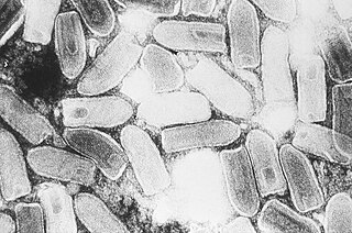
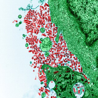
Indiana vesiculovirus, formerly Vesicular stomatitis Indiana virus is a virus in the family Rhabdoviridae; the well-known Rabies lyssavirus belongs to the same family. VSIV can infect insects, cattle, horses and pigs. It has particular importance to farmers in certain regions of the world where it infects cattle. This is because its clinical presentation is identical to the very important foot and mouth disease virus.

<i>Rhabdoviridae</i> Family of viruses in the order Mononegavirales

Rhabdoviridae is a family of negative-strand RNA viruses in the order Mononegavirales. Vertebrates, invertebrates, plants, fungi and protozoans serve as natural hosts. Diseases associated with member viruses include rabies encephalitis caused by the rabies virus, and flu-like symptoms in humans caused by vesiculoviruses. The name is derived from Ancient Greek rhabdos, meaning rod, referring to the shape of the viral particles. The family has 40 genera, most assigned to three subfamilies.

<i>Mononegavirales</i> Order of viruses

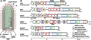
Mononegavirales is an order of negative-strand RNA viruses which have nonsegmented genomes. Some members that cause human disease in this order include Ebola virus, human respiratory syncytial virus, measles virus, mumps virus, Nipah virus, and rabies virus. Important pathogens of nonhuman animals and plants are also in the group. The order includes eleven virus families: Artoviridae, Bornaviridae, Filoviridae, Lispiviridae, Mymonaviridae, Nyamiviridae, Paramyxoviridae, Pneumoviridae, Rhabdoviridae, Sunviridae, and Xinmoviridae.

<span class="mw-page-title-main">Sin Nombre virus</span> Prototypical agent of hantavirus cardiopulmonary syndrome

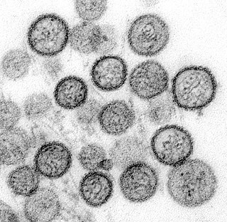
Sin Nombre virus (SNV) is the most common cause of hantavirus pulmonary syndrome (HPS) in North America. Sin Nombre virus is transmitted mainly by the eastern deer mouse. In its natural reservoir, SNV causes an asymptomatic, persistent infection and is spread through excretions, fighting, and grooming. Humans can become infected by inhaling aerosols that contain rodent saliva, urine, or feces, as well as through bites and scratches. In humans, infection leads to HPS, an illness characterized by an early phase of mild and moderate symptoms such as fever, headache, and fatigue, followed by sudden respiratory failure. The case fatality rate from infection is high, at 30–50%.

<i>Lyssavirus</i> Genus of viruses

Lyssavirus is a genus of RNA viruses in the family Rhabdoviridae, order Mononegavirales. Mammals, including humans, can serve as natural hosts. The genus Lyssavirus includes the causative agent of rabies.

<span class="mw-page-title-main">Rabies virus</span> Species of virus

Rabies virus, scientific name Rabies lyssavirus, is a neurotropic virus that causes rabies in animals, including humans. It can cause violence, hydrophobia, and fever. Rabies transmission can also occur through the saliva of animals and less commonly through contact with human saliva. Rabies lyssavirus, like many rhabdoviruses, has an extremely wide host range. In the wild it has been found infecting many mammalian species, while in the laboratory it has been found that birds can be infected, as well as cell cultures from mammals, birds, reptiles and insects. Rabies is reported in more than 150 countries and on all continents except Antarctica. The main burden of disease is reported in Asia and Africa, but some cases have been reported also in Europe in the past 10 years, especially in returning travellers.

<i>Lassa mammarenavirus</i> Type of viral hemorrhagic fever

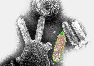
Lassa mammarenavirus (LASV) is an arenavirus that causes Lassa hemorrhagic fever, a type of viral hemorrhagic fever (VHF), in humans and other primates. Lassa mammarenavirus is an emerging virus and a select agent, requiring Biosafety Level 4-equivalent containment. It is endemic in West African countries, especially Sierra Leone, the Republic of Guinea, Nigeria, and Liberia, where the annual incidence of infection is between 300,000 and 500,000 cases, resulting in 5,000 deaths per year.

<i>Australian bat lyssavirus</i> Species of virus

Australian bat lyssavirus (ABLV), originally named Pteropid lyssavirus (PLV), is a enzootic virus closely related to the rabies virus. It was first identified in a 5-month-old juvenile black flying fox collected near Ballina in northern New South Wales, Australia, in January 1995 during a national surveillance program for the recently identified Hendra virus. ABLV is the seventh member of the genus Lyssavirus and the only Lyssavirus member present in Australia. ABLV has been categorized to the Phylogroup I of the Lyssaviruses.

Mokola lyssavirus, commonly called Mokola virus (MOKV), is an RNA virus related to rabies virus that has been sporadically isolated from mammals across sub-Saharan Africa. The majority of isolates have come from domestic cats exhibiting symptoms characteristically associated to rabies virus infection.

Seoul virus (SEOV) is one of the main causes of hemorrhagic fever with renal syndrome (HFRS). Seoul virus is transmitted by the brown rat and the black rat. In its natural reservoirs, SEOV causes an asymptomatic, persistent infection and is spread through excretions, fighting, and grooming. Humans can become infected by inhaling aerosols that contain rodent saliva, urine, or feces, as well as through bites and scratches. In humans, infection leads to HFRS, an illness characterized by general symptoms such as fever and headache, as well as the appearance of spots on the skin and renal symptoms such as kidney swelling, excess protein in urine, blood in urine, decreased urine production, and kidney failure. The case fatality rate from infection is 1–2%.

Snakehead rhabdovirus (SHRV) is a novirhabdovirus that affects warm water wild and pond-cultured fish of various species in Southeast Asia, including snakehead for which it is named.

<span class="mw-page-title-main">Rabies</span> Deadly viral disease, transmitted through animals

Rabies is a viral disease that causes encephalitis in humans and other mammals. It was historically referred to as hydrophobia because its victims panic when offered liquids to drink. Early symptoms can include fever and abnormal sensations at the site of exposure. These symptoms are followed by one or more of the following symptoms: nausea, vomiting, violent movements, uncontrolled excitement, fear of water, an inability to move parts of the body, confusion, and loss of consciousness. Once symptoms appear, the result is virtually always death. The time period between contracting the disease and the start of symptoms is usually one to three months but can vary from less than one week to more than one year. The time depends on the distance the virus must travel along peripheral nerves to reach the central nervous system.

Dobrava-Belgrade virus (DOBV) is the main cause of hemorrhagic fever with renal syndrome (HFRS) in southern Europe. In its natural reservoirs, DOBV causes a persistent, asymptomatic infection and is spread through excretions, fighting, and grooming. Humans can become infected by inhaling aerosols that contain rodent saliva, urine, or feces, as well as through bites and scratches. In humans, infection causes such as fever and headache, as well as the appearance of spots on the skin and renal symptoms such as kidney swelling, excess protein in urine, blood in urine, decreased urine production, and kidney failure. Acute respiratory distress syndrome occurs in about 10% of cases.

<span class="mw-page-title-main">Cryptic rabies</span>

Cryptic rabies refers to infection from unrecognized exposure to rabies virus. It is often phylogenetically traced to bats. It is most often seen in the southern United States. Silver-haired bats and tricolored bats are the two most common bat species associated with this form of infection, though both species are known to have less contact with humans than other bat species such as the big brown bat. That species is common throughout the United States and often roosts in buildings and homes where human contact is more likely.

Hantaan virus (HTNV) is the main cause of hemorrhagic fever with renal syndrome (HFRS) in East Asia. Hantaan virus is transmitted by the striped field mouse In its natural reservoir, HTNV causes a persistent, asymptomatic infection and is spread through excretions, fighting, and grooming. Humans can become infected by inhaling aerosols that contain rodent saliva, urine, or feces, as well as through bites and scratches. In humans, infection causes such as fever and headache, as well as the appearance of spots on the skin, hepatitis, and renal symptoms such as kidney swelling, excess protein in urine, blood in urine, decreased urine production, and kidney failure. Rarely, HTNV infection affects the pituitary gland and can cause empty sella syndrome. The case fatality rate from infection is up to 6.3%.

<span class="mw-page-title-main">Bat virome</span> Group of viruses associated with bats

The bat virome is the group of viruses associated with bats. Bats host a diverse array of viruses, including all seven types described by the Baltimore classification system: (I) double-stranded DNA viruses; (II) single-stranded DNA viruses; (III) double-stranded RNA viruses; (IV) positive-sense single-stranded RNA viruses; (V) negative-sense single-stranded RNA viruses; (VI) positive-sense single-stranded RNA viruses that replicate through a DNA intermediate; and (VII) double-stranded DNA viruses that replicate through a single-stranded RNA intermediate. The greatest share of bat-associated viruses identified as of 2020 are of type IV, in the family Coronaviridae.

European bat 1 lyssavirus(EBLV-1) is one of three rabies virus-like agents of the genus Lyssavirus found in serotine bats in Spain. Strains of EBLV-1 have been identified as EBLV-1a and EBLV-1b. EBLV-1a was isolated from bats found in the Netherlands and Russia, while EBLV-1b was found in bats in France, the Netherlands and Iberia. E. isabellinus bats are the EBLV-1b reservoir in the Iberian Peninsula. Between 1977 and 2010, 959 bat rabies cases of EBLV-1 were reported to the World Health Organization (WHO) Rabies Bulletin.

European bat 2 lyssavirus(EBLV-2) is one of three rabies-virus-like agents of the genus Lyssavirus found in Daubenton's bats in Great Britain. Human fatalities have occurred: the naturalist David McRae, who was bitten by a Daubenton's bat in Scotland, became infected with EBLV-2a and died in November 2002. It must now be assumed that the virus is present in bats in the UK. Testing of dead bats by MAFF/DEFRA over the last decade indicates that the overall incidence of infection is likely to be very low, although limited testing of live Daubenton's bats for antibodies suggests that exposure to EBLV-2 may be more widespread. Nevertheless, infected bat bites have caused human deaths so appropriate precautions against infection must be taken. The Department of Health’s recommendation is that people regularly handling bats should be vaccinated against rabies. Included in this category are all active bat workers and wardens, and those regularly taking in sick and injured bats. The Statutory Nature Conservation Organisations and the Bat Conservation Trust urge all those involved in bat work to ensure that they are fully vaccinated and that they receive regular boosters. Bats should not be handled by anyone who has not received these vaccinations. Even when fully vaccinated, people should avoid being bitten by wearing appropriate bite-proof gloves when handling bats. Any bat bite should be thoroughly cleansed with soap and water and advice should be sought from your doctor about the need for post-exposure treatment. Further information is available from the SNCOs, the Bat Conservation Trust or the Health Protection Agency (HPA) /Scottish Centre for Infection and Environmental Health (SCIEH).

Bat mumps orthorubulavirus, formerly Bat mumps rubulavirus (BMV), is a member of genus Orthorubulavirus, family Paramyxoviridae, and order Mononegavirales. Paramyxoviridae viruses were first isolated from bats using heminested PCR with degenerate primers. This process was then followed by Sanger sequencing. A specific location of this virus is not known because it was isolated from bats worldwide. Although multiple paramyxoviridae viruses have been isolated worldwide, BMV specifically has not been isolated thus far. However, BMV was detected in African fruit bats, but no infectious form has been isolated to date. It is known that BMV is transmitted through saliva in the respiratory system of bats. While the virus was considered its own species for a few years, phylogenetic analysis has since shown that it is a member of Mumps orthorubulavirus.

<i>Orthornavirae</i> Kingdom of viruses

Orthornavirae is a kingdom of viruses that have genomes made of ribonucleic acid (RNA), including genes which encode an RNA-dependent RNA polymerase (RdRp). The RdRp is used to transcribe the viral RNA genome into messenger RNA (mRNA) and to replicate the genome. Viruses in this kingdom share a number of characteristics which promote rapid evolution, including high rates of genetic mutation, recombination, and reassortment.

References

  1. Walker, Peter; et al. "mplementation of taxon-wide non-Latinized binomial species names in the family Rhabdoviridae" (PDF). International Committee on Taxonomy of Viruses (ICTV). Retrieved 12 March 2019.
  2. 1 2 "West Caucasian bat lyssavirus". www.genome.jp. Retrieved 2019-03-09.
  3. 1 2 Kuzmin, Ivan V.; Niezgoda, Michael; Franka, Richard; Agwanda, Bernard; Markotter, Wanda; Beagley, Janet C.; Urazova, Olga Yu; Breiman, Robert F.; Rupprecht, Charles E. (December 2008). "Possible Emergence of West Caucasian Bat Virus in Africa". Emerging Infectious Diseases. 14 (12): 1887–1889. doi:10.3201/eid1412.080750. ISSN   1080-6040. PMC   2634633 . PMID   19046512.
  4. 1 2 3 Rupprecht, Charles; Kuzmin, Ivan; Meslin, Francois (2017-02-23). "Lyssaviruses and rabies: current conundrums, concerns, contradictions and controversies". F1000Research. 6: 184. doi: 10.12688/f1000research.10416.1 . ISSN   2046-1402. PMC   5325067 . PMID   28299201.
  5. Gould, Allan R.; Kattenbelt, Jacqueline A.; Gumley, Sarah G.; Lunt, Ross A. (October 2002). "Characterisation of an Australian bat lyssavirus variant isolated from an insectivorous bat". Virus Research. 89 (1): 1–28. doi:10.1016/S0168-1702(02)00056-4. PMID   12367747.
  6. "Nucleoprotein", Wikipedia, 2019-02-10, retrieved 2019-03-12
  7. 1 2 "ProMED-mail post". www.promedmail.org. Retrieved 2019-03-09.
  8. "Virus Pathogen Database and Analysis Resource (ViPR) - Rhabdoviridae - Lyssavirus West Caucasian bat lyssavirus Strain UNKNOWN-NC_025377". www.viprbrc.org. Retrieved 2019-03-09.
  9. 1 2 Kuzmin, Ivan V.; Wu, Xianfu; Tordo, Noel; Rupprecht, Charles E. (September 2008). "Complete genomes of Aravan, Khujand, Irkut and West Caucasian bat viruses, with special attention to the polymerase gene and non-coding regions". Virus Research. 136 (1–2): 81–90. doi:10.1016/j.virusres.2008.04.021. ISSN   0168-1702. PMID   18514350.
  10. "Lyssavirus ~ ViralZone page". viralzone.expasy.org. Retrieved 2019-03-12.
  11. Jacob, Y.; Badrane, H.; Ceccaldi, P. E.; Tordo, N. (November 2000). "Cytoplasmic dynein LC8 interacts with lyssavirus phosphoprotein". Journal of Virology. 74 (21): 10217–10222. doi:10.1128/JVI.74.21.10217-10222.2000. ISSN   0022-538X. PMC   102062 . PMID   11024152.
  12. 1 2 3 4 Institute for International Cooperation in Animal Biologics; The Center for Food Security & Public Health (2004–2012). "Rabies and Rabies-Related Lyssaviruses" (PDF). CSFPH. Retrieved 2019-03-12.
  13. Warrell, D. A.; Warrell, M. J. (2004-03-20). "Rabies and other lyssavirus diseases". The Lancet. 363 (9413): 959–969. doi:10.1016/S0140-6736(04)15792-9. ISSN   0140-6736. PMID   15043965. S2CID   54326345.
  14. Weir, Dawn; Annand, Edward; Reid, Peter; Broder, Christopher (2014-02-19). "Recent Observations on Australian Bat Lyssavirus Tropism and Viral Entry". Viruses. 6 (2): 909–926. doi: 10.3390/v6020909 . ISSN   1999-4915. PMC   3939488 . PMID   24556791.
  15. 1 2 Hughes, G. J.; Kuzmin, I. V.; Schmitz, A.; Blanton, J.; Manangan, J.; Murphy, S.; Rupprecht, C. E. (2006-10-02). "Experimental infection of big brown bats (Eptesicus fuscus) with Eurasian bat lyssaviruses Aravan, Khujand, and Irkut virus". Archives of Virology. 151 (10): 2021–2035. doi: 10.1007/s00705-005-0785-0 . ISSN   0304-8608. PMID   16705370. S2CID   1626353.