Wide dynamic range neuron

Last updated
Gate Control Theory Firing of the inhibitory interneuron (responding to the non-painful stimuli) decreases the probability that the projection neuron (responsible for pain responses) will fire an action potential Gate control A firing.svg
Gate Control Theory Firing of the inhibitory interneuron (responding to the non-painful stimuli) decreases the probability that the projection neuron (responsible for pain responses) will fire an action potential

The wide dynamic range (WDR) neuron was first discovered by Mendell in 1966. [1] Early studies of this neuron established what is known as the gate control theory of pain. The basic concept is that non-painful stimuli block the pathways for painful stimuli, inhibiting possible painful responses. [2] This theory was supported by the fact that WDR neurons are responsible for responses to both painful and non-painful stimuli, and the idea that these neurons couldn't produce more than one of these responses simultaneously. WDR neurons respond to all types of somatosensory stimuli, make up the majority of the neurons found in the posterior grey column, and have the ability to produce long range responses including those responsible for pain and itch.

Contents

Anatomy and physiology

Spinal Cord Sectional Anatomy Spinal Cord Sectional Anatomy.png
Spinal Cord Sectional Anatomy

WDR neurons are found in the posterior grey column of the spinal cord. This area of the spinal cord houses two different types of neurons involved in the process of pain: WDR neurons and nociceptive specific neurons (NS). [3] As the name implies, NS neurons give specific short range responses. [4] WDR neurons are able to give long range responses for a large variety of stimuli giving them the ability to help identify the location and intensity of painful stimulation (sensory discrimination). [3] [5]

WDR neurons differ from most other neurons in that they are theorised to experience what is called a ‘wind up’. This allows for the intensity of their response to increase with an increased frequency of stimulus. [6] Most other neurons fire repeated action potentials of the same magnitude as a reaction to an increase in stimulus intensity. The intensity of the stimulus will only boost the frequency of action potentials, not their magnitude. However, WDR neurons exhibit increased action potential intensity with more presentations of a stimulus. [6] This allows for plasticity of synapses [6] and creates flexibility in the neuronal response. Though this may be of some benefit to the organism, this over excitation of the neurons can result in chronic pain. [3]

Role in pain responses

When there is a painful stimulus there are two pathways that can be taken. The nociceptive neurons in lamina 1 become compromised or the WDR neurons become compromised. [7] The WDR neurons can respond to electrical, mechanical, and thermal stimulation. [8] The dorsal cord has faulty plasticity, which encourages the development of neuropathic pain after an injury to a nerve. This allows for the over-excitation discussed previously, resulting in chronic pain. [9] The unique pain pathway of the WDR neurons allows information about the stimulus to be used to map out the intensity of the pain through sensory discrimination.

There are two main types of pain that we experience in our bodies: pain caused by damage of body tissue and pain caused by nerve damage. Nociceptive pain serves as a warning or signal for tissue damage and works to preserve the body’s equilibrium and functionality. This pain is signaled by the interworkings of both the peripheral and central nervous systems. [10] Another type of pain, known as neuropathic pain, is caused by a direct problem or disease that affects the nerves in the central nervous system. [10]

The sensory pathways the WDR neurons can play a role in. Touch Pain Pathways.png
The sensory pathways the WDR neurons can play a role in.

A subset of this neuropathic pain, known as chronic neuropathic pain, is characterized by its long lasting and high pain intensity. Although there is still a lot unknown about the direct cause of this chronic pain, it has been linked to WDR neurons. These neurons show significant activation by sympathetic stimulation while neurons, such as NS neurons, do not show the same level of activation. [11] The blocking of sympathetic pathways seemed to decrease pain and once unblocked, symptoms of pain persisted. This indicates that one of the many complex mechanisms contributing to this neuropathic chronic pain is the overstimulation of the WDR neurons by sympathetic stimulation. [10]

Another aspect that plays a role in neuropathic pain is the transient receptor channel called TRPA1. This channel is known to have influenced chronic pain injuries and diseases such as inflammation, diabetes, fibromyalgia, bronchitis, and emphysema. [12] WDR neurons are a huge part of the somatosensory system, helping to send and receive signals based on sensory changes in the body. The TRPA1 channel has been closely associated with temperature and pain sensation in primary afferent sensory neurons and are largely found in nociceptive sensory neurons in the dorsal root ganglia. [13] The inhibition of TRPA1 is known to contribute to different inflammatory and neuropathic diseases by amplifying the pain and hypersensitivity. [12] This is an area of study that is beneficial to continue to study and learn how to target and control aspects of chronic inflammatory and neuropathic diseases involved in sensory responses that WDR neurons play a role in.

Role in itch responses

Additionally, the itch pathway has also been linked with WDR neurons because itch and pain pathways are closely associated. As there are transient receptor channels present in the pain pathway, they are also present in the itch pathway. In the itch pathway, when the transient receptor channels are activated an itch response can be elicited. Itch responses can also be controlled by temperature changes (too high or too low), much like pain. This mechanism of control occurs when a stimulus is at an extremely low or extremely high temperature. The organism's sensitivity to the stimulus increases, meaning the pain or itch elicited will be greater at those temperatures than they would be at room temperature. [14] Though these pathways display many similarities, there are other mechanisms by which itch sensations can be controlled, such as those through nerve growth factor and substance-P. [15]

Brain imaging indicates similar activity in many areas of the brain such as prefrontal, supplementary motor areas, premotor cortex, anterior insular cortex, and many others when itch and pain regions are activated. [13] A better understanding of both these pathways will provide a greater understanding of WDR neurons.

Related Research Articles

Nociception is the sensory nervous system's process of encoding noxious stimuli. It deals with a series of events and processes required for an organism to receive a painful stimulus, convert it to a molecular signal, and recognize and characterize the signal in order to trigger an appropriate defense response.

<span class="mw-page-title-main">Itch</span> Sensation that causes the desire or reflex to scratch

Itch is a sensation that causes the desire or reflex to scratch. Itch has resisted many attempts to be classified as any one type of sensory experience. Itch has many similarities to pain, and while both are unpleasant sensory experiences, their behavioral response patterns are different. Pain creates a withdrawal reflex, whereas itch leads to a scratch reflex.

<span class="mw-page-title-main">Grey column</span>

The grey column refers to a somewhat ridge-shaped mass of grey matter in the spinal cord. This presents as three columns: the anterior grey column, the posterior grey column, and the lateral grey column, all of which are visible in cross-section of the spinal cord.

<span class="mw-page-title-main">Nociceptor</span> Sensory neuron that detects pain

A nociceptor is a sensory neuron that responds to damaging or potentially damaging stimuli by sending "possible threat" signals to the spinal cord and the brain. The brain creates the sensation of pain to direct attention to the body part, so the threat can be mitigated; this process is called nociception.

<span class="mw-page-title-main">Sensory neuron</span> Nerve cell that converts environmental stimuli into corresponding internal stimuli

Sensory neurons, also known as afferent neurons, are neurons in the nervous system, that convert a specific type of stimulus, via their receptors, into action potentials or graded potentials. This process is called sensory transduction. The cell bodies of the sensory neurons are located in the dorsal ganglia of the spinal cord.

<span class="mw-page-title-main">Hyperalgesia</span> Abnormally increased sensitivity to pain

Hyperalgesia is an abnormally increased sensitivity to pain, which may be caused by damage to nociceptors or peripheral nerves and can cause hypersensitivity to stimulus. Prostaglandins E and F are largely responsible for sensitizing the nociceptors. Temporary increased sensitivity to pain also occurs as part of sickness behavior, the evolved response to infection.

The withdrawal reflex is a spinal reflex intended to protect the body from damaging stimuli. The reflex rapidly coordinates the contractions of all the flexor muscles and the relaxations of the extensors in that limb causing sudden withdrawal from the potentially damaging stimulus. Spinal reflexes are often monosynaptic and are mediated by a simple reflex arc. A withdrawal reflex is mediated by a polysynaptic reflex resulting in the stimulation of many motor neurons in order to give a quick response.

Neuropathic pain is pain caused by damage or disease affecting the somatosensory system. Neuropathic pain may be associated with abnormal sensations called dysesthesia or pain from normally non-painful stimuli (allodynia). It may have continuous and/or episodic (paroxysmal) components. The latter resemble stabbings or electric shocks. Common qualities include burning or coldness, "pins and needles" sensations, numbness and itching.

<span class="mw-page-title-main">Referred pain</span> Pain perceived at a location other than the site of the painful stimulus

Referred pain, also called reflective pain, is pain perceived at a location other than the site of the painful stimulus. An example is the case of angina pectoris brought on by a myocardial infarction, where pain is often felt in the left side of neck, left shoulder, and back rather than in the thorax (chest), the site of the injury. The International Association for the Study of Pain has not officially defined the term; hence several authors have defined it differently.

Neuralgia is pain in the distribution of one or more nerves, as in intercostal neuralgia, trigeminal neuralgia, and glossopharyngeal neuralgia.

<span class="mw-page-title-main">Gate control theory</span>

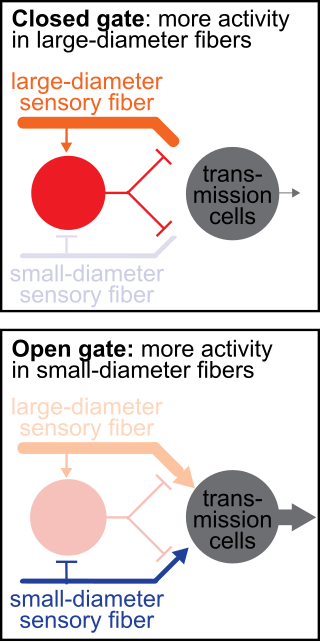
The gate control theory of pain asserts that non-painful input closes the nerve "gates" to painful input, which prevents pain sensation from traveling to the central nervous system.

<span class="mw-page-title-main">Allodynia</span> Medical condition

Allodynia is a condition in which pain is caused by a stimulus that does not normally elicit pain. For example, bad sunburn can cause temporary allodynia, and touching sunburned skin, or running cold or warm water over it, can be very painful. It is different from hyperalgesia, an exaggerated response from a normally painful stimulus. The term is from Ancient Greek άλλοςállos "other" and οδύνηodúnē "pain".

<span class="mw-page-title-main">Scratch reflex</span> Response to activation of sensory neurons

The scratch reflex is a response to activation of sensory neurons whose peripheral terminals are located on the surface of the body. Some sensory neurons can be activated by stimulation with an external object such as a parasite on the body surface. Alternatively, some sensory neurons can respond to a chemical stimulus that produces an itch sensation. During a scratch reflex, a nearby limb reaches toward and rubs against the site on the body surface that has been stimulated. The scratch reflex has been extensively studied to understand the functioning of neural networks in vertebrates. Despite decades of research, key aspects of the scratch reflex are still unknown, such as the neural mechanisms by which the reflex is terminated.

<span class="mw-page-title-main">Group C nerve fiber</span> One of three classes of nerve fiber in the central nervous system and peripheral nervous system

Group C nerve fibers are one of three classes of nerve fiber in the central nervous system (CNS) and peripheral nervous system (PNS). The C group fibers are unmyelinated and have a small diameter and low conduction velocity, whereas Groups A and B are myelinated. Group C fibers include postganglionic fibers in the autonomic nervous system (ANS), and nerve fibers at the dorsal roots. These fibers carry sensory information.

Mechanosensation is the transduction of mechanical stimuli into neural signals. Mechanosensation provides the basis for the senses of light touch, hearing, proprioception, and pain. Mechanoreceptors found in the skin, called cutaneous mechanoreceptors, are responsible for the sense of touch. Tiny cells in the inner ear, called hair cells, are responsible for hearing and balance. States of neuropathic pain, such as hyperalgesia and allodynia, are also directly related to mechanosensation. A wide array of elements are involved in the process of mechanosensation, many of which are still not fully understood.

<span class="mw-page-title-main">Rostral ventromedial medulla</span>

The rostral ventromedial medulla (RVM), or ventromedial nucleus of the spinal cord, is a group of neurons located close to the midline on the floor of the medulla oblongata (myelencephalon). The rostral ventromedial medulla sends descending inhibitory and excitatory fibers to the dorsal horn spinal cord neurons. There are 3 categories of neurons in the RVM: on-cells, off-cells, and neutral cells. They are characterized by their response to nociceptive input. Off-cells show a transitory decrease in firing rate right before a nociceptive reflex, and are theorized to be inhibitory. Activation of off-cells, either by morphine or by any other means, results in antinociception. On-cells show a burst of activity immediately preceding nociceptive input, and are theorized to be contributing to the excitatory drive. Neutral cells show no response to nociceptive input.

As long as humans have experienced pain, they have given explanations for its existence and sought soothing agents to dull or cease the painful sensation. Archaeologists have uncovered clay tablets dating back as far as 5,000 BC which reference the cultivation and use of the opium poppy to bring joy and cease pain. In 800 BC, the Greek writer Homer wrote in his epic, The Odyssey, of Telemachus, a man who used opium to soothe his pain and forget his worries. While some cultures researched analgesics and allowed or encouraged their use, others perceived pain to be a necessary, integral sensation. Physicians of the 19th century used pain as a diagnostic tool, theorizing that a greater amount of personally perceived pain was correlated to a greater internal vitality, and as a treatment in and of itself, inflicting pain on their patients to rid the patient of evil and unbalanced humors. This article focuses both on the history of how pain has been perceived across time and culture, but also how malleable an individual's perception of pain can be due to factors like situation, their visual perception of the pain, and previous history with pain.

Tactile induced analgesia is the phenomenon where concurrent touch and pain on the skin reduces the intensity of pain that is felt.

Sandra M. Garraway is a Canadian-American neuroscientist and assistant professor of physiology in the Department of Physiology at Emory University School of Medicine in Atlanta, Georgia. Garraway is the director of the Emory Multiplex Immunoassay Core (EMIC) where she assists researchers from both academia and industry to perform, analyze, and interpret their multiplexed immunoassays. Garraway studies the neural mechanisms of spinal nociceptive pain after spinal cord injury and as a postdoctoral researcher she discovered roles for both BDNF and ERK2 in pain sensitization and developed novel siRNA technology to inhibit ERK2 as a treatment for pain.

<span class="mw-page-title-main">Nociplastic pain</span> Symptom

Nociplastic pain or central sensitisation is a type of pain which is mechanically different from the normal nociceptive pain caused by inflammation and tissue damage or the neuropathic pain which results from nerve damage. It may occur in combination with the other types of pain or in isolation. Its location may be generalised or multifocal and it can be more intense than would be expected from any associated physical cause.

References

  1. Mendell, L. M. (1966-11-01). "Physiological properties of unmyelinated fiber projection to the spinal cord". Experimental Neurology. 16 (3): 316–332. doi:10.1016/0014-4886(66)90068-9. ISSN   0014-4886. PMID   5928985.
  2. Meyerson, Björn A.; Linderoth, Bengt (2006-04-01). "Mode of action of spinal cord stimulation in neuropathic pain". Journal of Pain and Symptom Management. 31 (4 Suppl): S6–12. doi: 10.1016/j.jpainsymman.2005.12.009 . ISSN   0885-3924. PMID   16647596.
  3. 1 2 3 Zhang, Tianhe (June 20, 2014). "Mechanisms and Models of Spinal Cord Stimulation for the Treatment of Neuropathic Pain". Brain Research. 1569: 19–31. doi:10.1016/j.brainres.2014.04.039. PMID   24802658. S2CID   377729.
  4. West, S.J. (August 6, 2015). "Circuitry and Plasticity of the Dorsal Horn - Toward a Better Understanding of Neuropathic Pain". Neuroscience. 300: 254–275. doi:10.1016/j.neuroscience.2015.05.020. PMID   25987204. S2CID   5368944.
  5. Craig, A.D. (March 6, 2017). "Pain Mechanisms: Labeled Lines Versus Convergence in Central Processing". Annual Review of Neuroscience. 26: 1–30. doi:10.1146/annurev.neuro.26.041002.131022. PMID   12651967.
  6. 1 2 3 D'Mello, R.; Dickenson, A. H. (2008-07-01). "Spinal cord mechanisms of pain". British Journal of Anaesthesia. 101 (1): 8–16. doi: 10.1093/bja/aen088 . ISSN   0007-0912. PMID   18417503.
  7. Inui, Koji (2012-11-01). "[Pain pathway]". Brain and Nerve = Shinkei Kenkyu No Shinpo. 64 (11): 1215–1224. ISSN   1881-6096. PMID   23131731.
  8. Lynn, R. B. (1992-05-27). "Mechanisms of esophageal pain". The American Journal of Medicine. 92 (5A): 11S–19S. doi:10.1016/0002-9343(92)80051-z. ISSN   0002-9343. PMID   1595755.
  9. West, S. J.; Bannister, K.; Dickenson, A. H.; Bennett, D. L. (2015-08-06). "Circuitry and plasticity of the dorsal horn--toward a better understanding of neuropathic pain". Neuroscience. 300: 254–275. doi:10.1016/j.neuroscience.2015.05.020. ISSN   1873-7544. PMID   25987204. S2CID   5368944.
  10. 1 2 3 Nickel, Florian T.; Seifert, Frank; Lanz, Stefan; Maihöfner, Christian (2012-02-01). "Mechanisms of neuropathic pain". European Neuropsychopharmacology. 22 (2): 81–91. doi:10.1016/j.euroneuro.2011.05.005. ISSN   0924-977X. PMID   21672666. S2CID   34762063.
  11. Roberts, W. J.; Foglesong, M. E. (1988-09-01). "Spinal recordings suggest that wide-dynamic-range neurons mediate sympathetically maintained pain". Pain. 34 (3): 289–304. doi:10.1016/0304-3959(88)90125-x. ISSN   0304-3959. PMID   3186277. S2CID   32428131.
  12. 1 2 Garrison, Sheldon R.; Stucky, Cheryl L. (2017-04-09). "The Dynamic TRPA1 Channel: A Suitable Pharmacological Pain Target?". Current Pharmaceutical Biotechnology. 12 (10): 1689–1697. doi:10.2174/138920111798357302. ISSN   1389-2010. PMC   3884818 . PMID   21466445.
  13. 1 2 Akiyama, Tasuku; Carstens, E. (2013-10-10). "Neural processing of itch". Neuroscience. 250: 697–714. doi:10.1016/j.neuroscience.2013.07.035. ISSN   0306-4522. PMC   3772667 . PMID   23891755.
  14. Patapoutain, Ardem (January 8, 2009). "Transient Receptor Potential Channels: Targeting Pain at the Source". Nat Rev Drug Discov. 8 (1): 55–68. doi:10.1038/nrd2757. PMC   2755576 . PMID   19116627.
  15. Lucaciu, Octavian C.; Connell, Gaelan P. (2013-07-01). "Itch sensation through transient receptor potential channels: a systematic review and relevance to manual therapy". Journal of Manipulative and Physiological Therapeutics. 36 (6): 385–393. doi:10.1016/j.jmpt.2013.05.018. ISSN   1532-6586. PMID   23896168.