AV nodal reentrant tachycardia

Last updated
AV-nodal reentrant tachycardia
Other namesAtrioventricular-nodal reentrant tachycardia
AV nodal reentrant tachycardia.png
An example of an ECG tracing typical of uncommon AV nodal reentrant tachycardia. Highlighted in yellow is the P wave that falls after the QRS complex.
Specialty Cardiology
Symptoms Palpitations, chest tightness, neck pulsation
Diagnostic method electrocardiogram, electrophysiological study
Differential diagnosis Atrioventricular reentrant tachycardia, focal atrial tachycardia, junctional ectopic tachycardia
Treatmentvagal manoeuvres, adenosine, ablation
Medication adenosine, calcium channel antagonists, beta blockers, flecainide

AV-nodal reentrant tachycardia (AVNRT) is a type of abnormal fast heart rhythm. It is a type of supraventricular tachycardia (SVT), meaning that it originates from a location within the heart above the bundle of His. AV nodal reentrant tachycardia is the most common regular supraventricular tachycardia. It is more common in women than men (approximately 75% of cases occur in females). The main symptom is palpitations. Treatment may be with specific physical maneuvers, medications, or, rarely, synchronized cardioversion. Frequent attacks may require radiofrequency ablation, in which the abnormally conducting tissue in the heart is destroyed.

Contents

AVNRT occurs when a reentrant circuit forms within or just next to the atrioventricular node. The circuit usually involves two anatomical pathways: the fast pathway and the slow pathway, which are both in the right atrium. The slow pathway (which is usually targeted for ablation) is located inferior and slightly posterior to the AV node, often following the anterior margin of the coronary sinus. The fast pathway is usually located just superior and posterior to the AV node. These pathways are formed from tissue that behaves very much like the AV node, and some authors regard them as part of the AV node.

The fast and slow pathways should not be confused with the accessory pathways that give rise to Wolff-Parkinson-White syndrome (WPW syndrome) or atrioventricular reciprocating tachycardia (AVRT). In AVNRT, the fast and slow pathways are located within the right atrium close to or within the AV node and exhibit electrophysiologic properties similar to AV nodal tissue. Accessory pathways that give rise to WPW syndrome and AVRT are located in the atrioventricular valvular rings. They provide a direct connection between the atria and ventricles, and have electrophysiologic properties similar to muscular heart tissue of the heart's ventricles.

Signs and symptoms

The main symptom of AVNRT is the sudden development of rapid regular palpitations. [1] These palpitations may be associated with a fluttering sensation in the neck, caused by near-simultaneous contraction of the atria and ventricles against a closed tricuspid valve leading to the pressure or atrial contraction being transmitted backwards into the venous system. [2] The rapid heart rate may lead to feelings of anxiety, and may therefore be mistaken for panic attacks. [2] In some cases, the onset of the fast heart is associated with a brief drop in blood pressure. When this happens, someone may experience dizziness or rarely lose consciousness (faint). [3] Someone with underlying coronary artery disease (narrowing of the arteries of the heart by atherosclerosis) who has a very rapid heart rate may experience chest pain similar to angina; this pain is band- or pressure-like around the chest and often radiates to the left arm and angle of the left jaw. [3]

Symptoms often occur without any specific trigger, although some find that their palpitations often occur after lifting heavy items or bending forwards. [1] The onset of palpitations is sudden, with the acceleration of the heart rate occurring within a single beat, and may be preceded by a feeling of the heart skipping a beat. The heart may continue to race for minutes or hours, but the eventual termination of the arrhythmia is as rapid as its onset. [1]

During AVNRT the heart rate is typically between 140 and 280 beats per minute. [3] Close inspection of the neck may reveal pulsation of the jugular vein in the form of "cannon A-waves" as the right atrium contracts against a closed tricuspid valve. [2]

Mechanisms

During typical AVNRT, electrical impulses travel down the slow pathway of the AV node and back up the fast pathway. De-Re entry (CardioNetworks ECGpedia).png
During typical AVNRT, electrical impulses travel down the slow pathway of the AV node and back up the fast pathway.

The fundamental mechanism of AVNRT is a presence of a dual atrioventricular node physiology (present in half of the population), which acts as a re-entrant circuit within the atrioventricular node. [4] This can take several forms. "Typical", "common", or "slow-fast" AVNRT uses the slow AV nodal pathway to conduct towards the ventricle (the anterograde limb of the circuit) and the fast AV nodal pathway to conduct to the atria (the retrograde limb). The re-entrant circuit can be reversed such that the fast AV nodal pathway is the anterograde limb and the slow AV nodal pathway is the retrograde limb, referred to as "atypical", "uncommon", or "fast-slow" AVNRT. Atypical AVNRT may also use the slow AV nodal pathway as the anterograde limb and left atrial fibres that approach the AV node from the left side of the inter-atrial septum as the retrograde limb, and is sometimes referred to as "slow-slow" AVNRT. [5]

Typical AVNRT

In typical AVNRT, the anterograde conduction is via the slow pathway and the retrograde conduction is via the fast pathway ("slow-fast" AVNRT).[ citation needed ]

Because the retrograde conduction is via the fast pathway, stimulation of the atria (which produces the inverted P wave) occurs very soon after stimulation of the ventricles (which causes the QRS complex). As a result, the time from the QRS complex to the P wave (the RP interval) is short, less than 50% of the time between consecutive QRS complexes. The RP interval is often so short that the inverted P waves may not be seen on the surface electrocardiogram (ECG) as they are buried within or immediately after the QRS complexes, appearing as a "pseudo R prime" wave in lead V1 or a "pseudo S" wave in the inferior leads. [6]

Atypical AVNRT

In atypical AVNRT, the anterograde conduction is via the fast pathway and the retrograde conduction is via the slow pathway ("fast-slow" AVNRT). [6]

Multiple slow pathways can exist so that both anterograde and retrograde conduction are over slow pathways. ("slow-slow" AVNRT).Because the retrograde conduction is via the slow pathway, stimulation of the atria will be delayed by the slow conduction tissue and will typically produce an inverted P wave that falls after the QRS complex on the surface ECG.[ citation needed ]

Diagnosis

If the symptoms are present while the person is receiving medical care (e.g., in an emergency department), an ECG may show typical changes that confirm the diagnosis i.e., QRS duration <120 ms, unless a heart block is suspected. [7] If the palpitations are recurrent, a doctor may request a Holter monitor (portable, wearable ECG recorder). Again, this will show the diagnosis if the recorder is attached at the time of the symptoms. In rare cases, disabling but infrequent episodes of palpitations may require the insertion of a small device under the skin that continuously record heart activity (an implantable loop recorder). All these ECG-based technologies also enable the distinction between AVNRT and other abnormal fast heart rhythms such as atrial fibrillation, atrial flutter, sinus tachycardia, ventricular tachycardia and tachyarrhythmias related to Wolff-Parkinson-White syndrome, all of which may have symptoms that are similar to AVNRT.[ citation needed ]

Blood tests commonly performed in people with palpitations are:[ citation needed ]

Treatment

Treatments for AVNRT aim to terminate episodes of tachycardia, and to prevent further episodes from occurring in the future. These treatments include physical manoeuvres, medication, and invasive procedures such as ablation. [8]

Arrhythmia termination

AVNRT termination following administration of adenosine De-Avnrt ecg (CardioNetworks ECGpedia).jpg
AVNRT termination following administration of adenosine

An episode of supraventricular tachycardia due to AVNRT can be terminated by any action that transiently blocks the AV node. Some of those with AVNRT may be able to stop their attack by using physical manoeuvres that increase the activity of the vagus nerve on the heart, specifically on the atrioventricular node. These manoeuvres include carotid sinus massage (pressure on the carotid sinus in the neck) and the Valsalva manoeuvre (increasing the pressure in the chest by attempting to exhale against a closed airway by bearing down or holding one's breath). [9]

Medications that slow or briefly halt electrical conduction through the AV node can terminate AVNRT, including adenosine, beta blockers, or non-dihydropyridine calcium channel blockers (such as verapamil or diltiazem). [9] Both adenosine and beta blockers may cause tightening of the airways, and are therefore used with caution in people who are known to have asthma. Less commonly used drugs for this purpose include antiarrhythmic drugs such as flecainide or amiodarone. [8]

If the fast heart rate is poorly tolerated (e.g. the development of heart failure symptoms, low blood pressure or coma) then AVNRT can be terminated electrically using a cardioversion. In this procedure, after administering a strong sedative or general anaesthetic, an electric shock is applied to the heart to restore a normal rhythm. [8]

Arrhythmia prevention

While preventative treatment may be very helpful at stopping the unpleasant symptoms associated with AVNRT, as this arrhythmia is a benign condition, preventative treatment is not essential. [8] Some of those who choose not to have further treatment will eventually become asymptomatic. [8] Those who wish to have further treatment can choose to take long term antiarrhythmic medication. The first line drugs are calcium channel antagonists and beta blockers, with second line agents including flecainide, amiodarone, and occasionally digoxin. These drugs are moderately effective at preventing further episodes but need to be taken long term. [8]

Alternatively, an invasive procedure called an electrophysiology (EP) study and catheter ablation can be used to confirm the diagnosis and potentially offer a cure. This procedure involves introducing wires or catheters into the heart through a vein in the leg. [2] The tip of one of these catheters can be used to heat or freeze the slow pathway of the AV node, destroying its ability to conduct electrical impulses, and preventing AVNRT. [10] The risks and benefits are weighed up before this is performed. Catheter ablation of the slow pathway, if successfully carried out, can potentially cure AVNRT with success rates of >95%, balanced against a small risk of complications including damaging the AV node and subsequently requiring a pacemaker. [8]

Related Research Articles

<span class="mw-page-title-main">Tachycardia</span> Heart rate that exceeds the normal resting rate

Tachycardia, also called tachyarrhythmia, is a heart rate that exceeds the normal resting rate. In general, a resting heart rate over 100 beats per minute is accepted as tachycardia in adults. Heart rates above the resting rate may be normal or abnormal.

<span class="mw-page-title-main">Wolff–Parkinson–White syndrome</span> Medical condition

Wolff–Parkinson–White syndrome (WPWS) is a disorder due to a specific type of problem with the electrical system of the heart involving an accessory pathway able to conduct electrical current between the atria and the ventricles, thus bypassing the atrioventricular node. About 60% of people with the electrical problem developed symptoms, which may include an abnormally fast heartbeat, palpitations, shortness of breath, lightheadedness, or syncope. Rarely, cardiac arrest may occur. The most common type of irregular heartbeat that occurs is known as paroxysmal supraventricular tachycardia.

<span class="mw-page-title-main">Atrioventricular node</span> Part of the electrical conduction system of the heart

The atrioventricular node or AV node electrically connects the heart's atria and ventricles to coordinate beating in the top of the heart; it is part of the electrical conduction system of the heart. The AV node lies at the lower back section of the interatrial septum near the opening of the coronary sinus, and conducts the normal electrical impulse from the atria to the ventricles. The AV node is quite compact.

<span class="mw-page-title-main">Atrial flutter</span> Medical condition

Atrial flutter (AFL) is a common abnormal heart rhythm that starts in the atrial chambers of the heart. When it first occurs, it is usually associated with a fast heart rate and is classified as a type of supraventricular tachycardia. Atrial flutter is characterized by a sudden-onset (usually) regular abnormal heart rhythm on an electrocardiogram (ECG) in which the heart rate is fast. Symptoms may include a feeling of the heart beating too fast, too hard, or skipping beats, chest discomfort, difficulty breathing, a feeling as if one's stomach has dropped, a feeling of being light-headed, or loss of consciousness.

<span class="mw-page-title-main">Cardiac conduction system</span> Aspect of heart function

The cardiac conduction system(CCS) (also called the electrical conduction system of the heart) transmits the signals generated by the sinoatrial node – the heart's pacemaker, to cause the heart muscle to contract, and pump blood through the body's circulatory system. The pacemaking signal travels through the right atrium to the atrioventricular node, along the bundle of His, and through the bundle branches to Purkinje fibers in the walls of the ventricles. The Purkinje fibers transmit the signals more rapidly to stimulate contraction of the ventricles.

<span class="mw-page-title-main">Supraventricular tachycardia</span> Abnormally fast heart rhythm

Supraventricular tachycardia (SVT) is an umbrella term for fast heart rhythms arising from the upper part of the heart. This is in contrast to the other group of fast heart rhythms – ventricular tachycardia, which start within the lower chambers of the heart. There are four main types of SVT: atrial fibrillation, atrial flutter, paroxysmal supraventricular tachycardia (PSVT), and Wolff–Parkinson–White syndrome. The symptoms of SVT include palpitations, feeling of faintness, sweating, shortness of breath, and/or chest pain.

<span class="mw-page-title-main">Atrioventricular block</span> Medical condition

Atrioventricular block is a type of heart block that occurs when the electrical signal traveling from the atria, or the upper chambers of the heart, to ventricles, or the lower chambers of the heart, is impaired. Normally, the sinoatrial node produces an electrical signal to control the heart rate. The signal travels from the SA node to the ventricles through the atrioventricular node. In an AV block, this electrical signal is either delayed or completely blocked. When the signal is completely blocked, the ventricles produce their own electrical signal to control the heart rate. The heart rate produced by the ventricles is much slower than that produced by the SA node.

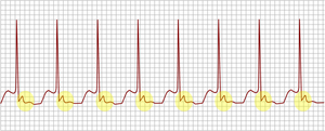
Premature atrial contraction (PAC), also known as atrial premature complexes (APC) or atrial premature beats (APB), are a common cardiac dysrhythmia characterized by premature heartbeats originating in the atria. While the sinoatrial node typically regulates the heartbeat during normal sinus rhythm, PACs occur when another region of the atria depolarizes before the sinoatrial node and thus triggers a premature heartbeat, in contrast to escape beats, in which the normal sinoatrial node fails, leaving a non-nodal pacemaker to initiate a late beat.

<span class="mw-page-title-main">Paroxysmal supraventricular tachycardia</span> Medical condition

Paroxysmal supraventricular tachycardia (PSVT) is a type of supraventricular tachycardia, named for its intermittent episodes of abrupt onset and termination. Often people have no symptoms. Otherwise symptoms may include palpitations, increased heart rate, feeling lightheaded, sweating, shortness of breath, and chest pain.

Lown–Ganong–Levine syndrome (LGL) is a pre-excitation syndrome of the heart. Those with LGL syndrome have episodes of abnormal heart racing with a short PR interval and normal QRS complexes seen on their electrocardiogram when in a normal sinus rhythm. LGL syndrome was originally thought to be due to an abnormal electrical connection between the atria and the ventricles, but is now thought to be due to accelerated conduction through the atrioventricular node in the majority of cases. The syndrome is named after Bernard Lown, William Francis Ganong, Jr., and Samuel A. Levine.

<span class="mw-page-title-main">Junctional rhythm</span> Medical condition

Junctional rhythm describes an abnormal heart rhythm resulting from impulses coming from a locus of tissue in the area of the atrioventricular node(AV node), the "junction" between atria and ventricles.

An accessory pathway is an additional electrical connection between two parts of the heart. These pathways can lead to abnormal heart rhythms or arrhythmias associated with symptoms of palpitations. Some pathways may activate a region of ventricular muscle earlier than would normally occur, referred to as pre-excitation, and this may be seen on an electrocardiogram. The combination of an accessory pathway that causes pre-excitation with arrhythmias is known as Wolff-Parkinson-White syndrome.

Re-entry ventricular arrhythmia is a type of paroxysmal tachycardia occurring in the ventricle where the cause of the arrhythmia is due to the electric signal not completing the normal circuit, but rather an alternative circuit looping back upon itself. There develops a self-perpetuating rapid and abnormal activation. Conditions necessary for re-entry include a combination of unidirectional block and slowed conduction. Circus movement may also occur on a smaller scale within the AV node, a large bypass tract is not necessary.

<span class="mw-page-title-main">Junctional tachycardia</span> Medical condition

Junctional tachycardia is a form of supraventricular tachycardia characterized by involvement of the AV node. It can be contrasted to atrial tachycardia. It is a tachycardia associated with the generation of impulses in a focus in the region of the atrioventricular node due to an A-V disassociation. In general, the AV junction's intrinsic rate is 40-60 bpm so an accelerated junctional rhythm is from 60-100bpm and then becomes junctional tachycardia at a rate of >100 bpm.

<span class="mw-page-title-main">Ectopic pacemaker</span> Cardiac condition

An ectopic pacemaker, also known as ectopic focus or ectopic foci, is an excitable group of cells that causes a premature heart beat outside the normally functioning SA node of the heart. It is thus a cardiac pacemaker that is ectopic, producing an ectopic beat. Acute occurrence is usually non-life-threatening, but chronic occurrence can progress into tachycardia, bradycardia or ventricular fibrillation. In a normal heart beat rhythm, the SA node usually suppresses the ectopic pacemaker activity due to the higher impulse rate of the SA node. However, in the instance of either a malfunctioning SA node or an ectopic focus bearing an intrinsic rate superior to SA node rate, ectopic pacemaker activity may take over the natural heart rhythm. This phenomenon is called an escape rhythm, the lower rhythm having escaped from the dominance of the upper rhythm. As a rule, premature ectopic beats indicate increased myocyte or conducting tissue excitability, whereas late ectopic beats indicate proximal pacemaker or conduction failure with an escape 'ectopic' beat.

<span class="mw-page-title-main">Junctional ectopic tachycardia</span> Medical condition

Junctional ectopic tachycardia (JET) is a rare syndrome of the heart that manifests in patients recovering from heart surgery. It is characterized by cardiac arrhythmia, or irregular beating of the heart, caused by abnormal conduction from or through the atrioventricular node. In newborns and infants up to 6 weeks old, the disease may also be referred to as His bundle tachycardia or congenital JET.

<span class="mw-page-title-main">PR interval</span> Period in electrocardiography

In electrocardiography, the PR interval is the period, measured in milliseconds, that extends from the beginning of the P wave until the beginning of the QRS complex ; it is normally between 120 and 200 ms in duration. The PR interval is sometimes termed the PQ interval.

<span class="mw-page-title-main">Atrioventricular reentrant tachycardia</span> Medical condition

Atrioventricular reentrant tachycardia (AVRT), or atrioventricular reciprocating tachycardia, is a type of abnormal fast heart rhythm and is classified as a type of supraventricular tachycardia (SVT). AVRT is most commonly associated with Wolff–Parkinson–White syndrome, but is also seen in permanent junctional reentrant tachycardia (PJRT). In AVRT, an accessory pathway allows electrical signals from the heart's ventricles to enter the atria and cause earlier than normal contraction, which leads to repeated stimulation of the atrioventricular node.

<span class="mw-page-title-main">Arrhythmia</span> Group of medical conditions characterized by irregular heartbeat

Arrhythmias, also known as cardiac arrhythmias, heart arrhythmias, or dysrhythmias, are irregularities in the heartbeat, including when it is too fast or too slow. A resting heart rate that is too fast – above 100 beats per minute in adults – is called tachycardia, and a resting heart rate that is too slow – below 60 beats per minute – is called bradycardia. Some types of arrhythmias have no symptoms. Symptoms, when present, may include palpitations or feeling a pause between heartbeats. In more serious cases, there may be lightheadedness, passing out, shortness of breath or chest pain. While most cases of arrhythmia are not serious, some predispose a person to complications such as stroke or heart failure. Others may result in sudden death.

<span class="mw-page-title-main">Permanent junctional reciprocating tachycardia</span> Medical condition

Permanent junctional reciprocating tachycardia(PJRT) is a rare cardiac arrhythmia. It is a supraventricular tachycardia, and a cause of atrioventricular reentrant tachycardia (AVRT). PJRT can cause chronic tachycardia that, untreated, leads to cardiomyopathy. The cause is an accessory pathway in the heart which conducts from the ventricles back to the atria. Unlike the accessory pathway in a more common cause of AVRT, Wolff–Parkinson–White syndrome, the accessory pathway in PJRT conducts slowly. This means that the associated tachycardia may be subclinical and only diagnosed at a late stage, after significant damage to the heart has been caused from prolonged and recurrent episodes of AVRT. While PJRT generally presents itself in infants, and often immediately after birth, few adults can suffer from a sudden onset of PJRT in which the degrading accessory pathway can more often than not be in a non-posteroseptal site.

References

  1. 1 2 3 Rosero, Spencer (2015), "A Brief Overview of Supraventricular Tachycardias", in Huang, MD, David T.; Prinzi, MD, Travis (eds.), Clinical Cardiac Electrophysiology in Clinical Practice, In Clinical Practice, Springer London, pp. 37–53, doi:10.1007/978-1-4471-5433-4_3, ISBN   978-1-4471-5432-7
  2. 1 2 3 4 Ayala-Paredes, Félix; Roux, Jean-Francois; Verdu, Mariano Badra (2014), Kibos, Ambrose S.; Knight, Bradley P.; Essebag, Vidal; Fishberger, Steven B. (eds.), "AVNRT Ablation: Significance of Anatomic Findings and Nodal Physiology", Cardiac Arrhythmias, Springer London, pp. 387–400, doi:10.1007/978-1-4471-5316-0_30, ISBN   978-1-4471-5315-3
  3. 1 2 3 Hafeez, Yamama; Armstrong, Tyler J. (2019), "Atrioventricular Nodal Reentry Tachycardia (AVNRT)", StatPearls, StatPearls Publishing, PMID   29763111 , retrieved 2019-08-15
  4. "Dual Atrioventricular Nodal Physiology - an overview | ScienceDirect Topics". www.sciencedirect.com. Retrieved 2022-11-14.
  5. "Atrioventricular Nodal Reentrant Tachycardia (AVNRT)". 2009-09-30.
  6. 1 2 Shen, Sharon; Knight, Bradley P. (2014), Kibos, Ambrose S.; Knight, Bradley P.; Essebag, Vidal; Fishberger, Steven B. (eds.), "How to Differentiate Between AVRT, AT, AVNRT, and Junctional Tachycardia Using the Baseline ECG and Intracardiac Tracings", Cardiac Arrhythmias, Springer London, pp. 199–208, doi:10.1007/978-1-4471-5316-0_15, ISBN   978-1-4471-5315-3
  7. Demosthenes G Katritsis; A John Camm (2010). "Atrioventricular nodal reentrant tachycardia". Circulation. 122 (8): 831–40. doi: 10.1161/CIRCULATIONAHA.110.936591 . PMID   20733110.
  8. 1 2 3 4 5 6 7 Page, Richard L.; Joglar, José A.; Caldwell, Mary A.; Calkins, Hugh; Conti, Jamie B.; Deal, Barbara J.; Estes, N. A. Mark; Field, Michael E.; Goldberger, Zachary D. (May 2016). "2015 ACC/AHA/HRS Guideline for the Management of Adult Patients With Supraventricular Tachycardia: A Report of the American College of Cardiology/American Heart Association Task Force on Clinical Practice Guidelines and the Heart Rhythm Society". Journal of the American College of Cardiology. 67 (13): e27–e115. doi: 10.1016/j.jacc.2015.08.856 . ISSN   1558-3597. PMID   26409259.
  9. 1 2 Brubaker, Sarah; Long, Brit; Koyfman, Alex (February 2018). "Alternative Treatment Options for Atrioventricular-Nodal-Reentry Tachycardia: An Emergency Medicine Review". The Journal of Emergency Medicine. 54 (2): 198–206. doi:10.1016/j.jemermed.2017.10.003. ISSN   0736-4679. PMID   29239759.
  10. Kumar, Darpan S.; Dewland, Thomas A.; Balaji, Seshadri; Henrikson, Charles A. (May 2017). "How to Approach Difficult Cases of AVNRT". Current Treatment Options in Cardiovascular Medicine. 19 (5): 34. doi:10.1007/s11936-017-0531-9. ISSN   1092-8464. PMID   28374333. S2CID   21354961.