Cone beam reconstruction

Last updated

In microtomography X-ray scanners, cone beam reconstruction is one of two common scanning methods, the other being Fan beam reconstruction. [1]
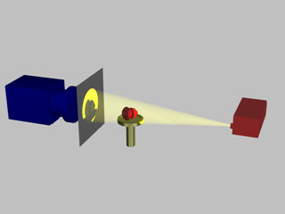
Cone beam set-up Cone beam image Cam 320x240.jpg
Cone beam set-up

Cone beam reconstruction uses a 2-dimensional approach for obtaining projection data. Instead of utilizing a single row of detectors, as fan beam methods do, a cone beam systems uses a standard charge-coupled device camera, focused on a scintillator material. The scintillator converts X-ray radiation to visible light, which is picked up by the camera and recorded. The method has enjoyed widespread implementation in microtomography, and is also used in several larger-scale systems.

An X-ray source is positioned across from the detector, with the object being scanned in between. (This is essentially the same setup used for an ordinary X-ray fluoroscope).

Projections from different angles are obtained in one of two ways. In one method, the object being scanned is rotated. This has the advantage of simplicity in implementation; a rotating stage results in little complexity. The second method involves rotating the X-ray source and camera around the object, as is done in ordinary CT scanning and SPECT imaging. This adds complexity, size and cost to the system, but removes the need to rotate the object.

Close-up to show the shadow image Cone beam image closeup 45 320x240.jpg
Close-up to show the shadow image

The method is referred to as cone-beam reconstruction because the X-rays are emitted from the source as a cone-shaped beam. In other words, it begins as a tight beam at the source, and expands as it moves away.

See also

Related Research Articles

CT scan Medical imaging procedure using X-rays to produce cross-sectional images

A CT scan or computed tomography scan is a medical imaging technique that uses computer-processed combinations of multiple X-ray measurements taken from different angles to produce tomographic (cross-sectional) images of a body, allowing the user to see inside the body without cutting. The personnel that perform CT scans are called radiographers or radiologic technologists.

Radiography

Radiography is an imaging technique using X-rays, gamma rays, or similar ionizing radiation and non-ionizing radiation to view the internal form of an object. Applications of radiography include medical radiography and industrial radiography. Similar techniques are used in airport security. To create an image in conventional radiography, a beam of X-rays is produced by an X-ray generator and is projected toward the object. A certain amount of the X-rays or other radiation is absorbed by the object, dependent on the object's density and structural composition. The X-rays that pass through the object are captured behind the object by a detector. The generation of flat two dimensional images by this technique is called projectional radiography. In computed tomography an X-ray source and its associated detectors rotate around the subject which itself moves through the conical X-ray beam produced. Any given point within the subject is crossed from many directions by many different beams at different times. Information regarding attenuation of these beams is collated and subjected to computation to generate two dimensional images in three planes which can be further processed to produce a three dimensional image.

Single-photon emission computed tomography Nuclear medicine tomographic imaging technique

Single-photon emission computed tomography is a nuclear medicine tomographic imaging technique using gamma rays. It is very similar to conventional nuclear medicine planar imaging using a gamma camera, but is able to provide true 3D information. This information is typically presented as cross-sectional slices through the patient, but can be freely reformatted or manipulated as required.

Tomography Imaging by sections or sectioning using a penetrative wave

Tomography is imaging by sections or sectioning through the use of any kind of penetrating wave. The method is used in radiology, archaeology, biology, atmospheric science, geophysics, oceanography, plasma physics, materials science, astrophysics, quantum information, and other areas of science. The word tomography is derived from Ancient Greek τόμος tomos, "slice, section" and γράφω graphō, "to write" or, in this context as well, " to describe." A device used in tomography is called a tomograph, while the image produced is a tomogram.

Gamma camera Camera to record gamma radiation

A gamma camera (γ-camera), also called a scintillation camera or Anger camera, is a device used to image gamma radiation emitting radioisotopes, a technique known as scintigraphy. The applications of scintigraphy include early drug development and nuclear medical imaging to view and analyse images of the human body or the distribution of medically injected, inhaled, or ingested radionuclides emitting gamma rays.

Technicare, formerly known as Ohio Nuclear, made CT, DR and MRI scanners and other medical imaging equipment. Its headquarters was in Solon, Ohio. Originally an independent company, it was later purchased by Johnson & Johnson. At the time, Invacare was also owned by Technicare. The company did not do well under Johnson & Johnson and in 1986, under economic pressure following unrelated losses from two Tylenol product tampering cases, J&J folded the company, selling the intellectual property and profitable service business to General Electric, a competitor.

Tomographic reconstruction is a type of multidimensional inverse problem where the challenge is to yield an estimate of a specific system from a finite number of projections. The mathematical basis for tomographic imaging was laid down by Johann Radon. A notable example of applications is the reconstruction of computed tomography (CT) where cross-sectional images of patients are obtained in non-invasive manner. Recent developments have seen the Radon transform and its inverse used for tasks related to realistic object insertion required for testing and evaluating computed tomography use in airport security.

Flat-field correction

Flat-field correction is a technique used to improve quality in digital imaging. It cancels the effects of image artifacts caused by variations in the pixel-to-pixel sensitivity of the detector and by distortions in the optical path. It is a standard calibration procedure in everything from personal digital cameras to large telescopes.

X-ray microtomography

X-ray microtomography, like tomography and X-ray computed tomography, uses X-rays to create cross-sections of a physical object that can be used to recreate a virtual model without destroying the original object. The prefix micro- is used to indicate that the pixel sizes of the cross-sections are in the micrometre range. These pixel sizes have also resulted in the terms high-resolution X-ray tomography, micro–computed tomography, and similar terms. Sometimes the terms high-resolution CT (HRCT) and micro-CT are differentiated, but in other cases the term high-resolution micro-CT is used. Virtually all tomography today is computed tomography.


Computed tomography laser mammography (CTLM) is the trademark of Imaging Diagnostic Systems, Inc. for its optical tomographic technique for female breast imaging.

Projectional radiography Formation of 2D images using X-rays

Projectional radiography, also known as conventional radiography, is a form of radiography and medical imaging that produces two-dimensional images by x-ray radiation. The image acquisition is generally performed by radiographers, and the images are often examined by radiologists. Both the procedure and any resultant images are often simply called "X-ray". Plain radiography generally refers to projectional radiography. Plain radiography can also refer to radiography without a radiocontrast agent or radiography that generates single static images, as contrasted to fluoroscopy, which are technically also projectional.

The 5DX was an automated X-ray inspection robot, which belonged to the set of automated test equipment robots and industrial robots utilizing machine vision. The 5DX was manufactured by Hewlett Packard, then later Agilent Technologies when HP was split into Hewlett Packard and Agilent Technologies in 1999. The 5DX performed a non-destructive structural test using X-ray laminography (tomography) to take 3D images of an assembled printed circuit board using 8-bit grayscale to indicate solder thickness. It was used in the assembled printed circuit board (PCB) electronics manufacturing industry to provide process feedback to a surface mount technology assembly line, as well as defect capture.

Flat-panel Volume CT is a technique under development to make computed tomography images with improved performance. The key difference between volume CT and traditional CT is that volume CT uses a two-dimensional x-ray detector orientation, to take multiple two-dimensional images. On the other hand, the conventional CT uses a one-dimensional x-ray detector orientation to take one-dimensional x-ray images.

Industrial computed tomography Computer-aided tomographic process

Industrial computed tomography (CT) scanning is any computer-aided tomographic process, usually X-ray computed tomography, that uses irradiation to produce three-dimensional internal and external representations of a scanned object. Industrial CT scanning has been used in many areas of industry for internal inspection of components. Some of the key uses for industrial CT scanning have been flaw detection, failure analysis, metrology, assembly analysis and reverse engineering applications. Just as in medical imaging, industrial imaging includes both nontomographic radiography and computed tomographic radiography.

Cone beam computed tomography medical imaging technique

Cone beam computed tomography is a medical imaging technique consisting of X-ray computed tomography where the X-rays are divergent, forming a cone.

Phase-contrast X-ray imaging

Phase-contrast X-ray imaging (PCI) or phase-sensitive X-ray imaging is a general term for different technical methods that use information concerning changes in the phase of an X-ray beam that passes through an object in order to create its images. Standard X-ray imaging techniques like radiography or computed tomography (CT) rely on a decrease of the X-ray beam's intensity (attenuation) when traversing the sample, which can be measured directly with the assistance of an X-ray detector. In PCI however, the beam's phase shift caused by the sample is not measured directly, but is transformed into variations in intensity, which then can be recorded by the detector.

Operation of computed tomography

X-ray computed tomography operates by using an X-ray generator that rotates around the object; X-ray detectors are positioned on the opposite side of the circle from the X-ray source.

Photon-counting computed tomography (CT) is a computed tomography technique currently under research and development, both within academia and by major vendors of CT systems. Photon-counting CT has the potential both to offer significant improvements to existing CT imaging techniques and to make possible completely novel applications. A photon-counting CT system employs a photon-counting detector (PCD) which registers the interactions of individual photons. By keeping track of the deposited energy in each interaction, the detector pixels of a PCD each record an approximate energy spectrum, making it a spectral or energy-resolved CT technique. In contrast, typical CT scanners use energy-integrating detectors (EIDs), where the total energy deposited in a pixel during a fixed period of time is registered. Typical CT detectors thus register only photon intensity, comparable to black-and-white photography, whereas photon-counting detectors register also spectral information, similar to colour photography.

History of computed tomography aspect of history

The history of X-ray computed tomography goes back to at least 1917 with the mathematical theory of the Radon transform In October 1963, William H. Oldendorf received a U.S. patent for a "radiant energy apparatus for investigating selected areas of interior objects obscured by dense material". The first commercially viable CT scanner was invented by Sir Godfrey Hounsfield in 1967.

Spectral imaging is an umbrella term for energy-resolved X-ray imaging in medicine. The technique makes use of the energy dependence of X-ray attenuation to either increase the contrast-to-noise ratio, or to provide quantitative image data and reduce image artefacts by so-called material decomposition. Dual-energy imaging, i.e. imaging at two energy levels, is a special case of spectral imaging and is still the most widely used terminology, but the terms "spectral imaging" and "spectral CT" have been coined to acknowledge the fact that photon-counting detectors have the potential for measurements at a larger number of energy levels.

References

  1. Chris C. Shaw (14 February 2014). Cone Beam Computed Tomography. Taylor & Francis. pp. 35–. ISBN   978-1-4398-4626-1.