Hip dysplasia (canine)

Last updated

Hip dysplasia in a Labrador Retriever Bilateral hip dysplasia.JPG
Hip dysplasia in a Labrador Retriever
Normal hips, for comparison Normal canine hips.JPG
Normal hips, for comparison

In dogs, hip dysplasia is an abnormal formation of the hip socket that, in its more severe form, can eventually cause lameness and arthritis of the joints. It is a genetic (polygenic) trait that is affected by environmental factors. It is common in many dog breeds, particularly the larger breeds, and is the most common single cause of arthritis of the hips. [1]

Contents

Overview

Normal hip anatomy

In the normal anatomy of the hip joint, the almost spherical end of the femur head (the caput, or caput ossis femoris) fits into the acetabulum (a concave socket located in the pelvis). The bony surfaces of the femur head and of the acetabulum are covered by cartilage. While bones provide the strength necessary to support body weight, cartilage ensures a smooth fit and a wide range of motion. Normal hip function can be affected by congenital conditions such as dysplasia, trauma, and by acquired diseases such as osteoarthritis and rheumatoid arthritis.

Dysplastic hip anatomy

The hip could have major contractions from dysplasias. The caput is not deeply and tightly held by the acetabulum. Instead of being a snug fit, it is a loose fit, or a partial fit. Secondly, the caput or acetabulum are not smooth and round, but are misshapen, causing abnormal wear and tear or friction within the joint as it moves. [2] The body reacts to this in several ways. First, the joint is continually repairing itself and laying down new cartilage. However, cartilage repair is a relatively slow process, the tissue being avascular, so the joint may suffer degradation due to the abnormal wear and tear, or may not support the body weight as intended. The joint becomes inflamed and a cycle of cartilage damage, inflammation and pain commences. This is a self-fueling process, in that the more the joint becomes damaged, the more damage it will cause to the surrounding tissues and bones. The inflammation also causes further damage. The bones of the joint may also develop osteoarthritis, visible on a radiograph as small outcrops of bone, which further degrade the joint. [3] Osteoarthritis is a degenerative disease marked by the breakdown of cartilage between joints resulting in painful bone-to-bone contact. [4]

The underlying deformity of the joint may get worse over time, or may remain static. A dog may have good radiographs and yet be in pain, or may have very poor radiographs and have no apparent pain issues. The hip condition is only one factor to determine the extent to which dysplasia is causing pain or affecting the quality of life. In mild to moderate dysplasia it is often the secondary effects of abnormal wear and tear or arthritis, rather than dysplasia itself, which is the direct causes of visible problems. [5]

Causes and effects

A Labrador Retriever standing with hind legs close together to compensate for hip dysplasia Hip dysplasia gait.jpg
A Labrador Retriever standing with hind legs close together to compensate for hip dysplasia

Hip dysplasia may be caused by a femur that does not fit correctly into the pelvic socket, or poorly developed muscles in the pelvic area. Large and giant breeds are most susceptible to hip dysplasia (possibly due to the body mass index (BMI) of the individual animal), [6] though many other breeds can suffer from it. The Orthopedic Foundation for Animals maintains a list of top 100 breeds affected. [7]

To reduce pain, the animal will typically reduce its movement of that hip. This may be visible as "bunny hopping", where both legs move together, or less dynamic movement (running, jumping), or stiffness. Since the hip cannot move fully, the body compensates by adapting its use of the spine, often causing spinal, stifle (a dog's knee joint), or soft tissue problems to arise.

The causes of hip dysplasia are considered heritable, but new research conclusively suggests that environment also plays a role. [8] To what degree the causality is genetic and what portion environmental is a topic of current debate. Neutering a dog, especially before the dog has reached an age of full developmental maturity, has been shown to almost double the chance he or she will develop hip dysplasia versus intact dogs or dogs that were neutered after reaching adulthood. [9] Other environmental influences include overweight condition, injury at a young age, overexertion on the hip joint at a young age, ligament tear at a young age, or repetitive motion on forming joint (e.g. jogging with puppy under the age of 1 year). As current studies progress, greater information may help provide procedures to effectively reduce the occurrence of this condition.

It is most common in medium-large pure bred dogs, such as Newfoundlands, German Shepherd Dogs, retrievers (such as Labradors, Tollers, or Goldens), Rottweilers and Mastiffs, but also occurs in some smaller breeds such as spaniels and pugs. [10]

Clinical detection and testing

Symptoms

Atrophy of thigh muscle after a two-year evolution of hip dysplasia Tchen splawnaedje hanche.jpg
Atrophy of thigh muscle after a two-year evolution of hip dysplasia

Dogs with hip dysplasia may exhibit the following signs and symptoms: [5]

Affected dogs can show clinical signs as early as seven months of age, but most do not until one to two years of age. [2] In part this is because the underlying hip problem may be mild or severe, worsening or stable, and the body may be more or less able to keep the joint in repair well enough to cope.

Diagnosis

Hip dysplasia is diagnosed with radiographs of the pelvis. There are several standardized systems for categorising dysplasia, set out by reputable bodies. The most widely used systems include the following:


It is also common to X-ray the spine and legs, as well as the hips, since soft tissues can be affected by the extra strain of a dysplastic hip, or there may be other undetected factors such as neurological issues (e.g. nerve damage) involved.

Other conditions with similar symptoms

The following conditions can give symptoms very similar to hip dysplasia, and should be ruled out during diagnosis:

A dog may misuse its rear legs, or adapt its gait, to compensate for pain in the forelimbs, notably osteoarthritis, osteochondritis (OCD) or shoulder or elbow dysplasia, as well as pain in the hocks and stifles or spinal issues. It is important to rule out other joint and bodily issues before concluding that only hip dysplasia is present. Even if some hip dysplasia is present, it is possible for other conditions to co-exist or be masked by it.

Treatment

There is no complete cure, although there are many options to alleviate the clinical signs. The aim of treatment is to enhance quality of life.

Most dogs with hip dysplasia do not need surgery as they do very well with a home physical therapy program, keeping trim and strong, and using pain medication as needed. [2]

If the problem cannot be controlled with medications, then often surgery is considered. There are traditionally two types of surgery - those which reshape the joint to reduce pain or help movement, and hip replacement which completely replaces the damaged hip with an artificial joint, similar to human hip replacements.

Non-surgical interventions

Non surgical intervention is dependent on many factors including age, weight, degree of hip laxity, lifestyle of the owner and their tolerance for the cost incurred for medication and physical therapy. [2]

Maintenance of minimal body weight

Weight control is often "the single most important thing that we can do to help a dog with arthritis," and "reducing the dog's weight is enough to control all of the symptoms of arthritis in many dogs." [14] With weight control, the goal is to prevent the dog from becoming overweight to reduce mechanical stresses applied to the hip joints. In general terms, the ribs should be easily palpated and there should be an indentation in front of the hip bones. [5]

Exercise routine

Reasonable exercise stimulates cartilage growth and reduces degeneration, [14] and also regular walks taken in the early stages of dysplasia can help prevent loss of muscle mass to the hips. Exercise should fit an individual dog's maximum intensity level with the goal to maintain muscle tone and cardiovascular function without causing pain, stiffness, and inflammation to the joint. Exercise also improves joint range of motion which in turn keeps the dog more comfortable. Swimming, because it is a non-weight bearing exercise, can be a very useful means of maintaining muscle tone and range of motion without placing concussive forces on the joint, though swimming is primarily a front-legged exercise. [5]

Medication

Medication can reduce pain and discomfort, and also reduce damaging inflammation. [14] Most often, medication consists of a non-steroidal anti-inflammatory drug (NSAID), which doubles as an anti-inflammatory and painkiller. Typical NSAIDs used for hip dysplasia include carprofen and meloxicam (often sold as Rimadyl and Metacam respectively). [15] Both are used to treat arthritis resulting from dysplasia, although other NSAIDs such as tepoxalin (Zubrin) and prednoleucotropin ("PLT", a combination of cinchophen and prednisolone) are sometimes prescribed.

Diet supplementation

Incorporating omega-3 fatty acids such as docosahexaenoic acid (DHA) and eicosapentaenoic acid (EPA) into the diet can result in improved symptoms of the disease. Omega-3 fatty acids can help decrease inflammation that occurs from osteoarthritis, as well as improve the locomotion of dogs who have the disease. EPA and DHA can be supplemented into the diet through fish oils.

Glucosamine and chondroitin sulfate are nutraceuticals that can also be added into the diet to help treat osteoarthritis caused by hip dysplasia. Both nutraceuticals help with improvement of cartilage, joint health and repairing of tissues. A glucosamine-based nutritional supplement may give the body additional raw materials used in joint repair. Glucosamine can take 3–4 weeks to start showing its effects, so the trial period for medication is usually at least 3–5 weeks. In vitro, glucosamine has been shown to have negative effects on cartilage cells. [16]

Another nutrient that can help improve the structural support of the body is vitamin C. Vitamin C contributes to the building blocks of collagen that can help to strengthen the joints.

Other interventions

Canine massage may alleviate discomfort, decrease muscle soreness, and spasms, and help move lymph and nutrients through the system.

Some attempts have been made to treat the pain caused by arthritic changes through the use of laser therapy, in particular class IV laser therapy. Well-controlled clinical trials are unfortunately lacking, and much of the evidence for these procedures remains anecdotal.

The use of pressure-reducing pet beds, ramps, stairs, and steps built with wood, plastic, metal, or foam prevent further damage. Hip hammocks have been tested and proven effective in aiding dogs suffering from hip dysplasia. [17]

Studies [18] and growing anecdotal evidence demonstrate that CBD [19] can help some dogs cope with the symptoms associated with Hip Dysplasia.

Surgical interventions

Surgical procedures attempt to modify, repair, or replace the hip joint in order to allow pain-free usage.

See also

Related Research Articles

<span class="mw-page-title-main">German Shepherd</span> German breed of shepherd dog

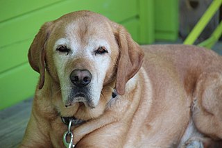
The German Shepherd, also known in Britain as an Alsatian, is a German breed of working dog of medium to large size. The breed was developed by Max von Stephanitz using various traditional German herding dogs from 1899.

<span class="mw-page-title-main">Acetabulum</span> Cavity where the thigh bone (femur) articulates with the pelvis

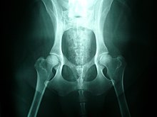
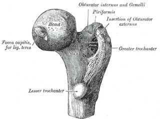
The acetabulum, also called the cotyloid cavity, is a concave surface of the pelvis. The head of the femur meets with the pelvis at the acetabulum, forming the hip joint.

<span class="mw-page-title-main">Osteoarthritis</span> Form of arthritis caused by degeneration of joints

Osteoarthritis (OA) is a type of degenerative joint disease that results from breakdown of joint cartilage and underlying bone. It is believed to be the fourth leading cause of disability in the world, affecting 1 in 7 adults in the United States alone. The most common symptoms are joint pain and stiffness. Usually the symptoms progress slowly over years. Other symptoms may include joint swelling, decreased range of motion, and, when the back is affected, weakness or numbness of the arms and legs. The most commonly involved joints are the two near the ends of the fingers and the joint at the base of the thumbs, the knee and hip joints, and the joints of the neck and lower back. The symptoms can interfere with work and normal daily activities. Unlike some other types of arthritis, only the joints, not internal organs, are affected.

An osteotomy is a surgical operation whereby a bone is cut to shorten or lengthen it or to change its alignment. It is sometimes performed to correct a hallux valgus, or to straighten a bone that has healed crookedly following a fracture. It is also used to correct a coxa vara, genu valgum, and genu varum. The operation is done under a general anaesthetic.

Arthroplasty is an orthopedic surgical procedure where the articular surface of a musculoskeletal joint is replaced, remodeled, or realigned by osteotomy or some other procedure. It is an elective procedure that is done to relieve pain and restore function to the joint after damage by arthritis or some other type of trauma.

<span class="mw-page-title-main">Hip</span> Anatomical region between the torso and the legs, holding the buttocks and genital region

In vertebrate anatomy, the hip, or coxa(pl.: coxae) in medical terminology, refers to either an anatomical region or a joint on the outer (lateral) side of the pelvis.

Osteochondrosis is a family of orthopedic diseases of the joint that occur in children, adolescents and rapidly growing animals, particularly pigs, horses, dogs, and broiler chickens. They are characterized by interruption of the blood supply of a bone, in particular to the epiphysis, followed by localized bony necrosis, and later, regrowth of the bone. This disorder is defined as a focal disturbance of endochondral ossification and is regarded as having a multifactorial cause, so no one thing accounts for all aspects of this disease.

<span class="mw-page-title-main">Elbow dysplasia</span> Medical condition in dogs

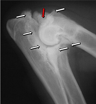
Elbow dysplasia is a condition involving multiple developmental abnormalities of the elbow-joint in the dog, specifically the growth of cartilage or the structures surrounding it. These abnormalities, known as 'primary lesions', give rise to osteoarthritic processes. Elbow dysplasia is a common condition of certain breeds of dogs.

<span class="mw-page-title-main">Hip replacement (animal)</span>

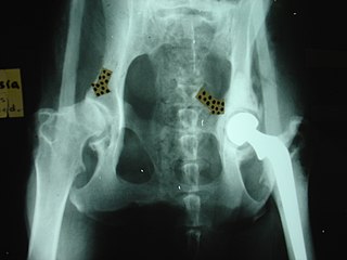
Hip replacement is a surgical procedure performed in dogs and cats as a salvage procedure, to alleviate severe pain in the hip due to, for example, hip dysplasia or irreparable bone fracture. The procedure replaces the head of the femur and the acetabulum with prosthetic implants. Because animals under about 40 pounds (18 kg) carry their own weight with little strain on each leg, hip modification surgeries are often sufficient to restore hip function in many cases. As a result, while hip replacement on animals can be seen in any animal of any size, from cats upwards, it is most often performed in the medium-large breeds of dogs.

PennHIP is a program which evaluates the quality of the hips in dogs. The program was established at the University of Pennsylvania School of Veterinary Medicine by Gail Smith in 1993, with the primary objective of reducing the prevalence of hip dysplasia in dogs. To assess a dog's hip joints, three radiographs (X-rays) are taken from different angles while the dog is under general anesthesia. Radiographs are submitted to the PennHIP for assessment, and are assigned a score, called a distraction index. Veterinarians must be trained members of the PennHIP Network in order to take radiographs for these assessments. The scheme is available through veterinarians in the United States and Canada. It was considered as the most evidence-based radiographic method to diagnose hip dysplasia.

<span class="mw-page-title-main">Femoral head ostectomy</span> Surgical removal of the head and neck of the femur

A femoral head ostectomy is a surgical operation to remove the head and neck from the femur. It is performed to alleviate pain, and is a salvage procedure, reserved for condition where pain can not be alleviated in any other way. It is common in veterinary surgery. Other names are excision arthroplasty of the femoral head and neck, Girdlestone's operation, Girdlestone procedure, and femoral head and neck ostectomy.

<span class="mw-page-title-main">Orthopedic pathology</span>

Orthopedic pathology, also known as bone pathology is a subspecialty of surgical pathology which deals with the diagnosis and feature of many bone diseases, specifically studying the cause and effects of disorders of the musculoskeletal system. It uses gross and microscopic findings along with the findings of in vivo radiological studies, and occasionally, specimen radiographs to diagnose diseases of the bones.

Protrusio acetabuli is an uncommon defect of the acetabulum, the socket that receives the femoral head to make the hip joint. The hip bone of the pelvic bone/girdle is composed of three bones, the ilium, the ischium and the pubis. In protrusio deformity, there is medial displacement of the femoral head in that the medial aspect of the femoral cortex is medial to the ilioischial line. The socket is too deep and may protrude into the pelvis.

Lameness is an abnormal gait or stance of an animal that is the result of dysfunction of the locomotor system. In the horse, it is most commonly caused by pain, but can be due to neurologic or mechanical dysfunction. Lameness is a common veterinary problem in racehorses, sport horses, and pleasure horses. It is one of the most costly health problems for the equine industry, both monetarily for the cost of diagnosis and treatment, and for the cost of time off resulting in loss-of-use.

<span class="mw-page-title-main">Hip dysplasia</span> Joint abnormality

Hip dysplasia is an abnormality of the hip joint where the socket portion does not fully cover the ball portion, resulting in an increased risk for joint dislocation. Hip dysplasia may occur at birth or develop in early life. Regardless, it does not typically produce symptoms in babies less than a year old. Occasionally one leg may be shorter than the other. The left hip is more often affected than the right. Complications without treatment can include arthritis, limping, and low back pain. Females are affected more often than males. Risk factors for hip dysplasia include female sex, family history, certain swaddling practices, and breech presentation whether an infant is delivered vaginally or by cesarean section. If one identical twin is affected, there is a 40% risk the other will also be affected. Screening all babies for the condition by physical examination is recommended. Ultrasonography may also be useful.

<span class="mw-page-title-main">Femoroacetabular impingement</span> Medical condition

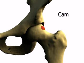
Femoroacetabular impingement (FAI) is a condition involving one or more anatomical abnormalities of the hip joint, which is a ball and socket joint. It is a common cause of hip pain and discomfort in young and middle-aged adults. It occurs when the ball shaped femoral head contacts the acetabulum abnormally or does not permit a normal range of motion in the acetabular socket. Damage can occur to the articular cartilage, or labral cartilage, or both. The condition may be symptomatic or asymptomatic. It may cause osteoarthritis of the hip. Treatment options range from conservative management to surgery.

<span class="mw-page-title-main">Hip arthroscopy</span>

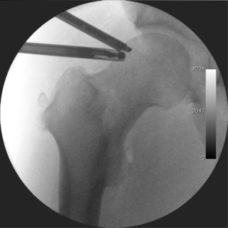
Hip arthroscopy refers to the viewing of the interior of the acetabulofemoral (hip) joint through an arthroscope and the treatment of hip pathology through a minimally invasive approach. This technique is sometimes used to help in the treatment of various joint disorders and has gained popularity because of the small incisions used and shorter recovery times when compared with conventional surgical techniques. Hip arthroscopy was not feasible until recently, new technology in both the tools used and the ability to distract the hip joint has led to a recent surge in the ability to do hip arthroscopy and the popularity of it.

<span class="mw-page-title-main">Hip pain</span>

Pain in the hip is the experience of pain in the muscles or joints in the hip/ pelvic region, a condition commonly arising from any of a number of factors. Sometimes it is closely associated with lower back pain.

<span class="mw-page-title-main">Polysulfated glycosaminoglycan</span> Injectable drug

Polysulfated glycosaminoglycan (PSGAG), sold under the brand name Adequan, is an injectable drug for dogs and horses that is used to alleviate the limpness, pain, and lowered range of motion caused by arthritis. It is made of repeat disaccharide units (comprising hexosamine and hexuronic acid), and is similar to glycosaminoglycans already present in the cartilage; PSGAG thus easily integrates itself there. In vitro studies have shown it to inhibit the enzymes that degrade cartilage and bone, as well as suppress inflammation and stimulate the synthesis of replacement cartilage. While it can cause an increased risk of bleeding, it is relatively safe and has a high LD50. PSGAG is one of the most widely prescribed joint treatments for horses.

<span class="mw-page-title-main">Senior dog diet</span>

Senior dog food diets are pet foods that are catered toward the senior or mature pet population. The senior dog population consists of dogs that are over the age of seven for most dog breeds, though in general large and giant breed dogs tend to reach this life stage earlier when compared to smaller breed dogs. Senior dog foods contain nutrients and characteristics that are used to improve the health of the aging dog. Aging in dogs causes many changes to occur physiologically that will require a change in nutrient composition of their diet.

References

  1. Workingdogs.com. "Canine hip dysplasia". Workingdogs.com. Retrieved August 18, 2013.
  2. 1 2 3 4 "Hip Dysplasia in Dogs". vca_corporate. Retrieved December 22, 2019.
  3. Randall, David (March 23, 2014). "Ask The Expert Dr. Randall - Hip Dysplasia In Dogs". Youtube. Flexpet. Retrieved November 10, 2015.
  4. Connor, J.R.; LePage, C.; Swift, B.A.; Yamashita, D.; Bendele, A.M.; Maul, D.; Kumar, S. (2009). "Protective effects of a cathepsin K inhibitor, SB-553484, in the canine partial medial meniscectomy model of osteoarthritis". Osteoarthritis and Cartilage. 17 (9): 1236–1243. doi: 10.1016/j.joca.2009.03.015 . PMID   19361586.
  5. 1 2 3 4 5 6 "What is canine hip dysplasia?". Orthopedic Foundation for Animals. 2018. Retrieved December 22, 2019.
  6. Comhaire, FH; Snaps, F (March 2008). "Comparison of two canine registry databases on the prevalence of hip dysplasia by breed and the relationship of dysplasia with body weight and height". American Journal of Veterinary Research. 69 (3): 330–3. doi: 10.2460/ajvr.69.3.330 . PMID   18312130.
  7. "Hip Dysplasia by Breed". Orthopedic Foundation for Animals. 2016. Archived from the original on October 19, 2010. Retrieved April 30, 2017.
  8. "A number of environmental factors can affect the incidence of hip dysplasia in dogs". Veths.no. March 26, 2012. Retrieved March 2, 2023.
  9. Torres; de la Riva, G; Hart, BL; Farver, TB; Oberbauer, AM; Messam, LLM; Willits, N; et al. (2013). "Neutering Dogs: Effects on Joint Disorders and Cancers in Golden Retrievers". PLOS ONE. 8 (2): e55937. Bibcode:2013PLoSO...855937T. doi: 10.1371/journal.pone.0055937 . PMC   3572183 . PMID   23418479.
  10. "Hip Dysplasia in Dogs: Causes, Symptoms & Treatment". American Kennel Club. Retrieved December 22, 2019.
  11. Kishimoto, Miori; Pae, Sa-Hun; Muroya, Naoyoshi; Watarai, Hirokazu; Anzai, Hiroshi; Lee, Ki-Ja; Shimizu, Junichiro; Sasaki, Motoki; Yamada, Kazutaka (2010). "The relationship between the size of caudolateral curvilinear osteophyte of the canine femoral neck and the radiographic view". Journal of Veterinary Science. 11 (1): 89. doi:10.4142/jvs.2010.11.1.89. PMC   2833436 .
  12. "Total Hip Replacement". Southern California Veterinary Referral Group. 2006. Archived from the original on June 13, 2006.
  13. Lenehan TM, Van Sickle DC (1985). "Chapter 84: Canine osteochondrosis". In Nunamaker DM, Newton CD (eds.). Textbook of small animal orthopaedics. Philadelphia: Lippincott. ISBN   978-0-397-52098-5.
  14. 1 2 3 "Arthritis in the Dog". Vetrica.com. February 9, 2004. Archived from the original on February 4, 2014. Retrieved August 18, 2013.
  15. "Helpful Medicine for Hip Dysplasia in Dogs. 10 Tips for Treating Hip Dysplasia without Surgery". Dr. Buzby's ToeGrips for Dogs. February 6, 2019. Retrieved December 22, 2019.
  16. Terry DE, Rees-Milton K, Smith P, Carran J, Pezeshki P, Woods C, Greer P, Anastassiades TP (2005). "N-acylation of glucosamine modulates chondrocyte growth, proteoglycan synthesis, and gene expression". J. Rheumatol. 32 (9): 1775–86. PMID   16142878.
  17. "Friday Find: Animal Assistance Products-For Pets Who Need a Little Extra Support". Fourgreensteps.com. February 17, 2012. Archived from the original on April 16, 2012. Retrieved August 18, 2013.
  18. Malfait, A M. "The nonpsychoactive cannabis constituent cannabidiol is an oral anti-arthritic therapeutic in murine collagen-induced arthritis". Pub Med. Retrieved October 12, 2023.
  19. Jones, Jason. "CBD For Dogs With Hip Dysplasia". Relievet. Retrieved October 12, 2023.