Related Research Articles
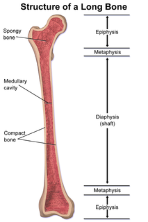
The epiphysis is the rounded end of a long bone, at its joint with adjacent bone(s). Between the epiphysis and diaphysis lies the metaphysis, including the epiphyseal plate. At the joint, the epiphysis is covered with articular cartilage; below that covering is a zone similar to the epiphyseal plate, known as subchondral bone.

In dogs, hip dysplasia is an abnormal formation of the hip socket that, in its more severe form, can eventually cause lameness and arthritis of the joints. It is a genetic (polygenic) trait that is affected by environmental factors. It is common in many dog breeds, particularly the larger breeds, and is the most common single cause of arthritis of the hips.
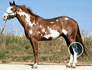
The hock, or gambrel, is the joint between the tarsal bones and tibia of a digitigrade or unguligrade quadrupedal mammal, such as a horse, cat, or dog. This joint may include articulations between tarsal bones and the fibula in some species, while in others the fibula has been greatly reduced and is only found as a vestigial remnant fused to the distal portion of the tibia. It is the anatomical homologue of the ankle of the human foot. While homologous joints occur in other tetrapods, the term is generally restricted to mammals, particularly long-legged domesticated species.

Synovial osteochondromatosis (SOC) is a rare disease that creates a benign change or proliferation in the synovium or joint-lining tissue, which changes to form bone-forming cartilage. In most occurrences, there is only one joint affected, either the knee, the hip, or the elbow. Rarely involves the TMJ.
Osteochondrosis is a family of orthopedic diseases of the joint that occur in children, adolescents and rapidly growing animals, particularly pigs, horses, dogs, and broiler chickens. They are characterized by interruption of the blood supply of a bone, in particular to the epiphysis, followed by localized bony necrosis, and later, regrowth of the bone. This disorder is defined as a focal disturbance of endochondral ossification and is regarded as having a multifactorial cause, so no one thing accounts for all aspects of this disease.
Osteitis is inflammation of bone. More specifically, it can refer to one of the following conditions:

Osteochondritis dissecans is a joint disorder primarily of the subchondral bone in which cracks form in the articular cartilage and the underlying subchondral bone. OCD usually causes pain during and after sports. In later stages of the disorder there will be swelling of the affected joint which catches and locks during movement. Physical examination in the early stages does only show pain as symptom, in later stages there could be an effusion, tenderness, and a crackling sound with joint movement.

Elbow dysplasia is a condition involving multiple developmental abnormalities of the elbow-joint in the dog, specifically the growth of cartilage or the structures surrounding it. These abnormalities, known as 'primary lesions', give rise to osteoarthritic processes. Elbow dysplasia is a common condition of certain breeds of dogs.

Synovial chondromatosis is a locally aggressive bone tumor of the cartilaginous type. It consists of several hyaline cartilaginous nodules and has the potential of becoming cancerous.

Scheuermann's disease is a self-limiting skeletal disorder of childhood. Scheuermann's disease describes a condition where the vertebrae grow unevenly with respect to the sagittal plane; that is, the posterior angle is often greater than the anterior. This uneven growth results in the signature "wedging" shape of the vertebrae, causing kyphosis. It is named after Danish surgeon Holger Scheuermann.

The femoral head is the highest part of the thigh bone (femur). It is supported by the femoral neck.

Franz König was a German surgeon. The son of a physician, he was born in Rotenburg an der Fulda.
Bone disease refers to the medical conditions which affect the bone.
In cell biology and pathophysiology, cellular adaptation refers to changes made by a cell in response to adverse or varying environmental changes. The adaptation may be physiologic (normal) or pathologic (abnormal). Four types of morphological adaptations include atrophy, hypertrophy, hyperplasia, and metaplasia.

Knee pain is pain in or around the knee.
Panner disease is an osteochondrosis of the capitellum of the elbow. Panner disease is primarily seen in boys between the ages of five and ten years old. Panner disease is often caused by excessive throwing due to valgus stress. The disease causes pain and stiffness in the affected elbow and may limit extension; the affected elbow is usually on the dominant arm the child uses. The disease may be associated with pitching and athletic activity. On radiographs, the capitellum may appear irregular with areas of radiolucency. Treatment is symptomatic, with a good prognosis. Treatment is minimal and includes restricting athletic activity to allow for the elbow to heal and for pain to be relieved. The disease is named after the Danish radiologist Hans Jessen Panner (1871–1930).
Orthopedic surgery is the branch of surgery concerned with conditions involving the musculoskeletal system. Orthopedic surgeons use both surgical and nonsurgical means to treat musculoskeletal injuries, sports injuries, degenerative diseases, infections, bone tumours, and congenital limb deformities. Trauma surgery and traumatology is a sub-specialty dealing with the operative management of fractures, major trauma and the multiply-injured patient.

The South German Coldblood is a breed of draught horse from southern Germany. It is distributed mainly in Bavaria. It is the most numerous of the four principal German draught horse breeds – the others being the Black Forest Horse, the Rhenish German Coldblood and the Schleswig Coldblood – and is the only one not listed as endangered by the FAO or by the Gesellschaft zur Erhaltung alter und gefährdeter Haustierrassen, the German national association for the conservation of historic and endangered domestic animal breeds.
References
- 1 2 3 " Osteochondritis " at Dorland's Medical Dictionary
- ↑ "CAL > Home". cal.vet.upenn.edu. Archived from the original on September 13, 2007. Retrieved August 20, 2015.
- ↑ Scheuermann's+disease at the US National Library of Medicine Medical Subject Headings (MeSH)
- ↑ Quoted from: Matthew Pead and Sue Guthrie. "Elbow Dysplasia in dogs – a new scheme explained" (PDF). British Veterinary Association (BVA). Archived from the original (PDF) on 2011-10-02. Retrieved 2010-07-16.