Panner disease

Last updated
Panner disease

Panner disease is an osteochondrosis of the capitellum of the elbow. [1] Panner disease is primarily seen in boys between the ages of five and ten years old. [2] Panner disease is often caused by excessive throwing due to valgus stress. The disease causes pain and stiffness in the affected elbow and may limit extension; the affected elbow is usually on the dominant arm the child uses. [3] The disease may be associated with pitching and athletic activity. On radiographs, the capitellum may appear irregular with areas of radiolucency. Treatment is symptomatic, with a good prognosis. Treatment is minimal and includes restricting athletic activity to allow for the elbow to heal and for pain to be relieved. [4] The disease is named after the Danish radiologist Hans Jessen Panner (1871–1930).

Contents

Symptoms

An individual with Panner disease most commonly experiences elbow pain near the capitellum. [2] Other symptoms include:

These symptoms worsen with physical activity such a throwing a ball or gymnastics for example. The symptoms begin unexpectedly and are often present for several days or weeks, and the symptoms tend to last even longer. [2]

Causes

Being an extremely rare disease, it is unknown as to what exactly causes Panner Disease. It is believed that the disease may be brought on by continuous overuse of the elbow and that puts pressure on the elbow and also strains the elbow in children during the period of rapid bone growth. The overuse of the elbow can be due to the involvement in sports such as baseball, handball, and gymnastics where these sports involve throwing or putting a lot of pressure on the joints. [2] [5] These repeated activities cause microtraumas and results in the affected elbow being swollen, irritated, and in pain. Panner Disease results when the blood supply to the capitellum is disrupted and therefore the cells within the growth plate of the capitellum die and it becomes flat due to the softening and collapsing of the surrounding bone.[ citation needed ]

Mechanism

The capitellum is the rounded knob on the end of the humerus, and it is held by the radius. Elbow-Dysplasia-Manifestations.svg
The capitellum is the rounded knob on the end of the humerus, and it is held by the radius.

Panner Disease affects the elbow of the arm. At the elbow, the humerus meets the ulna and the radius. The humerus is the long bone that runs from the shoulder to the elbow, and the ulna and radius are the two bones that make up the forearm. [6] The capitellum is the rounded knob on the end of the humerus and it is held by the radius due to the radius's cup-like shape. Panner Disease is part of a family of bone development diseases known as osteochondrosis. [6] [7] In osteochondrosis, the blood supply to an area of developing bone in the dominant elbow is temporarily disrupted by something that is not yet well understood. [6] Therefore, the tissues in the bone are not getting enough blood and they begin to go through necrosis, and they begin to die. Normally, bones grow by the expansion and uniting of the growth plates, but osteochondrosis disrupts this process and the result is cell death and the loss of newly formed tissue. The death of the tissues eventually leads to deterioration of the bone's growth plate. The bone's growth plate is defined as the area at the end of a developing bone where cartilage cells change into bone cells. [6] The bone tissue does regrow, but the necrosis can cause temporary problems in the affected area until the strenuous arm and elbow activity is significantly decreased or stopped for a period of time. [6]

It is believed that Panner Disease is a precursor to a similar condition called osteochondritis dissecans of the capitellum (OCD). [6] OCD is different from Panner disease because OCD occurs in older children and it does not involve the growth plate because by the time that OCD occurs the growth plates have already fused and the skeleton has finished growing.[ citation needed ]

Diagnosis

As the symptoms become prominent, the child will visit their pediatrician or family doctor to confirm whether or not the child has Panner Disease. When the child visits the doctor, the doctor will seek information about the child's age, sports participation, activity level, and what the child's dominant arm is. The affected elbow will be compared to the healthy elbow and any differences between the two will be noted. [5] The location of where the pain is in the elbow, and the child's range of motion and extension will also be determined to make an accurate diagnosis. To check the child's range of motion and extension limitation the child will be asked to move the arm of the affected elbow in various directions. The movement of the arm in various directions will allow the doctor to conclude how good the child is able to move the arm and the doctor will be able to determine if there is pain caused by the various directions of movement. [5]

To confirm the diagnosis, an x-ray or MRI scan will be done. The radiograph will enable the doctor to visualize irregularities and see the shape of the capitellum and also visualize the growth plate. In Panner Disease, the capitellum may appear flat and the bone growth plate will look irregular and fragmented. The areas where bone breakdown has occurred can also be visualized on the radiograph. [2] When the patient undergoes an MRI scan any irregularities of the capitellum will be able to be visualized, and the bone will be able to be visualized in more detail to determine the extent of swelling, if any. In the MRI results for Panner disease, there will be a decreased signal intensity of the capitellum on a T1 series and increased signal intensity on a T2 series. [2] [5]

Prevention

To prevent future instances of Panner Disease the child is instructed to cease all physical and sports activities that involve the use of the affected elbow until the symptoms are relieved[ citation needed ]

Treatment

The goal of treatment in Panner disease is to relieve pain. Treatment for Panner Disease is very minimal because in most children the bones repair their blood supply and rebuild themselves and this leads to the rebuilding of the growth plate and the capitellum returns to its normal shape. [6] The period of rebuilding and regrowth varies from child to child and can last anywhere between weeks to several months. To relieve the pain, the child is restricted from participating in sports and activities until the elbow is healed and to also rest the affected elbow. Rest will allow for the pain to be relieved and return of full elbow movement. It may also be recommended for children to apply an icepack or heat to the elbow to alleviate pain and swelling. If the child has great difficulty bending and straightening the arm then physical therapy may also be recommended. Occasionally, it is recommended for children to use nonsteroidal anti-inflammatory drugs (NSAIDs) or acetaminophen to also reduce pain and swelling. [4]

Prognosis

For treatment, Panner Disease heals well in children with rest and restriction of physical activity and sports using the affected arm. The prognosis is also good with treatment and the affected capitellum is remodeled. Irregularities of the capitellum and surrounding elbow area can both be seen by radiograph and MRI. When treatment is effective the flattened and fragmented capitellum is completely remodeled and returns to its normal circular shape, and also the high intensity signal on an MRI T2 series disappears. [5] These results indicate that the capitellum is completely remodeled and the child is able to return to normal physical and sports activities.[ citation needed ]

Research

Although the exact cause of Panner Disease is unknown, in recent research, it has been concluded that it may be associated with frequent throwing or other athletic activity. [4] In the same article that talks about varying osteochondrosis diseases, it is pointed out that Panner Disease always involves alteration of the capitellum, which can be visualized by radiography. In another research article, the research team aimed to summarize the best available evidence for diagnosis and treatment for Panner Disease. In the article it was found that the most common symptoms that patients with Panner Disease present with are elbow stiffness and swelling, limited range of motion, and limited elbow extension. [2] In alignment with the previously mentioned article, the team of researchers also concluded that Panner Disease involves irregularity of the capitellum, specifically that it appears flattened. Panner Disease often gets misdiagnosed as osteochondritis dissecans (OCD), and in this article they distinguish the difference between the two diseases are age difference and radiographic findings. [2] In alignment with the two previously discussed articles, another article that reports on three case studies of Panner Disease, states that the primary treatment that is used for Panner Disease is rest and restriction from all physical and athletic activity that involves the use of the upper extremities; the activity is suggested to be ceased until the symptoms are relieved. [5]

Related Research Articles

In human anatomy, the extensor carpi ulnaris is a skeletal muscle located on the ulnar side of the forearm. The extensor carpi ulnaris acts to extend and adduct at the carpus/wrist from anatomical position.

<span class="mw-page-title-main">Shoulder problem</span> Medical condition

Shoulder problems including pain, are one of the more common reasons for physician visits for musculoskeletal symptoms. The shoulder is the most movable joint in the body. However, it is an unstable joint because of the range of motion allowed. This instability increases the likelihood of joint injury, often leading to a degenerative process in which tissues break down and no longer function well.

<span class="mw-page-title-main">Wrist drop</span> Medical condition

Wrist drop is a medical condition in which the wrist and the fingers cannot extend at the metacarpophalangeal joints. The wrist remains partially flexed due to an opposing action of flexor muscles of the forearm. As a result, the extensor muscles in the posterior compartment remain paralyzed.

<span class="mw-page-title-main">Epiphysis</span>

An epiphysis is one of the rounded ends or tips of a long bone that ossify from a secondary center of ossification. Between the epiphysis and diaphysis lies the metaphysis, including the epiphyseal plate. At the joint, the epiphysis is covered with articular cartilage; below that covering is a zone similar to the epiphyseal plate, known as subchondral bone.

<span class="mw-page-title-main">Hip dysplasia (canine)</span> Joint abnormality in dogs

In dogs, hip dysplasia is an abnormal formation of the hip socket that, in its more severe form, can eventually cause lameness and arthritis of the joints. It is a genetic (polygenic) trait that is affected by environmental factors. It is common in many dog breeds, particularly the larger breeds, and is the most common single cause of arthritis of the hips.

<span class="mw-page-title-main">Osgood–Schlatter disease</span> Inflammation of the patellar ligament

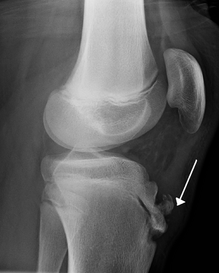
Osgood–Schlatter disease (OSD) is inflammation of the patellar ligament at the tibial tuberosity (apophysitis). It is characterized by a painful bump just below the knee that is worse with activity and better with rest. Episodes of pain typically last a few weeks to months. One or both knees may be affected and flares may recur.

<span class="mw-page-title-main">Enchondroma</span> Medical condition

Enchondroma is a type of benign bone tumor belonging to the group of cartilage tumors. There may be no symptoms, or it may present typically in the short tubular bones of the hands with a swelling, pain or pathological fracture.

<span class="mw-page-title-main">Synovial osteochondromatosis</span> Medical condition

Synovial osteochondromatosis (SOC) (synonyms include synovial chondromatosis, primary synovial chondromatosis, synovial chondrometaplasia) is a rare disease that creates a benign change or proliferation in the synovium or joint-lining tissue, which changes to form bone-forming cartilage. In most occurrences, there is only one joint affected, either the knee, the hip, or the elbow. Rarely involves the TMJ.

Osteochondrosis is a family of orthopedic diseases of the joint that occur in children, adolescents and rapidly growing animals, particularly pigs, horses, dogs, and broiler chickens. They are characterized by interruption of the blood supply of a bone, in particular to the epiphysis, followed by localized bony necrosis, and later, regrowth of the bone. This disorder is defined as a focal disturbance of endochondral ossification and is regarded as having a multifactorial cause, so no one thing accounts for all aspects of this disease.

<span class="mw-page-title-main">Köhler disease</span> Medical condition

Köhler disease is a rare bone disorder of the foot found in children between six and nine years of age. The disease typically affects boys, but it can also affect girls. It was first described in 1908 by Alban Köhler (1874–1947), a German radiologist. Dr. A. Köhler noted that children with foot pain displayed characteristics, within their x-rays, of irregularity in growth and development of the tarsal navicular bone in the foot. Furthermore, Köhler disease is known to affect five times more boys than girls and typically, only one foot is affected. The disease was then found to belong to a group of conditions called osteochondroses, which disturb bone growth at ossification centres which occurs during bone development.

<span class="mw-page-title-main">Pulled elbow</span> Medical condition

A pulled elbow, also known as a radial head subluxation, is when the ligament that wraps around the radial head slips off. Often a child will hold their arm against their body with the elbow slightly bent. They will not move the arm as this results in pain. Touching the arm, without moving the elbow, is usually not painful.

Osteochondritis is a painful type of osteochondrosis where the cartilage or bone in a joint is inflamed.

<span class="mw-page-title-main">Osteochondritis dissecans</span> Ischemic bone disease

Osteochondritis dissecans is a joint disorder primarily of the subchondral bone in which cracks form in the articular cartilage and the underlying subchondral bone. OCD usually causes pain during and after sports. In later stages of the disorder there will be swelling of the affected joint which catches and locks during movement. Physical examination in the early stages does only show pain as symptom, in later stages there could be an effusion, tenderness, and a crackling sound with joint movement.

<span class="mw-page-title-main">Elbow dysplasia</span> Medical condition in dogs

Elbow dysplasia is a condition involving multiple developmental abnormalities of the elbow-joint in the dog, specifically the growth of cartilage or the structures surrounding it. These abnormalities, known as 'primary lesions', give rise to osteoarthritic processes. Elbow dysplasia is a common condition of certain breeds of dogs.

<span class="mw-page-title-main">Synovial chondromatosis</span> Medical condition

Synovial chondromatosis is a locally aggressive bone tumor of the cartilaginous type. It consists of several hyaline cartilaginous nodules and has the potential of becoming cancerous.

<span class="mw-page-title-main">Chondroblastoma</span> Medical condition

Chondroblastoma is a rare, benign, locally aggressive bone tumor that typically affects the epiphyses or apophyses of long bones. It is thought to arise from an outgrowth of immature cartilage cells (chondroblasts) from secondary ossification centers, originating from the epiphyseal plate or some remnant of it.
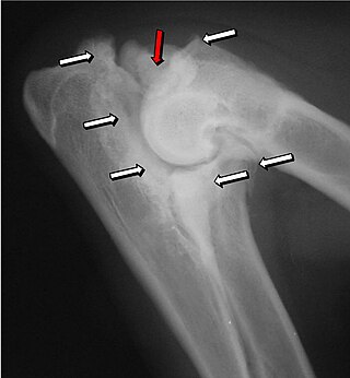
<span class="mw-page-title-main">Ulnar nerve entrapment</span> Medical condition

Ulnar nerve entrapment is a condition where the ulnar nerve becomes physically trapped or pinched, resulting in pain, numbness, or weakness, primarily affecting the little finger and ring finger of the hand. Entrapment may occur at any point from the spine at cervical vertebra C7 to the wrist; the most common point of entrapment is in the elbow. Prevention is mostly through correct posture and avoiding repetitive or constant strain. Treatment is usually conservative, including medication, activity modification, and exercise, but may sometimes include surgery. Prognosis is generally good, with mild to moderate symptoms often resolving spontaneously.

<span class="mw-page-title-main">Humerus fracture</span> Medical condition

A humerus fracture is a break of the humerus bone in the upper arm. Symptoms may include pain, swelling, and bruising. There may be a decreased ability to move the arm and the person may present holding their elbow. Complications may include injury to an artery or nerve, and compartment syndrome.

<span class="mw-page-title-main">Supracondylar humerus fracture</span> Medical condition

A supracondylar humerus fracture is a fracture of the distal humerus just above the elbow joint. The fracture is usually transverse or oblique and above the medial and lateral condyles and epicondyles. This fracture pattern is relatively rare in adults, but is the most common type of elbow fracture in children. In children, many of these fractures are non-displaced and can be treated with casting. Some are angulated or displaced and are best treated with surgery. In children, most of these fractures can be treated effectively with expectation for full recovery. Some of these injuries can be complicated by poor healing or by associated blood vessel or nerve injuries with serious complications.

<span class="mw-page-title-main">Medial epicondyle fracture of the humerus</span>

A medial epicondyle fracture is an avulsion injury to the medial epicondyle of the humerus; the prominence of bone on the inside of the elbow. Medial epicondyle fractures account for 10% elbow fractures in children. 25% of injuries are associated with a dislocation of the elbow.

References

  1. RESERVED, INSERM US14 -- ALL RIGHTS. "Orphanet: Panner disease". www.orpha.net. Retrieved 15 September 2017.
  2. 1 2 3 4 5 6 7 8 Claessen, Femke M. A. P.; Louwerens, Jan K. G.; Doornberg, Job N.; van Dijk, C. Niek; Eygendaal, Denise; van den Bekerom, Michel P. J. (2015-02-01). "Panner's disease: literature review and treatment recommendations". Journal of Children's Orthopaedics. 9 (1): 9–17. doi:10.1007/s11832-015-0635-2. ISSN   1863-2521. PMC   4340849 . PMID   25663360.
  3. "Panner disease | Lurie Children's, Chicago". www.luriechildrens.org. Retrieved 2017-11-07.
  4. 1 2 3 Atanda, Alfred; Shah, Suken A.; O'Brien, Kathleen (2011-02-01). "Osteochondrosis: common causes of pain in growing bones". American Family Physician. 83 (3): 285–291. ISSN   1532-0650. PMID   21302869.
  5. 1 2 3 4 5 6 Sakata, Ryosuke; Fujioka, Hiroyuki; Tomatsuri, Masaki; Kokubu, Takeshi; Mifune, Yutaka; Inui, Atsuyuki; Kurosaka, Masahiro (2015-04-22). "Treatment and Diagnosis of Panner's Disease. A Report of Three Cases". The Kobe Journal of Medical Sciences. 61 (2): E36–39. ISSN   1883-0498. PMID   26628012.
  6. 1 2 3 4 5 6 7 "Panner's Disease". kidshealth.org. Retrieved 2017-11-07.
  7. Wróblewski, Robert; Urban, Mariusz; Michalik, Dariusz; Zakrzewski, Piotr; Langner, Maciej; Pomianowski, Stanisław (January 2014). "Osteochondrosis of the capitellum of the humerus (Panner's disease, Osteochondritis Dissecans). Case study". Ortopedia, Traumatologia, Rehabilitacja. 16 (1): 79–90. doi: 10.5604/15093492.1097492 . ISSN   2084-4336. PMID   24728797.