Hydrostatic skeleton

Last updated

A hydrostatic skeleton or hydroskeleton is a type of skeleton supported by hydrostatic fluid pressure, [1] common among soft-bodied invertebrate animals colloquially referred to as "worms". While more advanced organisms can be considered hydrostatic, they are sometimes referred to as hydrostatic for their possession of a hydrostatic organ instead of a hydrostatic skeleton, where the two may have the same capabilities but are not the same. [1] As the prefix hydro- meaning "water", being hydrostatic means being fluid-filled. [2]

Contents

As a skeletal structure, a hydroskeleton possesses the ability to affect shape and movement, and involves two mechanical units: the muscle layers and the body wall. The muscular layers are longitudinal and circular, and part of the fluid-filled coelom within. Contractions of the circular muscles lengthen the organism's body, while contractions of the longitudinal muscles shorten the organism's body. Fluid within the organism is evenly concentrated so the forces of the muscle are spread throughout the whole organism and shape changes can persist. [2] These structural factors also persist in a hydrostatic organ.

A non-helical hydrostatic skeleton structure is the functional basis of the mammalian penis, [3] which fills the corpus cavernosa with blood to maintain physical rigidity during coitus. Helically reinforced hydrostatic skeleton structure is typical for flexible structures as in soft-bodied animals. [2]

Structure

Many animals with a wormlike cylindrical body have a hydrostatic skeleton with a flexible skin and a water-filled body cavity (coelom or pseudocoelom). They move by peristalsis, using opposed circular and longitudinal muscles, which act on the hydrostatic skeleton to change the body's shape. Bilaterian body plan.svg
Many animals with a wormlike cylindrical body have a hydrostatic skeleton with a flexible skin and a water-filled body cavity (coelom or pseudocoelom). They move by peristalsis, using opposed circular and longitudinal muscles, which act on the hydrostatic skeleton to change the body's shape.

Hydrostatic skeletons are typically arranged in a cylinder. Hydrostatic skeletons can be controlled by several different muscle types. Length can be adjusted by longitudinal muscle fibers parallel to the longitudinal axis. The muscle fibers may be found in continuous sheets or isolated bundles, and the diameter can be manipulated by three different muscle types: circular, radial, and transverse. [2] Circular musculature wraps around the circumference of the cylinder, radial musculature extends from the center of the cylinder towards the surface, and transverse musculature arrange in parallel and perpendicular sheets crossing the diameter of the cylinder. [2]

Within the cylinder lies fluid, most often water. The fluid is resistant to changes in volume. Contraction of circular, radial or transverse muscles increases the pressure within the cylinder, and results in an increase in length. Contraction of longitudinal muscles can shorten the cylinder. [2]

Change in shape is limited by connective tissue fibers. Connective fibers, often collagenous, are arranged in a helical shape within the wall of the hydrostatic skeleton. The helical shape formed by these fibers allows for elongation and shortening of the skeleton, while still remaining rigid to prevent torsion. As the shape of the cylinder changes, the pitch of the helix will change. The angle relative to the long axis will decrease during elongation and increase during shortening. [2]

Advantages and disadvantages

Organisms containing a hydrostatic skeleton have advantages and disadvantages. Their fluid shape allows them to move around easily while swimming and burrowing. They can fit through oddly shaped passages and hide themselves more effectively from predators. They are able to create a force when squeezing through rocks and create a “prying open” gesture. There is a lighteight, flexible component to them that allows this movement with very little muscle mass. [4]

These organisms are also able to heal faster than organisms that contain hard skeletons. Healing in these organisms varies from creature to creature. However, if the cavity needs to be refilled, the “fluid” can easily be refilled if it is water or blood. If the fluid is some other type of liquid, it can take longer, but it is still faster than healing a bone. The common earthworm is also able to regrow damaged parts of its body. [4]

These organisms have some relatively simple pathways for circulation and respiration. Also, these organisms have a cushion to allow protection for internal organs from shock. However, it does not protect internal organs from external damage very effectively. [4]

Because the hydrostatic skeletons have limited ability for attachment of limbs, the organisms are relatively simple and do not have many abilities to grab or latch onto things. Organisms with complete hydrostatic skeletons need to be in an environment that allows them to re-fill themselves with their fluid that is necessary for survival. This is why hydrostatic skeletons are common in marine life. They have a large amount of access to the necessary elements for survival. Terrestrial organisms that have hydrostatic skeletons generally have a lack of strength because they are not in a fluid environment. If one were to expand its body too much, it would collapse under its own weight. [4]

Organisms

The hemichordates are among the many marine animals with hydrostatic skeletons and peristaltic locomotion. Eichelwurm (cropped).jpg
The hemichordates are among the many marine animals with hydrostatic skeletons and peristaltic locomotion.

Hydrostatic skeletons are very common in invertebrates. A common example is the earthworm. Also, hydrostatic nature is common in marine life such as jellyfish and sea anemones. Earthworms have rings of muscles that are filled with fluid, making their entire body hydrostatic. A sea anemone has a hydrostatic head, with arms radiating out around the mouth. This structure is helpful in feeding and locomotion. [5]

An example of a simple Deuterostome containing a hydrostatic skeleton would be Enteropneusta, with the common name of acorn worm. This organism is classified as a Hemichordate, and they are marine worms that use their hydrostatic skeleton to tunnel and anchor themselves into the ground. This can be used for locomotion, but also can aid in the defense of the organism against outside forces as the worm can try to "hide" itself within the ocean floor. [5]

Vertebrates

The mammalian penis is a hydrostatic organ. The hydrostatic fluid, in this case blood, fills the penis during an erection. Unlike the hydrostatic skeletons of many invertebrates, which use the bending of the animal for locomotion, the penis must resist bending and shape changes during sexual intercourse. Instead of connective fibers arranged in a helical shape, the penis contains a layer called the corpus cavernosum. The corpus cavernosum contains connective fibers arranged both parallel and perpendicular to the longitudinal axis. These fibers remain folded when the penis is flaccid, but unfold as the penis fills with blood during an erection, which allows the penis to resist bending. The penises of turtles are structured similarly, although they evolved separately. [5]

Other vertebrates sometimes utilize a modified hydrostatic skeleton called a muscular hydrostat. [2] Muscular hydrostats do not contain a fluid-filled cavity. These structures are constructed of muscle and connective fibers, densely packed into a 3-D structure. In many cases, the muscular hydrostat can be manipulated in all three dimensions. This allows for more precise movement compared to a typical hydrostatic skeleton. While in typical hydrostatic skeletons, movement is generated by applying force to a fluid-filled cavity, muscular hydrostats generate movement by muscle contractions. When one muscle contracts and decreases in area, other muscles within the structure must expand in response. Helical muscles may be present, which can create torsion, an ability that is restricted in hydrostatic skeletons. Muscular hydrostats are found in mammalian, reptilian, and amphibian tongues. Mammalian tongues have the structure of a central core of muscle fibers surrounded by bundles of longitudinal muscles and alternating parallel sheets of transverse muscle fibers. Elephant trunks and tapir proboscises also utilize a muscular hydrostat. These structures are composed of longitudinal fibers surrounded by radial and helical fibers. [5]

See also

Related Research Articles

<span class="mw-page-title-main">Skeleton</span> Part of the body that forms the supporting structure

A skeleton is the structural frame that supports the body of most animals. There are several types of skeletons, including the exoskeleton, which is a rigid outer shell that holds up an organism's shape; the endoskeleton, a rigid internal frame to which the organs and soft tissues attach; and the hydroskeleton, a flexible internal structure supported by the hydrostatic pressure of body fluids.

<span class="mw-page-title-main">Skin</span> Soft outer covering organ of vertebrates

Skin is the layer of usually soft, flexible outer tissue covering the body of a vertebrate animal, with three main functions: protection, regulation, and sensation.

<span class="mw-page-title-main">Tissue (biology)</span> Group of similar cells performing a specific function

In biology, tissue is an assembly of similar cells and their extracellular matrix from the same embryonic origin that together carry out a specific function. Tissues occupy a biological organizational level between cells and a complete organ. Accordingly, organs are formed by the functional grouping together of multiple tissues.

<span class="mw-page-title-main">Peristalsis</span> Radially symmetrical contraction and relaxation of muscles

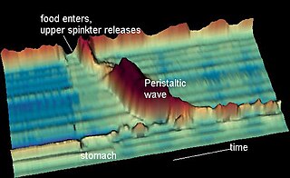
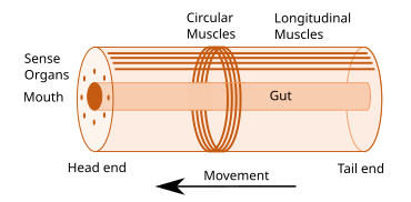
Peristalsis is a type of intestinal motility, characterized by radially symmetrical contraction and relaxation of muscles that propagate in a wave down a tube, in an anterograde direction. Peristalsis is progression of coordinated contraction of involuntary circular muscles, which is preceded by a simultaneous contraction of the longitudinal muscle and relaxation of the circular muscle in the lining of the gut.

The muscular system is an organ system consisting of skeletal, smooth, and cardiac muscle. It permits movement of the body, maintains posture, and circulates blood throughout the body. The muscular systems in vertebrates are controlled through the nervous system although some muscles can be completely autonomous. Together with the skeletal system in the human, it forms the musculoskeletal system, which is responsible for the movement of the body.

<span class="mw-page-title-main">Skeletal muscle</span> One of three major skeletal system types that connect to bones

Skeletal muscles are organs of the vertebrate muscular system and typically are attached by tendons to bones of a skeleton. The muscle cells of skeletal muscles are much longer than in the other types of muscle tissue, and are often known as muscle fibers. The muscle tissue of a skeletal muscle is striated – having a striped appearance due to the arrangement of the sarcomeres.

<span class="mw-page-title-main">Bulbospongiosus muscle</span> Superficial muscle of the perineum

The bulbospongiosus muscles are a subgroup of the superficial muscles of the perineum. They have a slightly different origin, insertion and function in males and females. In males, these muscles cover the bulb of the penis, while in females, they cover the vestibular bulbs.

<span class="mw-page-title-main">Human musculoskeletal system</span> Organ system that gives humans the capacity to move by using their muscular and skeletal systems

The human musculoskeletal system is an organ system that gives humans the ability to move using their muscular and skeletal systems. The musculoskeletal system provides form, support, stability, and movement to the body.

<span class="mw-page-title-main">Turbellaria</span> Class of flatworms

The Turbellaria are one of the traditional sub-divisions of the phylum Platyhelminthes (flatworms), and include all the sub-groups that are not exclusively parasitic. There are about 4,500 species, which range from 1 mm (0.039 in) to large freshwater forms more than 500 mm (20 in) long or terrestrial species like Bipalium kewense which can reach 600 mm (24 in) in length. All the larger forms are flat with ribbon-like or leaf-like shapes, since their lack of respiratory and circulatory systems means that they have to rely on diffusion for internal transport of metabolites. However, many of the smaller forms are round in cross section. Most are predators, and all live in water or in moist terrestrial environments. Most forms reproduce sexually and with few exceptions all are simultaneous hermaphrodites.

<span class="mw-page-title-main">Muscle contraction</span> Activation of tension-generating sites in muscle

Muscle contraction is the activation of tension-generating sites within muscle cells. In physiology, muscle contraction does not necessarily mean muscle shortening because muscle tension can be produced without changes in muscle length, such as when holding something heavy in the same position. The termination of muscle contraction is followed by muscle relaxation, which is a return of the muscle fibers to their low tension-generating state.

<span class="mw-page-title-main">Muscular hydrostat</span> Body part type that consists mainly of muscles with no skeletal support

A muscular hydrostat is a biological structure found in animals. It is used to manipulate items or to move its host about and consists mainly of muscles with no skeletal support. It performs its hydraulic movement without fluid in a separate compartment, as in a hydrostatic skeleton.

Myomeres are blocks of skeletal muscle tissue arranged in sequence, commonly found in aquatic chordates. Myomeres are separated from adjacent myomeres by connective fascia (myosepta) and most easily seen in larval fishes or in the olm. Myomere counts are sometimes used for identifying specimens, since their number corresponds to the number of vertebrae in the adults. Location varies, with some species containing these only near the tails, while some have them located near the scapular or pelvic girdles. Depending on the species, myomeres could be arranged in an epaxial or hypaxial manner. Hypaxial refers to ventral muscles and related structures while epaxial refers to more dorsal muscles. The horizontal septum divides these two regions in vertebrates from cyclostomes to gnathostomes. In terrestrial chordates, the myomeres become fused as well as indistinct, due to the disappearance of myosepta.

<span class="mw-page-title-main">Undulatory locomotion</span>

Undulatory locomotion is the type of motion characterized by wave-like movement patterns that act to propel an animal forward. Examples of this type of gait include crawling in snakes, or swimming in the lamprey. Although this is typically the type of gait utilized by limbless animals, some creatures with limbs, such as the salamander, forgo use of their legs in certain environments and exhibit undulatory locomotion. In robotics this movement strategy is studied in order to create novel robotic devices capable of traversing a variety of environments.

Role of skin in locomotion describes how the integumentary system is involved in locomotion. Typically the integumentary system can be thought of as skin, however the integumentary system also includes the segmented exoskeleton in arthropods and feathers of birds. The primary role of the integumentary system is to provide protection for the body. However, the structure of the skin has evolved to aid animals in their different modes of locomotion. Soft bodied animals such as starfish rely on the arrangement of the fibers in their tube feet for movement. Eels, snakes, and fish use their skin like an external tendon to generate the propulsive forces need for undulatory locomotion. Vertebrates that fly, glide, and parachute also have a characteristic fiber arrangements of their flight membranes that allows for the skin to maintain its structural integrity during the stress and strain experienced during flight.

Muscle architecture is the physical arrangement of muscle fibers at the macroscopic level that determines a muscle's mechanical function. There are several different muscle architecture types including: parallel, pennate and hydrostats. Force production and gearing vary depending on the different muscle parameters such as muscle length, fiber length, pennation angle, and the physiological cross-sectional area (PCSA).

<span class="mw-page-title-main">Anatomical terminology</span> Scientific terminology used by anatomists, zoologists, and health professionals

Anatomical terminology is a form of scientific terminology used by anatomists, zoologists, and health professionals such as doctors, physicians, and pharmacists.

<span class="mw-page-title-main">Gastrointestinal wall</span> Digestive system structure

The gastrointestinal wall of the gastrointestinal tract is made up of four layers of specialised tissue. From the inner cavity of the gut outwards, these are the mucosa, the submucosa, the muscular layer and the serosa or adventitia.

<span class="mw-page-title-main">Annelid</span> Phylum of segmented worms

The annelids, also known as the segmented worms, are a large phylum, with over 22,000 extant species including ragworms, earthworms, and leeches. The species exist in and have adapted to various ecologies – some in marine environments as distinct as tidal zones and hydrothermal vents, others in fresh water, and yet others in moist terrestrial environments.

<span class="mw-page-title-main">Francisco Torrent-Guasp</span>

Francisco ("Paco") Torrent-Guasp (Gandia, 1931 - Madrid, 2005). was a spanish cardiologist whose research focused on the anatomy and physiology of the human heart. His work led to the discovery and description of the ventricular myocardial band. His work can be found in reference books on anatomy and cardiac surgery.

<span class="mw-page-title-main">Anatomical terms of microanatomy</span> Anatomical terminology is used to describe microanatomical (or histological) structures

Anatomical terminology is used to describe microanatomical structures. This helps describe precisely the structure, layout and position of an object, and minimises ambiguity. An internationally accepted lexicon is Terminologia Histologica.

References

  1. 1 2 Kardong, Kenneth V. (2015). Vertebrates | Comparative Anatomy, Function, Evolution (7th ed.). Mc Graw Hill Education. pp. 426, 496. ISBN   978-0078023026.
  2. 1 2 3 4 5 6 7 8 Kier, William M. (2012-04-15). "The diversity of hydrostatic skeletons". Journal of Experimental Biology. 215 (8): 1247–1257. doi: 10.1242/jeb.056549 . PMID   22442361.
  3. Kelly, DA (April 2002). "The functional morphology of penile erection: tissue designs for increasing and maintaining stiffness" (PDF). Integrative and Comparative Biology. 42 (2): 216–221. doi: 10.1093/icb/42.2.216 . PMID   21708713.
  4. 1 2 3 4 "Everything Math's and Science". www.everythingmaths.co.za. Retrieved 2016-12-01.
  5. 1 2 3 4 "Hydrostatic Skeleton - The Infinite Spider". The Infinite Spider. 2015-02-10. Retrieved 2016-12-01.