Hypoplastic right heart syndrome

Last updated
Hypoplastic right heart syndrome
Hypoplastic Right Heart Syndrome.jpg
A newborn heart with hypoplastic right heart syndrome. Note the underdeveloped left side.
Specialty Cardiology
DurationLifetime
Diagnostic method Fetal echocardiogram [1]
Treatment Cardiac surgery [1]
Frequency60,000 cases per year, US [2]

Hypoplastic right heart syndrome or HRHS is a congenital heart defect in which the structures on the right side of the heart, particularly the right ventricle, are underdeveloped. This defect causes inadequate blood flow to the lungs, and thus a cyanotic infant.

Contents

Symptoms and signs

Common symptoms include a grayish-blue (cyanosis) coloration to the skin, lips, fingernails and other parts of the body. [3] [4] Other pronounced symptoms can be rapid or difficult breathing, poor feeding due to lack of energy, cold hands or feet, or being inactive and drowsy. Notably, patent ductus arteriosus and patent foramen ovale, normally dangerous defects, are necessary for a newborn with HRHS to survive. If either formation does close, the child will go into shock, signs of which can include cool or clammy skin, a weak or rapid pulse, and dilated pupils. [5]

Causes

It is mostly unknown what causes hypoplastic right heart syndrome in a given individual. [6] It is thought that a family history of similar heart defects can increase the risk, and there are some genes hypothesized to be factors. [2] However, it can also manifest in a child whose family is completely lacking in congenital heart abnormalities.

Pathogenesis

When the right side of the heart is more underdeveloped than the left side, this is known as hypoplastic right heart syndrome. HRHS is known for the pulmonary atresia valves, the tricuspid valve, right ventricle, and the pulmonary artery all failing to form properly. HRHS also causes the right ventricle to be a fair amount smaller than the left side. [3]

In people with hypoplastic right heart syndrome, the heart is not able to adequately pump blood to the lungs. The result of this is an inadequate supply of oxygenated blood to be circulated to the body. The severity of underdevelopment varies for each individual. A special team of pediatric cardiologists is required to develop a treatment plan.[ citation needed ]

Anatomy

A healthy heart has four chambers, each separated by valves that open and close to control blood flow between the chambers. When the heart beats, oxygen-poor blood enters the right atrium. The blood then flows into the right ventricle, where it pumps into the pulmonary artery to travel to the lungs for oxygen. Oxygen-rich blood returns to the left atrium, where it then travels into the left ventricle. The left ventricle pumps the oxygenated blood into the aorta to be circulated to the rest of the body. [7]

Crucial steps in heart formation are development of the ventricles and atrium formation, as well as septation and valve formation. Any disturbances of such processes may lead to various congenital heart diseases and defects that could be initiated by various genetic, epigenetic or environmental factors. The most common heart malformations from genetic or epigenetic problems are: stenosis of the aorta and pulmonary trunk, which is a narrowing of the vessels, atrial and/or ventricular septal defect, tricuspid atresia, and hypoplastic left and right heart syndrome. When an individual has hypoplastic right or left heart syndrome, it means that more than one of these problems have occurred together.[ citation needed ]

Diagnosis

If a cardiac anomaly is suspected in a routine ultrasound during pregnancy, often a perinatologist (maternal-fetal specialist) will perform a fetal echocardiogram (noninvasive ultrasound of the fetus heart), which may be able to confirm a diagnosis of HRHS. This can help with possible options for treatment. [1] [8] If the child is born without a diagnosis, an ECG will most likely be performed after the child starts showing symptoms.

Treatment

There is no cure for hypoplastic right heart syndrome. A three-stage series of surgical procedures is commonly used to treat the condition. The surgeries redirect the blood flow within the heart and allows the left ventricle to do the work for the underdeveloped right side of the heart. The three surgeries are spread out over the patients first few years of life. The first procedure, usually either the Norwood procedure or the Blalock–Thomas–Taussig shunt, is typically done within the first few days or weeks of life. The second procedure, called the Glenn procedure, is usually performed between four and twelve months of age. The last surgery, known as the Fontan procedure, is typically performed around the age of 18 months and older. The final result of these surgeries is to redirect the superior and inferior vena cavae into the pulmonary artery, bypassing the right atrium. [ citation needed ]

In a stage 1 Norwood procedure for hypoplastic right heart, the main pulmonary artery is separated from the left and right portions of the pulmonary artery and joined with the upper portion of the aorta. The proximal pulmonary artery is connected to the aortic arch, while the narrowed segment of the pulmonary trunk is repaired. An aortopulmonary shunt is created to connect the aorta to the main pulmonary artery to provide pulmonary blood flow to the lungs. The Glenn procedure disconnects the superior vena cava from the heart and connects it to the right pulmonary artery so deoxygenated blood from the upper body goes directly to the lungs. The Fontan procedure, done usually after the patient is two years old, disconnects the inferior vena cava from the heart and connects it directly with the other pulmonary artery so that deoxygenated blood from the lower body then is sent directly to the lungs.[ citation needed ]

Follow-up care

With a series of operations or even a heart transplant, a newborn can be treated but not be cured. Young individuals who have undergone reconstructive surgery must refer to a cardiologist who is experienced in congenital heart diseases, [9] People who have been diagnosed with HRHS must limit the physical activity they participate in to their own lowered endurance level. [10]

Prevalence

Hypoplastic right heart syndrome is both less common and less severe than hypoplastic left heart syndrome. Within the United States it occurs in 1 in 60,000 births, [2] as opposed to HLHS, which occurs in 1 in 4,300 births. [3] [1] [11] HRHS requires prenatal diagnosis since it often necessitates immediate and emergency treatment. Pregnant women whose pregnancy is complicated with this anomaly should be referred to a level 3 hospital with pediatric cardiology and pediatric cardiothoracic surgical team.[ citation needed ]

It can be associated with aortic stenosis. [12]

Related Research Articles

<span class="mw-page-title-main">Heart valve</span> A flap of tissue that prevent backflow of blood around the heart

A heart valve is a biological one-way valve that allows blood to flow in one direction through the chambers of the heart. Four valves are usually present in a mammalian heart and together they determine the pathway of blood flow through the heart. A heart valve opens or closes according to differential blood pressure on each side.

<span class="mw-page-title-main">Aortic valve</span> Valve in the human heart between the left ventricle and the aorta

The aortic valve is a valve in the heart of humans and most other animals, located between the left ventricle and the aorta. It is one of the four valves of the heart and one of the two semilunar valves, the other being the pulmonary valve. The aortic valve normally has three cusps or leaflets, although in 1–2% of the population it is found to congenitally have two leaflets. The aortic valve is the last structure in the heart the blood travels through before stopping the flow through the systemic circulation.

<span class="mw-page-title-main">Congenital heart defect</span> Defect in the structure of the heart that is present at birth

A congenital heart defect (CHD), also known as a congenital heart anomaly, congenital cardiovascular malformation, and congenital heart disease, is a defect in the structure of the heart or great vessels that is present at birth. A congenital heart defect is classed as a cardiovascular disease. Signs and symptoms depend on the specific type of defect. Symptoms can vary from none to life-threatening. When present, symptoms are variable and may include rapid breathing, bluish skin (cyanosis), poor weight gain, and feeling tired. CHD does not cause chest pain. Most congenital heart defects are not associated with other diseases. A complication of CHD is heart failure.

A cyanotic heart defect is any congenital heart defect (CHD) that occurs due to deoxygenated blood bypassing the lungs and entering the systemic circulation, or a mixture of oxygenated and unoxygenated blood entering the systemic circulation. It is caused by structural defects of the heart such as right-to-left or bidirectional shunting, malposition of the great arteries, or any condition which increases pulmonary vascular resistance. The result may be the development of collateral circulation.

<span class="mw-page-title-main">Fontan procedure</span> Surgical procedure used in children with univentricular hearts

The Fontan procedure or Fontan–Kreutzer procedure is a palliative surgical procedure used in children with univentricular hearts. It involves diverting the venous blood from the inferior vena cava (IVC) and superior vena cava (SVC) to the pulmonary arteries. The procedure varies for differing congenital heart pathologies. For example in tricuspid atresia, the procedure can be done where the blood does not pass through the morphologic right ventricle; i.e., the systemic and pulmonary circulations are placed in series with the functional single ventricle. Whereas in hypoplastic left heart syndrome, the heart is more reliant on the more functional right ventricle to provide blood flow to the systemic circulation. The procedure was initially performed in 1968 by Francis Fontan and Eugene Baudet from Bordeaux, France, published in 1971, simultaneously described in 1971 by Guillermo Kreutzer from Buenos Aires, Argentina, and finally published in 1973.

The Rastelli procedure is an open heart surgical procedure developed by Italian physician and cardiac surgery researcher, Giancarlo Rastelli, in 1967 at the Mayo Clinic, and involves using a pulmonary or aortic homograft conduit to relieve pulmonary obstruction in double outlet right ventricle with pulmonary stenosis.

<span class="mw-page-title-main">Hypoplastic left heart syndrome</span> Type of congenital heart defect

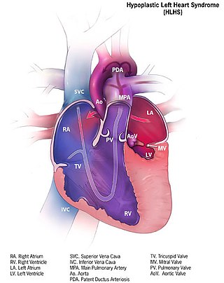
Hypoplastic left heart syndrome (HLHS) is a rare congenital heart defect in which the left side of the heart is severely underdeveloped and incapable of supporting the systemic circulation. It is estimated to account for 2-3% of all congenital heart disease. Early signs and symptoms include poor feeding, cyanosis, and diminished pulse in the extremities. The etiology is believed to be multifactorial resulting from a combination of genetic mutations and defects resulting in altered blood flow in the heart. Several structures can be affected including the left ventricle, aorta, aortic valve, or mitral valve all resulting in decreased systemic blood flow.

<span class="mw-page-title-main">Pulmonary atresia</span> Medical condition

Pulmonary atresia is a congenital malformation of the pulmonary valve in which the valve orifice fails to develop. The valve is completely closed thereby obstructing the outflow of blood from the heart to the lungs. The pulmonary valve is located on the right side of the heart between the right ventricle and pulmonary artery. In a normal functioning heart, the opening to the pulmonary valve has three flaps that open and close.

<span class="mw-page-title-main">Tricuspid atresia</span> Medical condition

Tricuspid atresia is a form of congenital heart disease whereby there is a complete absence of the tricuspid valve. Therefore, there is an absence of right atrioventricular connection. This leads to a hypoplastic (undersized) or absent right ventricle. This defect is contracted during prenatal development, when the heart does not finish developing. It causes the systemic circulation to be filled with relatively deoxygenated blood. The causes of tricuspid atresia are unknown.

Levo-Transposition of the great arteries is an acyanotic congenital heart defect in which the primary arteries are transposed, with the aorta anterior and to the left of the pulmonary artery; the morphological left and right ventricles with their corresponding atrioventricular valves are also transposed.

<span class="mw-page-title-main">Norwood procedure</span> Surgery performed on the heart

The Norwood procedure is the first of three surgeries intended to create a new functional systemic circuit in patients with hypoplastic left heart syndrome and other complex heart defects with single ventricle physiology. The first successful Norwood procedure involving the use of a cardiopulmonary bypass was reported by Dr. William Imon Norwood, Jr. and colleagues in 1981.

Crisscross heart is a type of congenital heart defect where the right atrium is closely associated with the left ventricle in space, and the left atrium is closely associated with the right ventricle.

<span class="mw-page-title-main">Double inlet left ventricle</span> Medical condition

A double inlet left ventricle (DILV) or "single ventricle", is a congenital heart defect appearing in 5 in 100,000 newborns, where both the left atrium and the right atrium feed into the left ventricle. The right ventricle is hypoplastic or does not exist.

The following outline is provided as an overview of and topical guide to cardiology, the branch of medicine dealing with disorders of the human heart. The field includes medical diagnosis and treatment of congenital heart defects, coronary artery disease, heart failure, valvular heart disease and electrophysiology. Physicians who specialize in cardiology are called cardiologists.

<span class="mw-page-title-main">Bidirectional Glenn procedure</span>

The bidirectional Glenn (BDG) shunt, or bidirectional cavopulmonary anastomosis, is a surgical technique used in pediatric cardiac surgery procedure used to temporarily improve blood oxygenation for patients with a congenital cardiac defect resulting in a single functional ventricle. Creation of a bidirectional shunt reduces the amount of blood volume that the heart needs to pump at the time of surgical repair with the Fontan procedure.

<span class="mw-page-title-main">Ventricular outflow tract obstruction</span> Medical condition

A ventricular outflow tract obstruction is a heart condition in which either the right or left ventricular outflow tract is blocked or obstructed. These obstructions represent a spectrum of disorders. Majority of these cases are congenital, but some are acquired throughout life.

Fetal aortic stenosis is a disorder that occurs when the fetus’ aortic valve does not fully open during development. The aortic valve is a one way valve that is located between the left ventricle and the aorta, keeping blood from leaking back into the ventricle. It has three leaflets that separate when the ventricle contracts to allow blood to move from the ventricle to the aorta. These leaflets come together when the ventricle relaxes.

The Damus–Kaye–Stansel (DKS) procedure is a cardiovascular surgical procedure used as part of the repair of some congenital heart defects. This procedure joins the pulmonary artery and the aorta in situations where the systemic circulation is obstructed. It is commonly used when a patient has the combination of a small left ventricle and a transposition of the great arteries (TGA); in this case, the procedure allows blood to flow from the left ventricle to the aorta.

The Yasui procedure is a pediatric heart operation used to bypass the left ventricular outflow tract (LVOT) that combines the aortic repair of the Norwood procedure and a shunt similar to that used in the Rastelli procedure in a single operation. It is used to repair defects that result in the physiology of hypoplastic left heart syndrome even though both ventricles are functioning normally. These defects are common in DiGeorge syndrome and include interrupted aortic arch and LVOT obstruction (IAA/LVOTO); aortic atresia-severe stenosis with ventricular septal defect (AA/VSD); and aortic atresia with interrupted aortic arch and aortopulmonary window. This procedure allows the surgeon to keep the left ventricle connected to the systemic circulation while using the pulmonary valve as its outflow valve, by connecting them through the ventricular septal defect. The Yasui procedure includes a modified Damus–Kaye–Stansel procedure to connect the aortic and pulmonary roots, allowing the coronary arteries to remain perfused. It was first described in 1987.

References

  1. 1 2 3 4 "Hypoplastic Right Heart Syndrome (HRHS): Diagnosis & Treatment | SSM Health".
  2. 1 2 3 Dimopoulos, Aggeliki (Jan 20, 2017). "Rare Copy Number Variants in a Population Based Investigation of Hypoplastic Right Heart Syndrome". Birth Defects Research. 109 (1): 8–15. doi:10.1002/bdra.23586. PMC   5388571 . PMID   28009100.
  3. 1 2 3 CHD-UK, Hypoplastic Right heart Syndrome (HRHS) Archived 2019-05-26 at the Wayback Machine , 2007-2015. 25 April 2015.
  4. "Hypoplastic right heart syndrome". Genetic and Rare Diseases Information Center (GARD). Retrieved 17 April 2018.
  5. Mayo Foundation for Medical Education and Research, Hypoplastic Left Heart Syndrome, 1998-2015. 12 April 2015.
  6. "Hypoplastic Right Heart Syndrome | Winchester Hospital". www.winchesterhospital.org. Retrieved 2023-04-26.
  7. Heart Contraction and Blood Flow Archived 2014-10-07 at the Wayback Machine . National Institutes of Health.
  8. Barrett, Heidi (January 11, 2016). "Hypoplasia of the Right Ventricle".
  9. Reviewed by David J. Goldberg, MD, About Hypoplastic Left Heart Syndrome (HLHS), 2013-2015. Web. 12 April 2015
  10. American Heart Association, Single Ventricle Defects, 2015. 12 April 2015.
  11. Paulick J, Tennstedt C, Schwabe M, Körner H, Bommer C, Chaoui R (May 2004). "Prenatal diagnosis of an isochromosome 5p in a fetus with increased nuchal translucency thickness and pulmonary atresia with hypoplastic right heart at 14 weeks". Prenat. Diagn. 24 (5): 371–4. doi:10.1002/pd.877. PMID   15164412. S2CID   11614779.
  12. Sharma J, Friedman D, Schiller M, Flynn P, Alonso ML (December 1997). "Aortic stenosis in hypoplastic right heart syndrome, associated with interstitial deletion of chromosome 2". Int. J. Cardiol. 62 (3): 199–202. doi: 10.1016/S0167-5273(97)00255-6 . PMID   9476678.